|
Genitourinary Tract Injury
The genitourinary tract, or simply the urinary tract, consists of the kidneys, ureters, bladder, and the urethra. The kidney is the most frequently injured.{{Cite book, title=Smith & Tanagho's General Urology, last=McAnich, first=Jack, last2=Lue, first2=Tom, publisher=Lange, year=2013, isbn=, location=, pages=Chapter 18 Injuries to the kidney commonly occur after automobile or sports-related accidents. A blunt force is involved in 80-85% of injuries. Major decelerations can result in vascular injuries near the kidney's hilum. Gunshots and knife wounds and fractured ribs can result in penetrating injuries to the kidney. Pelvic fractures can damage the urethra and bladder. Presentation Comorbidity In 90% of bladder injuries there is a concurrent pelvic fractures. Pelvic bone fragments penetrate and perforate the bladder. Perforations can be either extraperitoneal or intraperitoneal. Intraperitoneal perforations allow for urine to enter the peritoneal cavity. Symptoms typically d ... [...More Info...] [...Related Items...] OR: [Wikipedia] [Google] [Baidu] |
Urinary Tract
The urinary system, also known as the urinary tract or renal system, consists of the kidneys, ureters, bladder, and the urethra. The purpose of the urinary system is to eliminate waste from the body, regulate blood volume and blood pressure, control levels of electrolytes and metabolites, and regulate blood pH. The urinary tract is the body's drainage system for the eventual removal of urine. The kidneys have an extensive blood supply via the renal arteries which leave the kidneys via the renal vein. Each kidney consists of functional units called nephrons. Following filtration of blood and further processing, wastes (in the form of urine) exit the kidney via the ureters, tubes made of smooth muscle fibres that propel urine towards the urinary bladder, where it is stored and subsequently expelled from the body by urination ( voiding). The female and male urinary system are very similar, differing only in the length of the urethra. Urine is formed in the kidneys through a ... [...More Info...] [...Related Items...] OR: [Wikipedia] [Google] [Baidu] |
Hemodynamics
Hemodynamics or haemodynamics are the dynamics of blood flow. The circulatory system is controlled by homeostatic mechanisms of autoregulation, just as hydraulic circuits are controlled by control systems. The hemodynamic response continuously monitors and adjusts to conditions in the body and its environment. Hemodynamics explains the physical laws that govern the flow of blood in the blood vessels. Blood flow ensures the transportation of nutrients, hormones, metabolic waste products, oxygen, and carbon dioxide throughout the body to maintain cell-level metabolism, the regulation of the pH, osmotic pressure and temperature of the whole body, and the protection from microbial and mechanical harm. Blood is a non-Newtonian fluid, and is most efficiently studied using rheology rather than hydrodynamics. Because blood vessels are not rigid tubes, classic hydrodynamics and fluids mechanics based on the use of classical viscometers are not capable of explaining haemodynamics. The ... [...More Info...] [...Related Items...] OR: [Wikipedia] [Google] [Baidu] |
Acute Kidney Injury
Acute kidney injury (AKI), previously called acute renal failure (ARF), is a sudden decrease in kidney function that develops within 7 days, as shown by an increase in serum creatinine or a decrease in urine output, or both. Causes of AKI are classified as either prerenal (due to decreased blood flow to the kidney), intrinsic renal (due to damage to the kidney itself), or postrenal (due to blockage of urine flow). Prerenal causes of AKI include sepsis, dehydration, excessive blood loss, cardiogenic shock, heart failure, cirrhosis, and certain medications like ACE inhibitors or NSAIDs. Intrinsic renal causes of AKI include glomerulonephritis, lupus nephritis, acute tubular necrosis, certain antibiotics, and chemotherapeutic agents. Postrenal causes of AKI include kidney stones, bladder cancer, neurogenic bladder, enlargement of the prostate, narrowing of the urethra, and certain medications like anticholinergics. The diagnosis of AKI is made based on a person's signs a ... [...More Info...] [...Related Items...] OR: [Wikipedia] [Google] [Baidu] |
Bulb Of Penis
Just before each crus of the penis A penis (plural ''penises'' or ''penes'' () is the primary sexual organ that male animals use to inseminate females (or hermaphrodites) during copulation. Such organs occur in many animals, both vertebrate and invertebrate, but males d ... meets its fellow, it presents a slight enlargement, which Georg Ludwig Kobelt named the bulb of the corpus cavernosum penis. The bulb of penis is also known as the urethral bulb. The bulb is homologous to the vestibular bulbs in females. Additional images File:Gray1142.png, Male urethra. File:Gray1158.png, Diagram of the arteries of the penis. References External links * * Mammal male reproductive system Human penis anatomy {{genitourinary-stub ... [...More Info...] [...Related Items...] OR: [Wikipedia] [Google] [Baidu] |
Voiding Cystourethrography
In urology, voiding cystourethrography (VCUG) is a frequently performed technique for visualizing a person's urethra and urinary bladder while the person urinates (voids). It is used in the diagnosis of vesicoureteral reflux (kidney reflux), among other disorders. The technique consists of catheterizing the person in order to fill the bladder with a radiocontrast agent, typically diatrizoic acid. Under fluoroscopy (real time x-rays) the radiologist watches the contrast enter the bladder and looks at the anatomy of the patient. If the contrast moves into the ureters and back into the kidneys, the radiologist makes the diagnosis of vesicoureteral reflux, and gives the degree of severity a score. The exam ends when the person voids while the radiologist is watching under fluoroscopy. Consumption of fluid promotes excretion of contrast media after the procedure. It is important to watch the contrast during voiding, because this is when the bladder has the most pressure, and it is mos ... [...More Info...] [...Related Items...] OR: [Wikipedia] [Google] [Baidu] |
Prostate
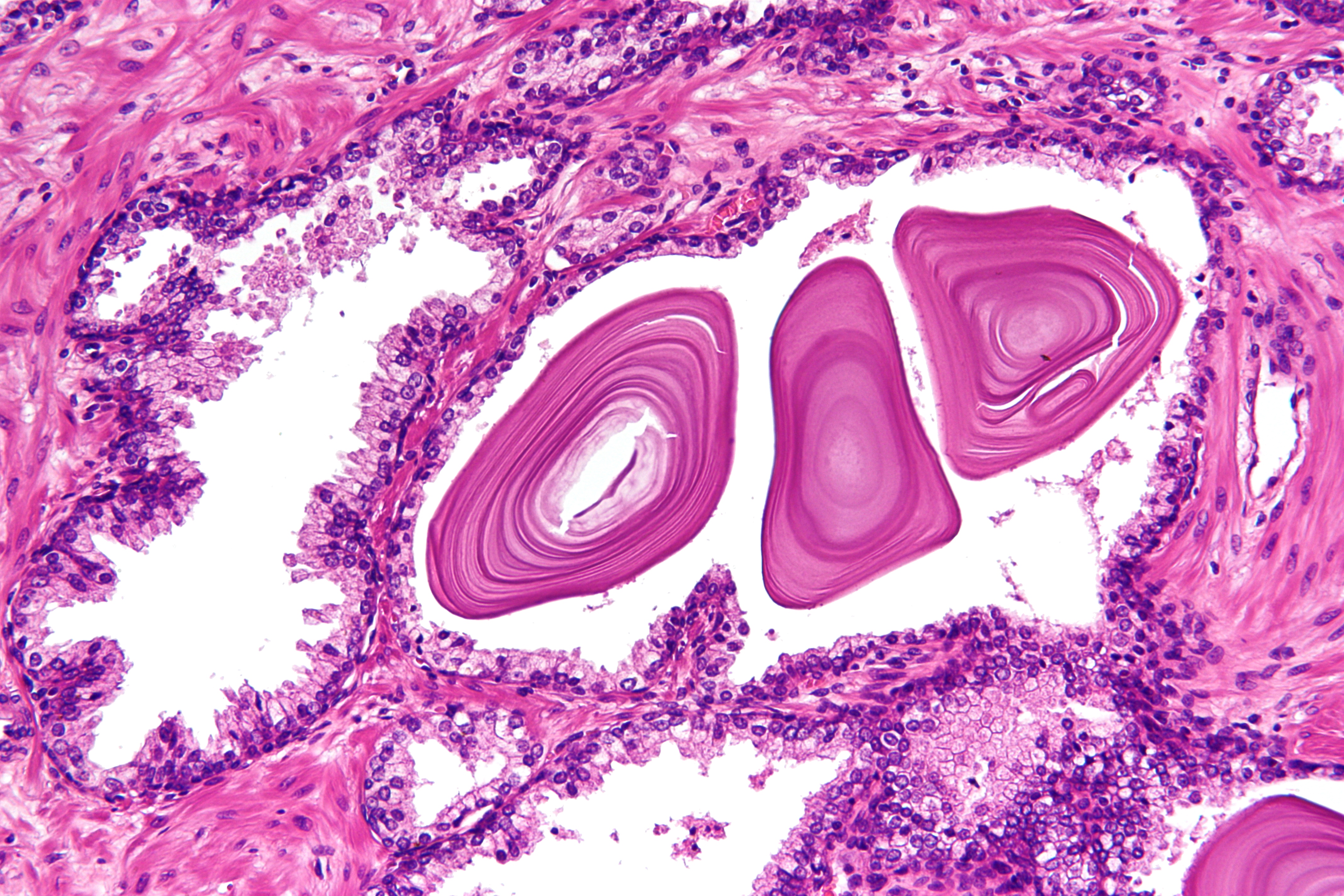
The prostate is both an accessory gland of the male reproductive system and a muscle-driven mechanical switch between urination and ejaculation. It is found only in some mammals. It differs between species anatomically, chemically, and physiologically. Anatomically, the prostate is found below the bladder, with the urethra passing through it. It is described in gross anatomy as consisting of lobes and in microanatomy by zone. It is surrounded by an elastic, fibromuscular capsule and contains glandular tissue as well as connective tissue. The prostate glands produce and contain fluid that forms part of semen, the substance emitted during ejaculation as part of the male sexual response. This prostatic fluid is slightly alkaline, milky or white in appearance. The alkalinity of semen helps neutralize the acidity of the vaginal tract, prolonging the lifespan of sperm. The prostatic fluid is expelled in the first part of ejaculate, together with most of the sperm, because of th ... [...More Info...] [...Related Items...] OR: [Wikipedia] [Google] [Baidu] |
Membranous Urethra
The membranous urethra or intermediate part of male urethra is the shortest, least dilatable, and, with the exception of the urinary meatus, the narrowest part of the urethra. It extends downward and forward, with a slight anterior concavity, between the apex of the prostate and the bulb of the urethra, perforating the urogenital diaphragm about 2.5 cm below and behind the pubic symphysis. The hinder part of the urethral bulb lies in apposition with the inferior fascia of the urogenital diaphragm, but its upper portion diverges somewhat from this fascia: the anterior wall of the membranous urethra is thus prolonged for a short distance in front of the urogenital diaphragm; it measures about 2 cm in length, while the posterior wall which is between the two fasciæ of the diaphragm is only 1.25 cm long. The anatomical variation in membranous urethral length measurements in men have been reported to range from 0.5 cm to 3.4 cm. The membranous portion of ... [...More Info...] [...Related Items...] OR: [Wikipedia] [Google] [Baidu] |
Parenchyma
Parenchyma () is the bulk of functional substance in an animal organ or structure such as a tumour. In zoology it is the name for the tissue that fills the interior of flatworms. Etymology The term ''parenchyma'' is New Latin from the word παρέγχυμα ''parenchyma'' meaning 'visceral flesh', and from παρεγχεῖν ''parenchyma'' meaning 'to pour in' from παρα- ''para-'' 'beside' + ἐν ''en-'' 'in' + χεῖν ''chyma'' 'to pour'. Originally, Erasistratus and other anatomists used it to refer to certain human tissues. Later, it was also applied to plant tissues by Nehemiah Grew. Structure The parenchyma is the ''functional'' parts of an organ, or of a structure such as a tumour in the body. This is in contrast to the stroma, which refers to the ''structural'' tissue of organs or of structures, namely, the connective tissues. Brain The brain parenchyma refers to the functional tissue in the brain that is made up of the two types of brain cell, neurons ... [...More Info...] [...Related Items...] OR: [Wikipedia] [Google] [Baidu] |
Angiography
Angiography or arteriography is a medical imaging technique used to visualize the inside, or lumen, of blood vessels and organs of the body, with particular interest in the arteries, veins, and the heart chambers. Modern angiography is performed by injecting a radio-opaque contrast agent into the blood vessel and imaging using X-ray based techniques such as fluoroscopy. The word itself comes from the Greek words ἀγγεῖον ''angeion'' 'vessel' and γράφειν ''graphein'' 'to write, record'. The film or image of the blood vessels is called an ''angiograph'', or more commonly an ''angiogram''. Though the word can describe both an arteriogram and a venogram, in everyday usage the terms angiogram and arteriogram are often used synonymously, whereas the term venogram is used more precisely. The term angiography has been applied to radionuclide angiography and newer vascular imaging techniques such as CO2 angiography, CT angiography and MR angiography. The term ''iso ... [...More Info...] [...Related Items...] OR: [Wikipedia] [Google] [Baidu] |
Cystography
In radiology and urology, a cystography (also known as cystogram) is a procedure used to visualise the urinary bladder. Using a urinary catheter, radiocontrast is instilled in the bladder, and X-ray imaging is performed. Cystography can be used to evaluate bladder cancer, vesicoureteral reflux, bladder polyps, and hydronephrosis. It requires less radiation than pelvic CT, although it is less sensitive and specific than MRI or CT. In adult cases, the patient is typically instructed to void three times, after which a post voiding image is obtained to see how much urine is left within the bladder (residual urine), which is useful to evaluate bladder contraction dysfunction. A final radiograph of the kidneys after the procedure is finished is performed to evaluate for occult vesicoureteral reflux that was not seen during the procedure itself. CT cystography CT cystography is performed by filling up the urinary bladder using diluted iodinated contrast to visualise any bladder inj ... [...More Info...] [...Related Items...] OR: [Wikipedia] [Google] [Baidu] |
Bladder Neck
The urinary bladder, or simply bladder, is a hollow organ in humans and other vertebrates that stores urine from the kidneys before disposal by urination. In humans the bladder is a distensible organ that sits on the pelvic floor. Urine enters the bladder via the ureters and exits via the urethra. The typical adult human bladder will hold between 300 and (10.14 and ) before the urge to empty occurs, but can hold considerably more. The Latin phrase for "urinary bladder" is ''vesica urinaria'', and the term ''vesical'' or prefix ''vesico -'' appear in connection with associated structures such as vesical veins. The modern Latin word for "bladder" – ''cystis'' – appears in associated terms such as cystitis (inflammation of the bladder). Structure In humans, the bladder is a hollow muscular organ situated at the base of the pelvis. In gross anatomy, the bladder can be divided into a broad , a body, an apex, and a neck. The apex (also called the vertex) is directed for ... [...More Info...] [...Related Items...] OR: [Wikipedia] [Google] [Baidu] |
Urinary Meatus
The urinary meatus, (, ) also known as the external urethral orifice, is the opening of the urethra. It is the point where urine exits the urethra in both sexes and where semen exits the urethra in males. The meatus has varying degrees of sensitivity to touch. The meatus is located on the glans of the penis or in the vulval vestibule. In human males The male external urethral orifice is the external opening or urinary meatus, normally located at the tip of the glans penis, at its junction with the frenular delta. It presents as a vertical slit, possibly bounded on either side by two small labia-like projections, and continues longitudinally along the front aspect of the glans, which facilitates the flow of urine micturition. In some cases, the opening may be more rounded. This can occur naturally or may also occur as a side effect of excessive skin removal during circumcision. The meatus is a sensitive part of the male reproductive system. In human females The female exte ... [...More Info...] [...Related Items...] OR: [Wikipedia] [Google] [Baidu] |

