|
Genioglossus
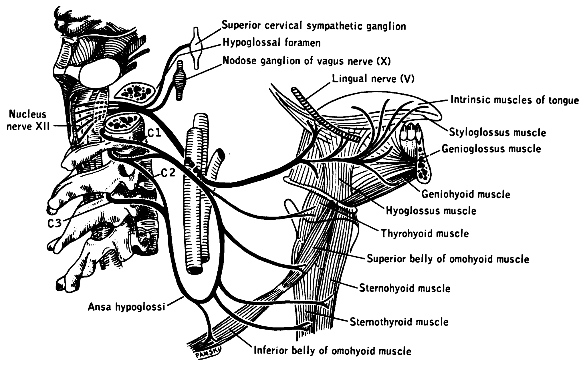
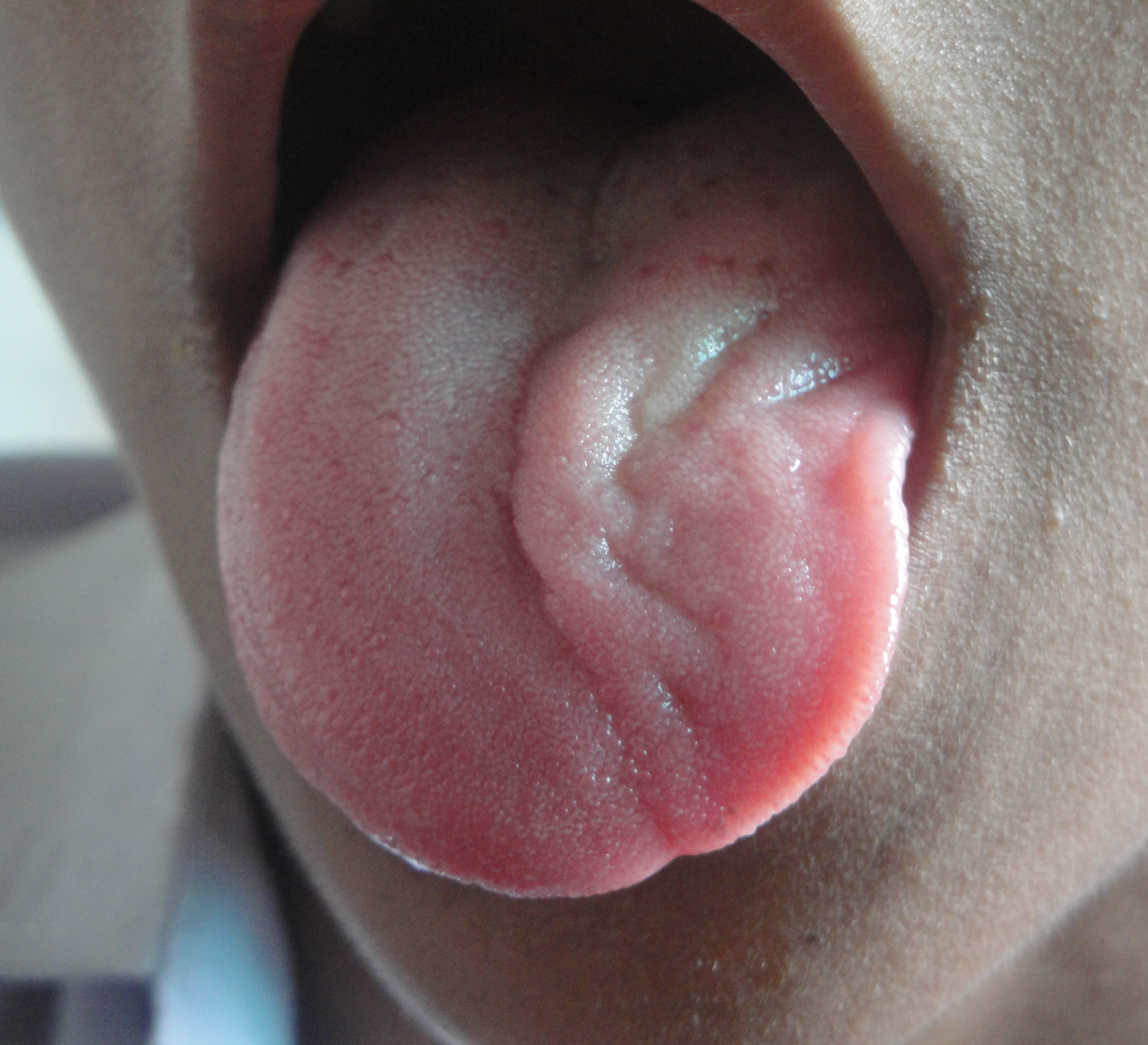
The genioglossus is one of the paired extrinsic muscles of the tongue. The genioglossus is the major muscle responsible for protruding (or sticking out) the tongue. Structure Genioglossus is the fan-shaped extrinsic tongue muscle that forms the majority of the body of the tongue. It arises from the mental spine of the mandible and its insertions are the hyoid bone and the bottom of the tongue. The genioglossus is innervated by the hypoglossal nerve, as are all muscles of the tongue except for the palatoglossus. Blood is supplied to the sublingual branch of the lingual artery, a branch of the external carotid artery. The canine genioglossus muscle has been divided into horizontal and oblique compartments. Function The left and right genioglossus muscles protrude the tongue and deviate it towards the opposite side. When acting together, the muscles depress the center of the tongue at its back. Clinical significance Contraction of the genioglossus stabilizes and enlarges the port ... [...More Info...] [...Related Items...] OR: [Wikipedia] [Google] [Baidu] |
Hypoglossal Nerve
The hypoglossal nerve, also known as the twelfth cranial nerve, cranial nerve XII, or simply CN XII, is a cranial nerve that innervates all the extrinsic and intrinsic muscles of the tongue except for the palatoglossus, which is innervated by the vagus nerve. CN XII is a nerve with a solely motor function. The nerve arises from the hypoglossal nucleus in the medulla as a number of small rootlets, passes through the hypoglossal canal and down through the neck, and eventually passes up again over the tongue muscles it supplies into the tongue. The nerve is involved in controlling tongue movements required for speech and swallowing, including sticking out the tongue and moving it from side to side. Damage to the nerve or the neural pathways which control it can affect the ability of the tongue to move and its appearance, with the most common sources of damage being injury from trauma or surgery, and motor neuron disease. The first recorded description of the nerve is by Herop ... [...More Info...] [...Related Items...] OR: [Wikipedia] [Google] [Baidu] |
Mental Spine
A mental spine is a small projection of bone on the posterior aspect of the mandible in the midline. There are usually four mental spines: two superior and two inferior. Collectively they are also known as the ''genial tubercle'',"Genial tubercle." The American Heritage Stedman's Medical Dictionary. Houghton Mifflin Company, 2002. Accessed: 22 Oct. 2007. ''genial apophysis'' and the Latin name ''spinae mentalis''. The inferior mental spines are the points of origin of the geniohyoid muscle,"Genial tubercle." Mosby's Dental Dictionary. Elsevier, Inc., 2004. Accessed: 22 Oct. 2007. one of the suprahyoid muscles, and the superior mental spines are the origin of the genioglossus muscle, one of the muscles of the tongue. Mental spines are important landmarks in clinical practice. Structure Mental spines are found on the posterior aspect of the mandible (lower jaw bone) in the midline. They usually surround spinous mental foramina in the midline. Variation Mental spines may be foun ... [...More Info...] [...Related Items...] OR: [Wikipedia] [Google] [Baidu] |
Tongue
The tongue is a muscular organ in the mouth of a typical tetrapod. It manipulates food for mastication and swallowing as part of the digestive process, and is the primary organ of taste. The tongue's upper surface (dorsum) is covered by taste buds housed in numerous lingual papillae. It is sensitive and kept moist by saliva and is richly supplied with nerves and blood vessels. The tongue also serves as a natural means of cleaning the teeth. A major function of the tongue is the enabling of speech in humans and vocalization in other animals. The human tongue is divided into two parts, an oral part at the front and a pharyngeal part at the back. The left and right sides are also separated along most of its length by a vertical section of fibrous tissue (the lingual septum) that results in a groove, the median sulcus, on the tongue's surface. There are two groups of muscles of the tongue. The four intrinsic muscles alter the shape of the tongue and are not attached to bone. ... [...More Info...] [...Related Items...] OR: [Wikipedia] [Google] [Baidu] |
Hyoid
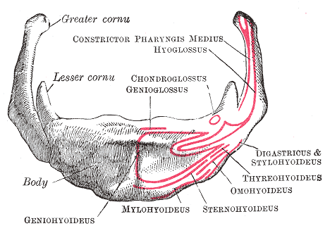
The hyoid bone (lingual bone or tongue-bone) () is a horseshoe-shaped bone situated in the anterior midline of the neck between the chin and the thyroid cartilage. At rest, it lies between the base of the mandible and the third cervical vertebra. Unlike other bones, the hyoid is only distantly articulated to other bones by muscles or ligaments. It is the only bone in the human body that is not connected to any other bones nearby. The hyoid is anchored by muscles from the anterior, posterior and inferior directions, and aids in tongue movement and swallowing. The hyoid bone provides attachment to the muscles of the floor of the mouth and the tongue above, the larynx below, and the epiglottis and pharynx behind. Its name is derived . Structure The hyoid bone is classed as an irregular bone and consists of a central part called the body, and two pairs of horns, the greater and lesser horns. Body The body of the hyoid bone is the central part of the hyoid bone. *At the fron ... [...More Info...] [...Related Items...] OR: [Wikipedia] [Google] [Baidu] |
Lingual Artery
The lingual artery arises from the external carotid artery between the superior thyroid artery and facial artery. It can be located easily in the tongue. Structure The lingual artery first branches off from the external carotid artery. It runs obliquely upward and medially to the greater horns of the hyoid bone. It then curves downward and forward, forming a loop which is crossed by the hypoglossal nerve. It then passes beneath the digastric muscle and stylohyoid muscle running horizontally forward, beneath the hyoglossus. This takes it through the sublingual space. Finally, ascending almost perpendicularly to the tongue, it turns forward on its lower surface as far as the tip of the tongue, now called the deep lingual artery ( profunda linguae). Branches The lingual artery gives 4 main branches: the deep lingual artery, the sublingual artery, the suprahyoid branch, and the dorsal lingual branch. Deep lingual artery The deep lingual artery (or ranine artery) is the term ... [...More Info...] [...Related Items...] OR: [Wikipedia] [Google] [Baidu] |
Hyoid Bone
The hyoid bone (lingual bone or tongue-bone) () is a horseshoe-shaped bone situated in the anterior midline of the neck between the chin and the thyroid cartilage. At rest, it lies between the base of the mandible and the third cervical vertebra. Unlike other bones, the hyoid is only distantly articulated to other bones by muscles or ligaments. It is the only bone in the human body that is not connected to any other bones nearby. The hyoid is anchored by muscles from the anterior, posterior and inferior directions, and aids in tongue movement and swallowing. The hyoid bone provides attachment to the muscles of the floor of the mouth and the tongue above, the larynx below, and the epiglottis and pharynx behind. Its name is derived . Structure The hyoid bone is classed as an irregular bone and consists of a central part called the body, and two pairs of horns, the greater and lesser horns. Body The body of the hyoid bone is the central part of the hyoid bone. *At the ... [...More Info...] [...Related Items...] OR: [Wikipedia] [Google] [Baidu] |
Lingual Artery
The lingual artery arises from the external carotid artery between the superior thyroid artery and facial artery. It can be located easily in the tongue. Structure The lingual artery first branches off from the external carotid artery. It runs obliquely upward and medially to the greater horns of the hyoid bone. It then curves downward and forward, forming a loop which is crossed by the hypoglossal nerve. It then passes beneath the digastric muscle and stylohyoid muscle running horizontally forward, beneath the hyoglossus. This takes it through the sublingual space. Finally, ascending almost perpendicularly to the tongue, it turns forward on its lower surface as far as the tip of the tongue, now called the deep lingual artery ( profunda linguae). Branches The lingual artery gives 4 main branches: the deep lingual artery, the sublingual artery, the suprahyoid branch, and the dorsal lingual branch. Deep lingual artery The deep lingual artery (or ranine artery) is the term ... [...More Info...] [...Related Items...] OR: [Wikipedia] [Google] [Baidu] |
Symphysis Menti
In human anatomy, the facial skeleton of the skull the external surface of the mandible is marked in the median line by a faint ridge, indicating the mandibular symphysis (Latin: ''symphysis menti'') or line of junction where the two lateral halves of the mandible typically fuse at an early period of life (1-2 years). It is not a true symphysis as there is no cartilage between the two sides of the mandible. This ridge divides below and encloses a triangular eminence, the mental protuberance, the base of which is depressed in the center but raised on either side to form the mental tubercle. The lowest (most inferior) end of the mandibular symphysis — the point of the chin — is called the "menton". It serves as the origin for the geniohyoid and the genioglossus muscles. Other animals Solitary mammalian carnivores that rely on a powerful canine bite to subdue their prey have a strong mandibular symphysis, while pack hunters delivering shallow bites have a weaker one. When filter ... [...More Info...] [...Related Items...] OR: [Wikipedia] [Google] [Baidu] |
Human Mandible
In anatomy, the mandible, lower jaw or jawbone is the largest, strongest and lowest bone in the human facial skeleton. It forms the lower jaw and holds the lower teeth in place. The mandible sits beneath the maxilla. It is the only movable bone of the skull (discounting the ossicles of the middle ear). It is connected to the temporal bones by the temporomandibular joints. The bone is formed in the fetus from a fusion of the left and right mandibular prominences, and the point where these sides join, the mandibular symphysis, is still visible as a faint ridge in the midline. Like other symphyses in the body, this is a midline articulation where the bones are joined by fibrocartilage, but this articulation fuses together in early childhood.Illustrated Anatomy of the Head and Neck, Fehrenbach and Herring, Elsevier, 2012, p. 59 The word "mandible" derives from the Latin word ''mandibula'', "jawbone" (literally "one used for chewing"), from '' mandere'' "to chew" and ''-bula' ... [...More Info...] [...Related Items...] OR: [Wikipedia] [Google] [Baidu] |
Geniohyoideus
The geniohyoid muscle is a narrow muscle situated superior to the medial border of the mylohyoid muscle. It is named for its passage from the chin ("genio-" is a standard prefix for "chin") to the hyoid bone. Structure It arises from the inferior mental spine, on the back of the mandibular symphysis, and runs backward and slightly downward, to be inserted into the anterior surface of the body of the hyoid bone. It lies in contact with its fellow of the opposite side. It thus belongs to the suprahyoid muscles. The muscle is supplied by branches of the lingual artery. Innervation The geniohyoid muscle is innervated by fibres from the first cervical spinal nerve travelling alongside the hypoglossal nerve. Although the first three cervical nerves give rise to the ansa cervicalis, the geniohyoid muscle is said to be innervated by the first cervical nerve, as some of its efferent fibers do not contribute to ansa cervicalis. Variations It may be blended with the one on opposite side ... [...More Info...] [...Related Items...] OR: [Wikipedia] [Google] [Baidu] |
Anaesthesia
Anesthesia is a state of controlled, temporary loss of sensation or awareness that is induced for medical or veterinary purposes. It may include some or all of analgesia (relief from or prevention of pain), paralysis (muscle relaxation), amnesia (loss of memory), and unconsciousness. An individual under the effects of anesthetic drugs is referred to as being anesthetized. Anesthesia enables the painless performance of procedures that would otherwise cause severe or intolerable pain in a non-anesthetized individual, or would otherwise be technically unfeasible. Three broad categories of anesthesia exist: * General anesthesia suppresses central nervous system activity and results in unconsciousness and total lack of sensation, using either injected or inhaled drugs. * Sedation suppresses the central nervous system to a lesser degree, inhibiting both anxiety and creation of long-term memories without resulting in unconsciousness. * Regional and local anesthesia, which blocks ... [...More Info...] [...Related Items...] OR: [Wikipedia] [Google] [Baidu] |


