|
GORASP2
Golgi reassembly-stacking protein of 55 k Da (GRASP55) also known as golgi reassembly-stacking protein 2 (GORASP2) is a protein that in humans is encoded by the ''GORASP2'' gene. It was identified by its homology with GRASP65 and the protein's amino acid sequence was determined by analysis of a molecular clone of its complementary DNA. The first ( N-terminus) 212 amino acid residues of GRASP55 are highly homologous to those of GRASP65, but the remainder of the 454 amino acid residues are highly diverged from GRASP65. The conserved region is known as the GRASP domain, and it is conserved among GRASPs of a wide variety of eukaryotes, but not plants. The C-terminus portion of the molecule is called the ''SPR domain'' ( serine, proline-rich). GRASP55 is more closely related to homologues in other species, suggesting that GRASP55 is ancestral to GRASP65. GRASP55 is found associated with the medial and trans cisternae of the Golgi apparatus. Function GRASP55 is involved in es ... [...More Info...] [...Related Items...] OR: [Wikipedia] [Google] [Baidu] |
BLZF1
Golgin-45 is a protein that in humans is encoded by the ''BLZF1'' gene. Interactions BLZF1 has been shown to interact with GORASP2 Golgi reassembly-stacking protein of 55 k Da (GRASP55) also known as golgi reassembly-stacking protein 2 (GORASP2) is a protein that in humans is encoded by the ''GORASP2'' gene. It was identified by its homology with GRASP65 and the protein's .... References Further reading * * * * * * * * External links * * Transcription factors {{gene-1-stub ... [...More Info...] [...Related Items...] OR: [Wikipedia] [Google] [Baidu] |
TGF Alpha
Transforming growth factor alpha (TGF-α) is a protein that in humans is encoded by the TGFA gene. As a member of the epidermal growth factor (EGF) family, TGF-α is a mitogenic polypeptide. The protein becomes activated when binding to receptors capable of protein kinase activity for cellular signaling. TGF-α is a transforming growth factor that is a ligand for the epidermal growth factor receptor, which activates a signaling pathway for cell proliferation, differentiation and development. This protein may act as either a transmembrane-bound ligand or a soluble ligand. This gene has been associated with many types of cancers, and it may also be involved in some cases of cleft lip/palate. Synthesis TGF-α is synthesized internally as part of a 160 (human) or 159 (rat) amino acid transmembrane precursor.Ferrer, I.; Alcantara, S.; Ballabriga, J.; Olive, M.; Blanco, R.; Rivera, R.; Carmona, M.; Berruezo, M.; Pitarch, S.; Planas, A. Transforming growth factor- α (TGF-α) and epide ... [...More Info...] [...Related Items...] OR: [Wikipedia] [Google] [Baidu] |
Kilo-
Kilo is a decimal unit prefix in the metric system denoting multiplication by one thousand (103). It is used in the International System of Units, where it has the symbol k, in lowercase. The prefix ''kilo'' is derived from the Greek word (), meaning "thousand". In 19th century English it was sometimes spelled chilio, in line with a puristic opinion by Thomas Young. As an opponent of suggestions to introduce the metric system in Britain, he qualified the nomenclature adopted in France as barbarous. Examples * one kilogram (kg) is 1000 grams * one kilometre (km) is 1000 metres * one kilojoule (kJ) is 1000 joules * one kilolitre (kL) is 1000 litres * one kilobaud (kBd) is 1000 baud * one kilohertz (kHz) is 1000 hertz * one kilobit (kb) is 1000 bits * one kilobyte (kB) is 1000 bytes * one kiloohm is (kΩ) is 1000 ohms * one kilosecond (ks) is 1000 seconds *one kilotonne (kt) is 1000 tonnes By extension, currencies are also sometimes preceded by the prefix kilo-: * one kil ... [...More Info...] [...Related Items...] OR: [Wikipedia] [Google] [Baidu] |
Phosphorylation
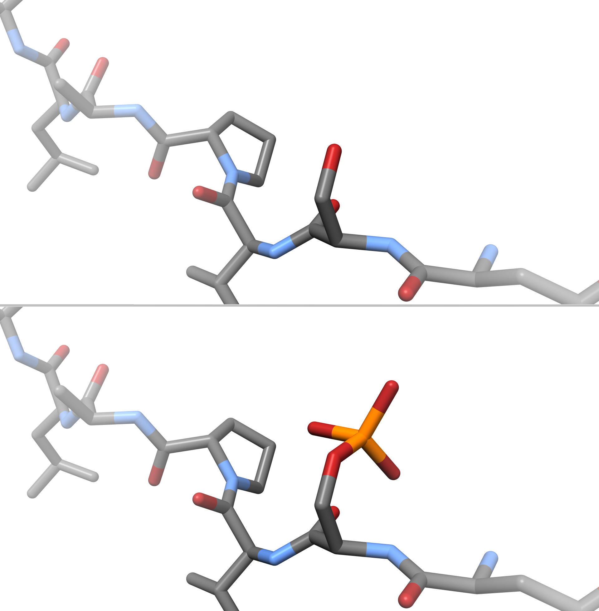
In chemistry, phosphorylation is the attachment of a phosphate group to a molecule or an ion. This process and its inverse, dephosphorylation, are common in biology and could be driven by natural selection. Text was copied from this source, which is available under a Creative Commons Attribution 4.0 International License. Protein phosphorylation often activates (or deactivates) many enzymes. Glucose Phosphorylation of sugars is often the first stage in their catabolism. Phosphorylation allows cells to accumulate sugars because the phosphate group prevents the molecules from diffusing back across their transporter. Phosphorylation of glucose is a key reaction in sugar metabolism. The chemical equation for the conversion of D-glucose to D-glucose-6-phosphate in the first step of glycolysis is given by :D-glucose + ATP → D-glucose-6-phosphate + ADP : ΔG° = −16.7 kJ/mol (° indicates measurement at standard condition) Hepatic cells are freely permeable to glucose, and ... [...More Info...] [...Related Items...] OR: [Wikipedia] [Google] [Baidu] |
Mitosis
In cell biology, mitosis () is a part of the cell cycle in which replicated chromosomes are separated into two new nuclei. Cell division by mitosis gives rise to genetically identical cells in which the total number of chromosomes is maintained. Therefore, mitosis is also known as equational division. In general, mitosis is preceded by S phase of interphase (during which DNA replication occurs) and is often followed by telophase and cytokinesis; which divides the cytoplasm, organelles and cell membrane of one cell into two new cells containing roughly equal shares of these cellular components. The different stages of mitosis altogether define the mitotic (M) phase of an animal cell cycle—the division of the mother cell into two daughter cells genetically identical to each other. The process of mitosis is divided into stages corresponding to the completion of one set of activities and the start of the next. These stages are preprophase (specific to plant cells), prophase ... [...More Info...] [...Related Items...] OR: [Wikipedia] [Google] [Baidu] |
Lipidation
Lipid-anchored proteins (also known as lipid-linked proteins) are proteins located on the surface of the cell membrane that are covalently attached to lipids embedded within the cell membrane. These proteins insert and assume a place in the bilayer structure of the membrane alongside the similar fatty acid tails. The lipid-anchored protein can be located on either side of the cell membrane. Thus, the lipid serves to anchor the protein to the cell membrane. They are a type of proteolipids. The lipid groups play a role in protein interaction and can contribute to the function of the protein to which it is attached. Furthermore, the lipid serves as a mediator of membrane associations or as a determinant for specific protein-protein interactions. For example, lipid groups can play an important role in increasing molecular hydrophobicity. This allows for the interaction of proteins with cellular membranes and protein domains. In a dynamic role, lipidation can sequester a protein away f ... [...More Info...] [...Related Items...] OR: [Wikipedia] [Google] [Baidu] |
Rab (G-protein)
The Rab family of proteins is a member of the Ras superfamily of small G proteins. Approximately 70 types of Rabs have now been identified in humans. Rab proteins generally possess a GTPase fold, which consists of a six-stranded beta sheet which is flanked by five alpha helices. Rab GTPases regulate many steps of membrane trafficking, including vesicle formation, vesicle movement along actin and tubulin networks, and membrane fusion. These processes make up the route through which cell surface proteins are trafficked from the Golgi to the plasma membrane and are recycled. Surface protein recycling returns proteins to the surface whose function involves carrying another protein or substance inside the cell, such as the transferrin receptor, or serves as a means of regulating the number of a certain type of protein molecules on the surface. Function Rab proteins are peripheral membrane proteins, anchored to a membrane via a lipid group covalently linked to an amino acid. Specifi ... [...More Info...] [...Related Items...] OR: [Wikipedia] [Google] [Baidu] |
Lipid Bilayer
The lipid bilayer (or phospholipid bilayer) is a thin polar membrane made of two layers of lipid molecules. These membranes are flat sheets that form a continuous barrier around all cells. The cell membranes of almost all organisms and many viruses are made of a lipid bilayer, as are the nuclear membrane surrounding the cell nucleus, and membranes of the membrane-bound organelles in the cell. The lipid bilayer is the barrier that keeps ions, proteins and other molecules where they are needed and prevents them from diffusing into areas where they should not be. Lipid bilayers are ideally suited to this role, even though they are only a few nanometers in width, because they are impermeable to most water-soluble (hydrophilic) molecules. Bilayers are particularly impermeable to ions, which allows cells to regulate salt concentrations and pH by transporting ions across their membranes using proteins called ion pumps. Biological bilayers are usually composed of amphiphilic phosphol ... [...More Info...] [...Related Items...] OR: [Wikipedia] [Google] [Baidu] |
Myristoylation
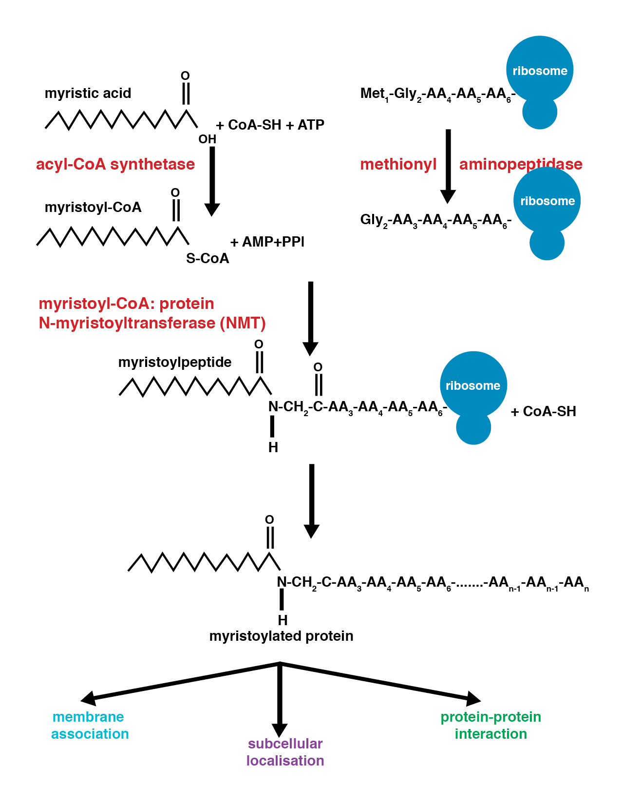
Myristoylation is a lipidation modification where a myristoyl group, derived from myristic acid, is covalently attached by an amide bond to the alpha-amino group of an N-terminus, N-terminal glycine residue. Myristic acid is a 14-carbon saturated fatty acid (14:0) with the systematic name of ''n''-Tetradecanoic acid. This modification can be added either co-translationally or Posttranslational modification, post-translationally. N-myristoyltransferase 1, N-myristoyltransferase (NMT) catalyzes the myristic acid addition reaction in the cytoplasm of cells. This lipidation event is the most found type of fatty acylation and is common among many organisms including animals, plants, fungi, protozoans and viruses. Myristoylation allows for weak protein–protein and protein–lipid interactions and plays an essential role in membrane targeting, protein–protein interactions and functions widely in a variety of signal transduction pathways. Discovery In 1982, Koiti Titani's lab id ... [...More Info...] [...Related Items...] OR: [Wikipedia] [Google] [Baidu] |
Protein–protein Interaction
Protein–protein interactions (PPIs) are physical contacts of high specificity established between two or more protein molecules as a result of biochemical events steered by interactions that include electrostatic forces, hydrogen bonding and the hydrophobic effect. Many are physical contacts with molecular associations between chains that occur in a cell or in a living organism in a specific biomolecular context. Proteins rarely act alone as their functions tend to be regulated. Many molecular processes within a cell are carried out by molecular machines that are built from numerous protein components organized by their PPIs. These physiological interactions make up the so-called interactomics of the organism, while aberrant PPIs are the basis of multiple aggregation-related diseases, such as Creutzfeldt–Jakob and Alzheimer's diseases. PPIs have been studied with many methods and from different perspectives: biochemistry, quantum chemistry, molecular dynamics, signal trans ... [...More Info...] [...Related Items...] OR: [Wikipedia] [Google] [Baidu] |


