|
Gluteus Medius Muscle
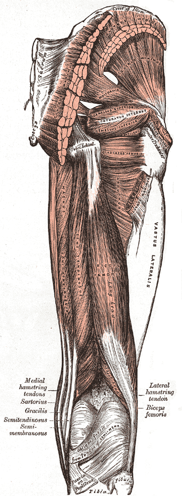
The gluteus medius, one of the three gluteal muscles, is a broad, thick, radiating muscle. It is situated on the outer surface of the pelvis. Its posterior third is covered by the gluteus maximus, its anterior two-thirds by the gluteal aponeurosis, which separates it from the superficial fascia and integument. Structure The gluteus medius muscle starts, or "originates", on the outer surface of the ilium between the iliac crest and the posterior gluteal line above, and the anterior gluteal line below; the gluteus medius also originates from the gluteal aponeurosis that covers its outer surface. The fibers of the muscle converge into a strong flattened tendon that inserts on the lateral surface of the greater trochanter. More specifically, the muscle's tendon inserts into an oblique ridge that runs downward and forward on the lateral surface of the greater trochanter. Relations A bursa separates the tendon of the muscle from the surface of the trochanter over which it glid ... [...More Info...] [...Related Items...] OR: [Wikipedia] [Google] [Baidu] |
Ilium (bone)
The ilium () (plural ilia) is the uppermost and largest part of the hip bone, and appears in most vertebrates including mammals and birds, but not bony fish. All reptiles have an ilium except snakes, although some snake species have a tiny bone which is considered to be an ilium. The ilium of the human is divisible into two parts, the body and the wing; the separation is indicated on the top surface by a curved line, the arcuate line, and on the external surface by the margin of the acetabulum. The name comes from the Latin (''ile'', ''ilis''), meaning "groin" or "flank". Structure The ilium consists of the body and wing. Together with the ischium and pubis, to which the ilium is connected, these form the pelvic bone, with only a faint line indicating the place of union. The body ( la, corpus) forms less than two-fifths of the acetabulum; and also forms part of the acetabular fossa. The internal surface of the body is part of the wall of the lesser pelvis and gives or ... [...More Info...] [...Related Items...] OR: [Wikipedia] [Google] [Baidu] |
Posterior Gluteal Line
The gluteal lines are three curved lines outlined from three bony ridges on the exterior surface of the ilium in the gluteal region. They are the anterior gluteal line; the inferior gluteal line, and the posterior gluteal line. The gluteus minimus, medius, and maximus are muscles that arise from the gluteal lines. Location Anterior gluteal line The anterior gluteal line is the middle curved gluteal line on the hip bone. It is the longest of the three gluteal lines, begins at the iliac crest, about 4 cm. behind its anterior extremity, and, taking a curved direction downward and backward, ends at the upper part of the greater sciatic notch. The space between the anterior and posterior gluteal lines and the crest is concave, and gives origin to the gluteus medius muscle. Near the middle of this line a nutrient foramen is often seen. Posterior gluteal line Posterior gluteal line, also known as the superior curved line, the shortest of the three gluteal lines, begins at the iliac ... [...More Info...] [...Related Items...] OR: [Wikipedia] [Google] [Baidu] |
Muscles Of The Gluteus
Skeletal muscles (commonly referred to as muscles) are organs of the vertebrate muscular system and typically are attached by tendons to bones of a skeleton. The muscle cells of skeletal muscles are much longer than in the other types of muscle tissue, and are often known as muscle fibers. The muscle tissue of a skeletal muscle is striated – having a striped appearance due to the arrangement of the sarcomeres. Skeletal muscles are voluntary muscles under the control of the somatic nervous system. The other types of muscle are cardiac muscle which is also striated and smooth muscle which is non-striated; both of these types of muscle tissue are classified as involuntary, or, under the control of the autonomic nervous system. A skeletal muscle contains multiple fascicles – bundles of muscle fibers. Each individual fiber, and each muscle is surrounded by a type of connective tissue layer of fascia. Muscle fibers are formed from the fusion of developmental myoblasts in a pr ... [...More Info...] [...Related Items...] OR: [Wikipedia] [Google] [Baidu] |
Hip Medial Rotators
In vertebrate anatomy, hip (or "coxa"Latin ''coxa'' was used by Celsus in the sense "hip", but by Pliny the Elder in the sense "hip bone" (Diab, p 77) in medical terminology) refers to either an anatomical region or a joint. The hip region is located lateral and anterior to the gluteal region, inferior to the iliac crest, and overlying the greater trochanter of the femur, or "thigh bone". In adults, three of the bones of the pelvis have fused into the hip bone or acetabulum which forms part of the hip region. The hip joint, scientifically referred to as the acetabulofemoral joint (''art. coxae''), is the joint between the head of the femur and acetabulum of the pelvis and its primary function is to support the weight of the body in both static (e.g., standing) and dynamic (e.g., walking or running) postures. The hip joints have very important roles in retaining balance, and for maintaining the pelvic inclination angle. Pain of the hip may be the result of numerous causes, ... [...More Info...] [...Related Items...] OR: [Wikipedia] [Google] [Baidu] |
Hip Lateral Rotators
In vertebrate anatomy, hip (or "coxa"Latin ''coxa'' was used by Celsus in the sense "hip", but by Pliny the Elder in the sense "hip bone" (Diab, p 77) in medical terminology) refers to either an anatomical region or a joint. The hip region is located lateral and anterior to the gluteal region, inferior to the iliac crest, and overlying the greater trochanter of the femur, or "thigh bone". In adults, three of the bones of the pelvis have fused into the hip bone or acetabulum which forms part of the hip region. The hip joint, scientifically referred to as the acetabulofemoral joint (''art. coxae''), is the joint between the head of the femur and acetabulum of the pelvis and its primary function is to support the weight of the body in both static (e.g., standing) and dynamic (e.g., walking or running) postures. The hip joints have very important roles in retaining balance, and for maintaining the pelvic inclination angle. Pain of the hip may be the result of numerous causes, i ... [...More Info...] [...Related Items...] OR: [Wikipedia] [Google] [Baidu] |
Hip Abductors
In vertebrate anatomy, hip (or "coxa"Latin ''coxa'' was used by Celsus in the sense "hip", but by Pliny the Elder in the sense "hip bone" (Diab, p 77) in medical terminology) refers to either an anatomical region or a joint. The hip region is located lateral and anterior to the gluteal region, inferior to the iliac crest, and overlying the greater trochanter of the femur, or "thigh bone". In adults, three of the bones of the pelvis have fused into the hip bone or acetabulum which forms part of the hip region. The hip joint, scientifically referred to as the acetabulofemoral joint (''art. coxae''), is the joint between the head of the femur and acetabulum of the pelvis and its primary function is to support the weight of the body in both static (e.g., standing) and dynamic (e.g., walking or running) postures. The hip joints have very important roles in retaining balance, and for maintaining the pelvic inclination angle. Pain of the hip may be the result of numerous causes, i ... [...More Info...] [...Related Items...] OR: [Wikipedia] [Google] [Baidu] |
Trendelenburg Gait
Trendelenburg gait, named after Friedrich Trendelenburg, is an abnormal gait. It is caused by weakness or ineffective action of the gluteus medius muscle and the gluteus minimus muscle. Gandbhir and Rayi point out that the biomechanical action involved comprises a Class 3 lever, where the lower limb's weight is the load, the hip joint is the fulcrum, and the lateral glutei, which attach to the antero-lateral surface of the greater trochanter of the femur, provide the effort. The causes can thus be categorized systematically as failures of this lever system at various points. Signs and symptoms During the stance phase, or when standing on one leg, the weakened abductor muscles allow the pelvis to tilt down on the opposite side. To compensate, the trunk lurches to the weakened side to attempt to maintain a level pelvis throughout the gait cycle. When the hip abductor muscles (gluteus medius and minimus) are weak or ineffective, the stabilizing effect of these muscles during ga ... [...More Info...] [...Related Items...] OR: [Wikipedia] [Google] [Baidu] |
Hip Bone
The hip bone (os coxae, innominate bone, pelvic bone or coxal bone) is a large flat bone, constricted in the center and expanded above and below. In some vertebrates (including humans before puberty) it is composed of three parts: the ilium, ischium, and the pubis. The two hip bones join at the pubic symphysis and together with the sacrum and coccyx (the pelvic part of the spine) comprise the skeletal component of the pelvis – the pelvic girdle which surrounds the pelvic cavity. They are connected to the sacrum, which is part of the axial skeleton, at the sacroiliac joint. Each hip bone is connected to the corresponding femur (thigh bone) (forming the primary connection between the bones of the lower limb and the axial skeleton) through the large ball and socket joint of the hip. Structure The hip bone is formed by three parts: the ilium, ischium, and pubis. At birth, these three components are separated by hyaline cartilage. They join each other in a Y-shaped portion of car ... [...More Info...] [...Related Items...] OR: [Wikipedia] [Google] [Baidu] |
Trendelenburg's Sign
Trendelenburg's sign is found in people with weak or paralyzed abductor muscles of the hip, namely gluteus medius and gluteus minimus. It is named after the German surgeon Friedrich Trendelenburg. It is often incorrectly referenced as the Trendelenburg test which is a test for vascular insufficiency in the lower extremities. The Trendelenburg sign is said to be positive if, when standing on one leg (the 'stance leg'), the pelvis severely drops on the side opposite to the stance leg (the 'swing limb'). The muscle weakness is present on the side of the stance leg. If the patient compensates for this weakness (by tilting their trunk/thorax to the affected side), then the pelvis will be raised, rather than dropped, on the side opposite to the stance leg. Ergo, in the same situation, the patient's hip may be dropped or raised, dependent upon whether the patient is actively compensating, as above, or not. Compensation shifts the centre of gravity to the affected side, and also decrea ... [...More Info...] [...Related Items...] OR: [Wikipedia] [Google] [Baidu] |
Piriformis Muscle
The piriformis muscle () is a flat, pyramidally-shaped muscle in the gluteal region of the lower limbs. It is one of the six muscles in the lateral rotator group. The piriformis muscle has its origin upon the front surface of the sacrum, and inserts onto the greater trochanter of the femur. Depending upon the given position of the leg, it acts either as external (lateral) rotator of the thigh or as abductor of the thigh. It is innervated by the piriformis nerve. Structure The piriformis is a flat muscle, and is pyramidal in shape. Origin The piriformis muscle originates from the anterior (front) surface of the sacrum by three fleshy digitations attached to the second, third, and fourth sacral vertebra. It also arises from the superior margin of the greater sciatic notch, the gluteal surface of the ilium (near the posterior inferior iliac spine), the sacroiliac joint capsule, and (sometimes) the sacrotuberous ligament (more specifically, the superior part of the ... [...More Info...] [...Related Items...] OR: [Wikipedia] [Google] [Baidu] |
Bursa (anatomy)
( grc-gre, Προῦσα, Proûsa, Latin: Prusa, ota, بورسه, Arabic:بورصة) is a city in northwestern Turkey and the administrative center of Bursa Province. The fourth-most populous city in Turkey and second-most populous in the Marmara Region, Bursa is one of the industrial centers of the country. Most of Turkey's automotive production takes place in Bursa. As of 2019, the Metropolitan Province was home to 3,056,120 inhabitants, 2,161,990 of whom lived in the 3 city urban districts ( Osmangazi, Yildirim and Nilufer) plus Gursu and Kestel, largely conurbated. Bursa was the first major and second overall capital of the Ottoman State between 1335 and 1363. The city was referred to as (, meaning "God's Gift" in Ottoman Turkish, a name of Persian origin) during the Ottoman period, while a more recent nickname is ("") in reference to the parks and gardens located across its urban fabric, as well as to the vast and richly varied forests of the surrounding re ... [...More Info...] [...Related Items...] OR: [Wikipedia] [Google] [Baidu] |
Greater Trochanter
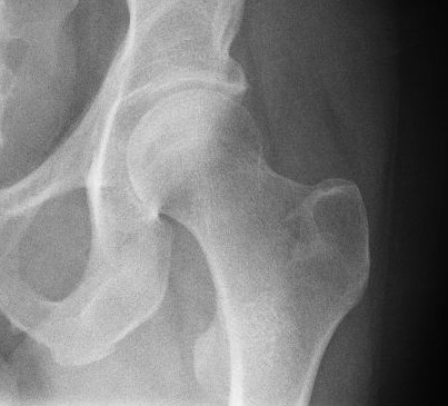
The greater trochanter of the femur is a large, irregular, quadrilateral eminence and a part of the skeletal system. It is directed lateral and medially and slightly posterior. In the adult it is about 2–4 cm lower than the femoral head.Standring, Susan, editor. ''Gray’s Anatomy: The Anatomical Basis of Clinical Practice''. Forty-First edition, Elsevier Limited, 2016, p. 1327. Because the pelvic outlet in the female is larger than in the male, there is a greater distance between the greater trochanters in the female. It has two surfaces and four borders. It is a traction epiphysis. Surfaces The ''lateral surface'', quadrilateral in form, is broad, rough, convex, and marked by a diagonal impression, which extends from the postero-superior to the antero-inferior angle, and serves for the insertion of the tendon of the gluteus medius. Above the impression is a triangular surface, sometimes rough for part of the tendon of the same muscle, sometimes smooth for the interpo ... [...More Info...] [...Related Items...] OR: [Wikipedia] [Google] [Baidu] |


