|
Glenohumeral Ligaments
In human anatomy, the glenohumeral ligaments (GHL) are three ligaments on the anterior side of the glenohumeral joint (i.e. between the glenoid cavity of the scapula and the head of the humerus; colloquially called the shoulder joint). Reinforcing the anterior glenohumeral joint capsule, the superior, middle, and inferior glenohumeral ligaments play different roles in the stability of the head of the humerus depending on arm position and degree of rotation. Location The ligaments may be best seen by opening the capsule at the back of the joint and removing the head of the humerus:''Gray's Anatomy (1918), see infobox * One on the medial side of the joint passes from the medial edge of the glenoid cavity to the lower part of the lesser tubercle of the humerus. * A second at the lower part of the joint extends from the under edge of the glenoid cavity to the under part of the anatomical neck of the humerus. * A third at the upper part of the joint is fixed above to the apex of the g ... [...More Info...] [...Related Items...] OR: [Wikipedia] [Google] [Baidu] |
Glenoid Cavity
The glenoid fossa of the scapula or the glenoid cavity is a bone part of the shoulder. The word ''glenoid'' is pronounced or (both are common) and is from el, gléne, "socket", reflecting the shoulder joint's ball-and-socket form. It is a shallow, pyriform articular surface, which is located on the lateral angle of the scapula. It is directed laterally and forward and articulates with the head of the humerus; it is broader below than above and its vertical diameter is the longest. This cavity forms the glenohumeral joint along with the humerus. This type of joint is classified as a synovial, ball and socket joint. The humerus is held in place within the glenoid cavity by means of the long head of the biceps tendon. This tendon originates on the superior margin of the glenoid cavity and loops over the shoulder, bracing humerus against the cavity. The rotator cuff also reinforces this joint more specifically with the supraspinatus tendon to hold the head of the humerus in the ... [...More Info...] [...Related Items...] OR: [Wikipedia] [Google] [Baidu] |
Coracoid Process
The coracoid process (from Greek κόραξ, raven) is a small hook-like structure on the lateral edge of the superior anterior portion of the scapula (hence: coracoid, or "like a raven's beak"). Pointing laterally forward, it, together with the acromion, serves to stabilize the shoulder joint. It is palpable in the deltopectoral groove between the deltoid and pectoralis major muscles. Structure The coracoid process is a thick curved process attached by a broad base to the upper part of the neck of the scapula; it runs at first upward and medialward; then, becoming smaller, it changes its direction, and projects forward and lateralward. Anatomically it is divided into intervals of: base of coracoid process, angle of coracoid process, shaft and the apex of the coracoid process. The coracoglenoid notch is an indentation localized between the coracoid process and the glenoid. As the coracoid process projects laterally, it house underneath it the subcoracoid space. The ''ascend ... [...More Info...] [...Related Items...] OR: [Wikipedia] [Google] [Baidu] |
Coracoacromial Ligament
The coracoacromial ligament is a strong triangular ligament between the coracoid process and the acromion. It protects the head of the humerus. Its acromial attachment may be repositioned to the clavicle during reconstructive surgery of the acromioclavicular joint (shoulder joint). Structure The coracoacromial ligament originates from the summit of the acromion of the scapula, just in front of the articular surface for the clavicle. It inserts by its broad base along the whole length of the lateral border of the coracoid process of the scapula. The clavicle and under surface of the deltoid muscle are above it. The tendon of the supraspinatus muscle (and its bursa) are below it. Its lateral border is continuous with a dense lamina that passes beneath the deltoid muscle upon the tendons of the supraspinatus and infraspinatus muscle. The ligament is sometimes described as consisting of two marginal bands and a thinner intervening portion, the two bands being attached respectivel ... [...More Info...] [...Related Items...] OR: [Wikipedia] [Google] [Baidu] |
Bicipital Groove
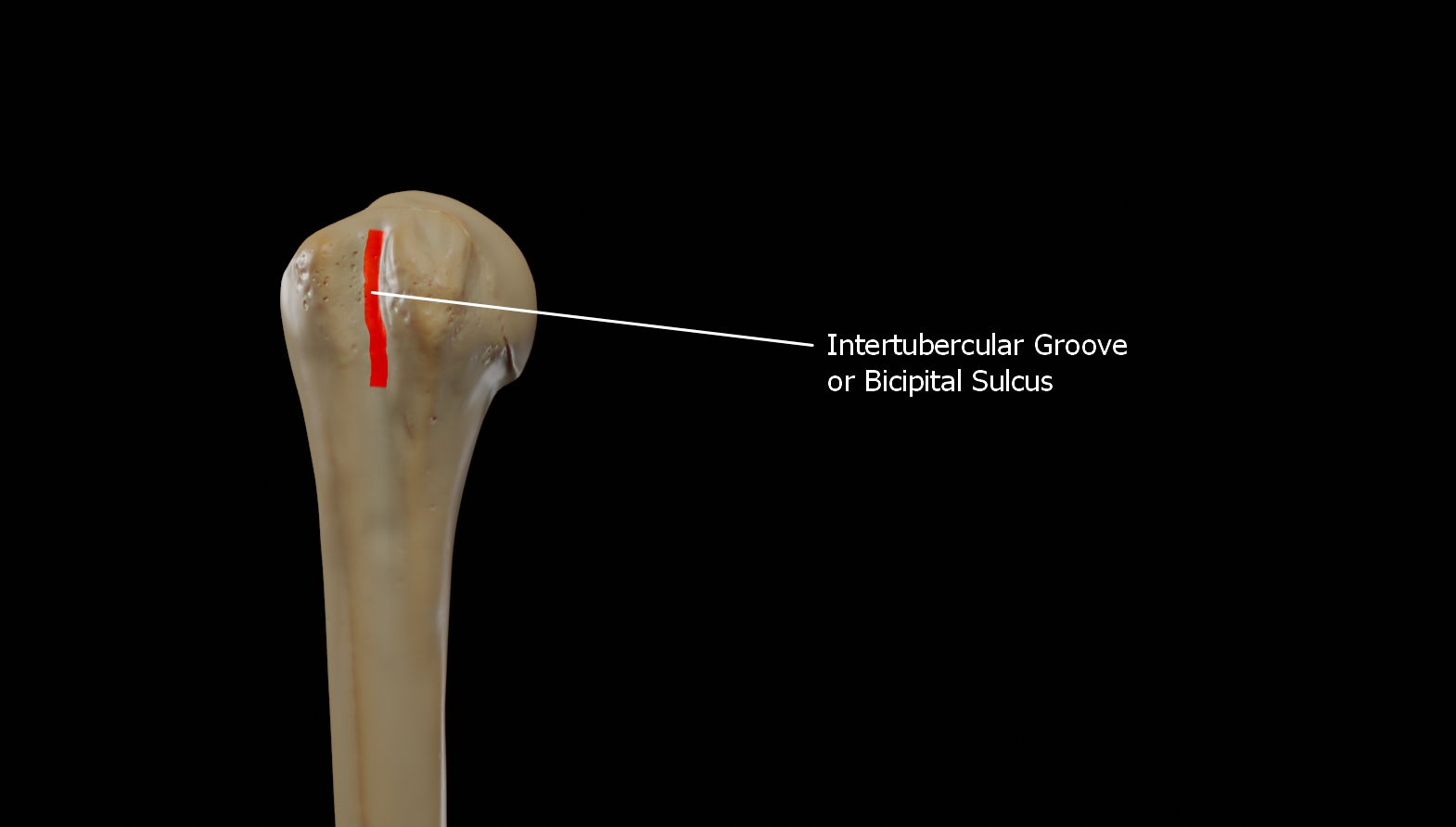
The bicipital groove (intertubercular groove, sulcus intertubercularis) is a deep groove on the humerus that separates the greater tubercle from the lesser tubercle. It allows for the long tendon of the biceps brachii muscle to pass. Structure The bicipital groove separates the greater tubercle from the lesser tubercle. It is usually around 8 cm long and 1 cm wide in adults. It lodges the long tendon of the biceps brachii muscle between the tendon of the pectoralis major muscle on the lateral lip and the tendon of the teres major muscle on the medial lip. It also transmits a branch of the anterior humeral circumflex artery to the shoulder joint. The insertion of the latissimus dorsi muscle is found along the floor of the bicipital groove. The teres major muscle inserts on the medial lip of the groove. It runs obliquely downward, and ends near the junction of the upper with the middle third of the bone. It is the lateral wall of the axilla. Function The bicipital groove all ... [...More Info...] [...Related Items...] OR: [Wikipedia] [Google] [Baidu] |
Greater Tubercle
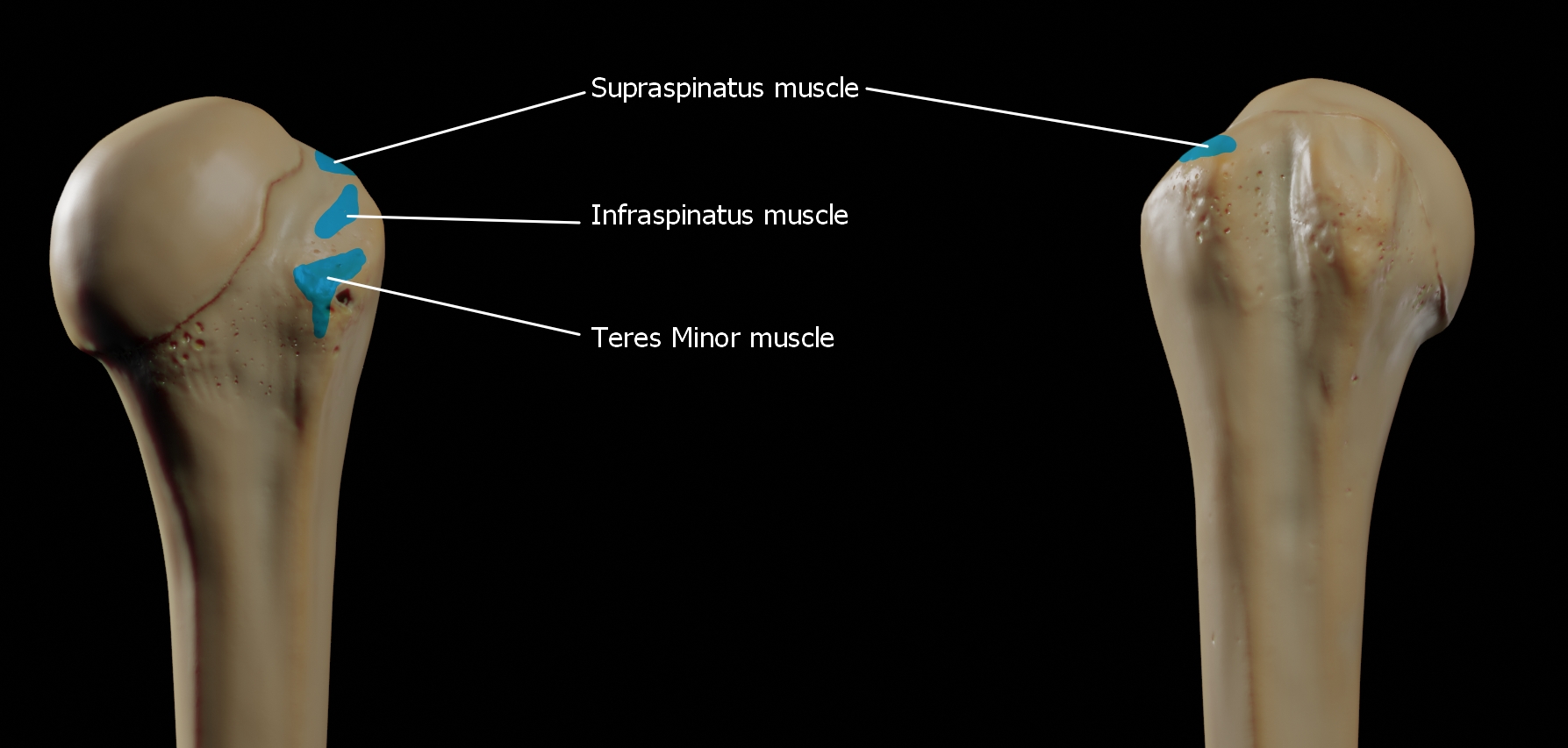
The greater tubercle of the humerus is the outward part the upper end of that bone, adjacent to the large rounded prominence of the humerus head. It provides attachment points for the supraspinatus, infraspinatus, and teres minor muscles, three of the four muscles of the rotator cuff, a muscle group that stabilizes the shoulder joint. In doing so the tubercle acts as a location for the transfer of forces from the rotator cuff muscles to the humerus. Structure The upper surface of the greater tubercle is rounded, and marked by three flat impressions: * the highest ("superior facet") gives insertion to the supraspinatus muscle. * the middle ("middle facet") gives insertion to the infraspinatus muscle. * the lowest ("inferior facet"), and the body of the bone for about 2.5 cm, gives insertion to the teres minor muscle. The lateral surface of the greater tubercle is convex, rough, and continuous with the lateral surface of the body of the humerus. It can be described a ... [...More Info...] [...Related Items...] OR: [Wikipedia] [Google] [Baidu] |
Abduction (kinesiology)
Motion, the process of movement, is described using specific anatomical terms. Motion includes movement of organs, joints, limbs, and specific sections of the body. The terminology used describes this motion according to its direction relative to the anatomical position of the body parts involved. Anatomists and others use a unified set of terms to describe most of the movements, although other, more specialized terms are necessary for describing unique movements such as those of the hands, feet, and eyes. In general, motion is classified according to the anatomical plane it occurs in. ''Flexion'' and ''extension'' are examples of ''angular'' motions, in which two axes of a joint are brought closer together or moved further apart. ''Rotational'' motion may occur at other joints, for example the shoulder, and are described as ''internal'' or ''external''. Other terms, such as ''elevation'' and ''depression'', describe movement above or below the horizontal plane. Many anatomic ... [...More Info...] [...Related Items...] OR: [Wikipedia] [Google] [Baidu] |
Teres Major
The teres major muscle is a muscle of the upper limb. It attaches to the scapula and the humerus and is one of the seven scapulohumeral muscles. It is a thick but somewhat flattened muscle. The teres major muscle (from Latin ''teres'', meaning "rounded") is positioned above the latissimus dorsi muscle and assists in the Anatomical terms of motion#Flexion and extension, extension and Anatomical terms of motion#Rotation, medial rotation of the humerus. This muscle is commonly confused as a rotator cuff muscle, but it is not because it does not attach to the capsule of the shoulder joint, unlike the teres minor muscle for example. Structure The teres major muscle originates on the dorsal surface of the Scapula, inferior angle and the lower part of the Scapula, lateral border of the scapula. The fibers of teres major insert into the medial lip of the Bicipital groove, intertubercular sulcus of the humerus. Relations The tendon, at its insertion, lies behind that of the latissimus ... [...More Info...] [...Related Items...] OR: [Wikipedia] [Google] [Baidu] |
Pectoralis Major
The pectoralis major () is a thick, fan-shaped or triangular convergent muscle, situated at the chest of the human body. It makes up the bulk of the chest muscles and lies under the breast. Beneath the pectoralis major is the pectoralis minor, a thin, triangular muscle. The pectoralis major's primary functions are flexion, adduction, and internal rotation of the humerus. The pectoral major may colloquially be referred to as "pecs", "pectoral muscle", or "chest muscle", because it is the largest and most superficial muscle in the chest area. Structure It arises from the anterior surface of the sternal half of the clavicle from breadth of the half of the anterior surface of the sternum, as low down as the attachment of the cartilage of the sixth or seventh rib; from the cartilages of all the true ribs, with the exception, frequently, of the first or seventh, and from the aponeurosis of the abdominal external oblique muscle. From this extensive origin the fibers converge toward the ... [...More Info...] [...Related Items...] OR: [Wikipedia] [Google] [Baidu] |
Biceps Brachii
The biceps or biceps brachii ( la, musculus biceps brachii, "two-headed muscle of the arm") is a large muscle that lies on the front of the upper arm between the shoulder and the elbow. Both heads of the muscle arise on the scapula and join to form a single muscle belly which is attached to the upper forearm. While the biceps crosses both the shoulder and elbow joints, its main function is at the elbow where it flexes the forearm and supinates the forearm. Both these movements are used when opening a bottle with a corkscrew: first biceps screws in the cork (supination), then it pulls the cork out (flexion). Structure The biceps is one of three muscles in the anterior compartment of the upper arm, along with the brachialis muscle and the coracobrachialis muscle, with which the biceps shares a nerve supply. The biceps muscle has two heads, the short head and the long head, distinguished according to their origin at the coracoid process and supraglenoid tubercle of the sca ... [...More Info...] [...Related Items...] OR: [Wikipedia] [Google] [Baidu] |
Anatomical Neck Of The Humerus .
The anatomical neck of the humerus is obliquely directed, forming an obtuse angle with the body of the humerus. It represents the fused epiphyseal plate. Structure The anatomical neck divides the head of the humerus from the greater and lesser tubercles of the humerus It gives attachment to the capsular ligament of the shoulder joint except at the upper inferior-medial aspects. It is best marked in the lower half of its circumference; in the upper half it is represented by a narrow groove separating the head of the humerus from the two tubercles, the greater tubercle and the lesser tubercle. It affords attachment to the articular capsule of the shoulder-joint, and is perforated by numerous vascular foramina In anatomy and osteology, a foramen (; in [...More Info...] [...Related Items...] OR: [Wikipedia] [Google] [Baidu] |
Humerus
The humerus (; ) is a long bone in the arm that runs from the shoulder to the elbow. It connects the scapula and the two bones of the lower arm, the radius and ulna, and consists of three sections. The humeral upper extremity consists of a rounded head, a narrow neck, and two short processes (tubercles, sometimes called tuberosities). The body is cylindrical in its upper portion, and more prismatic below. The lower extremity consists of 2 epicondyles, 2 processes (trochlea & capitulum), and 3 fossae (radial fossa, coronoid fossa, and olecranon fossa). As well as its true anatomical neck, the constriction below the greater and lesser tubercles of the humerus is referred to as its surgical neck due to its tendency to fracture, thus often becoming the focus of surgeons. Etymology The word "humerus" is derived from la, humerus, umerus meaning upper arm, shoulder, and is linguistically related to Gothic ''ams'' shoulder and Greek ''ōmos''. Structure Upper extremity The upper or pr ... [...More Info...] [...Related Items...] OR: [Wikipedia] [Google] [Baidu] |
Lesser Tubercle Of The Humerus
The lesser tubercle of the humerus, although smaller, is more prominent than the greater tubercle: it is situated in front, and is directed medially and anteriorly. The projection of the lesser tubercle is anterior from the junction that is found between the anatomical neck and the shaft of the humerus and easily identified due to the intertubercular sulcus (Bicipital groove). Above and in front it presents an impression for the insertion of the tendon of the subscapularis The subscapularis is a large triangular muscle which fills the subscapular fossa and inserts into the lesser tubercle of the humerus and the front of the capsule of the shoulder-joint. Structure It arises from its medial two-thirds and Som .... Additional images File:Gray326.png, The left shoulder and acromioclavicular joints, and the proper ligaments of the scapula. File:Human arm bones diagram.svg, Human arm bones diagram References External links * * * Diagram at uwlax.edu Bones of ... [...More Info...] [...Related Items...] OR: [Wikipedia] [Google] [Baidu] |


