|
Glandular Papillae
In animals, a gland is a group of cells in an animal's body that synthesizes substances (such as hormones) for release into the bloodstream ( endocrine gland) or into cavities inside the body or its outer surface ( exocrine gland). Structure Development Every gland is formed by an ingrowth from an epithelial surface. This ingrowth may in the beginning possess a tubular structure, but in other instances glands may start as a solid column of cells which subsequently becomes tubulated. As growth proceeds, the column of cells may split or give off offshoots, in which case a compound gland is formed. In many glands, the number of branches is limited, in others (salivary, pancreas) a very large structure is finally formed by repeated growth and sub-division. As a rule, the branches do not unite with one another, but in one instance, the liver, this does occur when a reticulated compound gland is produced. In compound glands the more typical or secretory epithelium is found form ... [...More Info...] [...Related Items...] OR: [Wikipedia] [Google] [Baidu] |
Submandibular Gland
The paired submandibular glands (historically known as submaxillary glands) are major salivary glands located beneath the floor of the mouth. They each weigh about 15 grams and contribute some 60–67% of unstimulated saliva secretion; on stimulation their contribution decreases in proportion as the parotid secretion rises to 50%. The average length of the normal human submandibular salivary gland is approximately 27mm, while the average width is approximately 14.3mm. Structure Lying superior to the digastric muscles, each submandibular gland is divided into superficial and deep lobes, which are separated by the mylohyoid muscle: * The superficial lobe comprises most of the gland, with the mylohyoid muscle runs under it * The deep lobe is the smaller part Secretions are delivered into the submandibular duct on the deep portion after which they hook around the posterior edge of the mylohyoid muscle and proceed on the superior surface laterally. The excretory ducts are then cross ... [...More Info...] [...Related Items...] OR: [Wikipedia] [Google] [Baidu] |
Thymus Gland
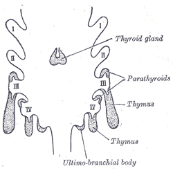
The thymus is a specialized primary lymphoid organ of the immune system. Within the thymus, thymus cell lymphocytes or ''T cells'' mature. T cells are critical to the adaptive immune system, where the body adapts to specific foreign invaders. The thymus is located in the upper front part of the chest, in the anterior superior mediastinum, behind the sternum, and in front of the heart. It is made up of two lobes, each consisting of a central medulla and an outer cortex, surrounded by a capsule. The thymus is made up of immature T cells called thymocytes, as well as lining cells called epithelial cells which help the thymocytes develop. T cells that successfully develop react appropriately with MHC immune receptors of the body (called ''positive selection'') and not against proteins of the body (called ''negative selection''). The thymus is largest and most active during the neonatal and pre-adolescent periods. By the early teens, the thymus begins to decrease in size and act ... [...More Info...] [...Related Items...] OR: [Wikipedia] [Google] [Baidu] |
Sebaceous Gland
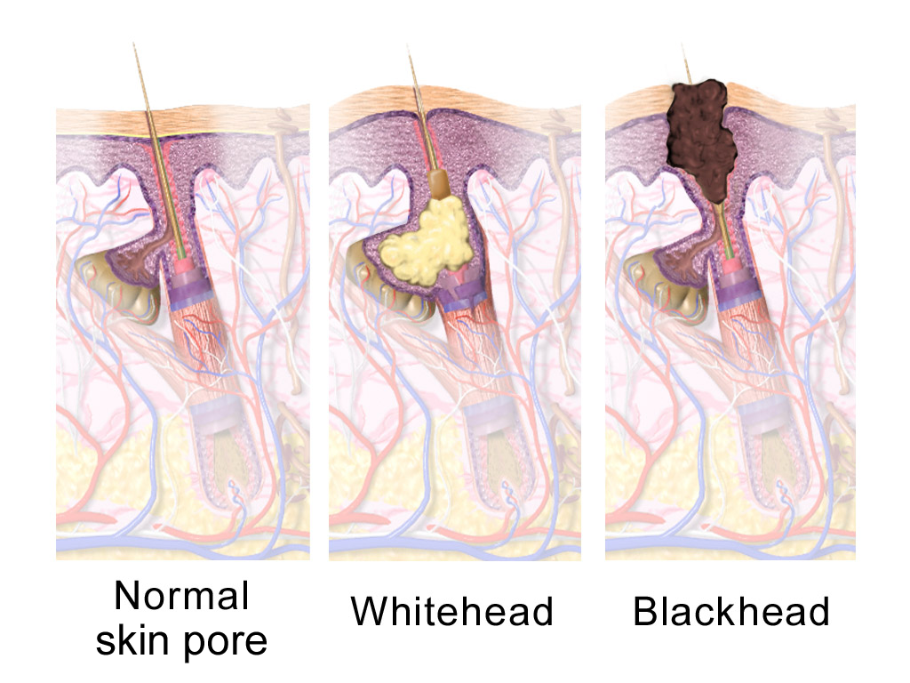
A sebaceous gland is a microscopic exocrine gland in the skin that opens into a hair follicle to secrete an oily or waxy matter, called sebum, which lubricates the hair and skin of mammals. In humans, sebaceous glands occur in the greatest number on the face and scalp, but also on all parts of the skin except the palms of the hands and soles of the feet. In the eyelids, meibomian glands, also called tarsal glands, are a type of sebaceous gland that secrete a special type of sebum into tears. Surrounding the female nipple, areolar glands are specialized sebaceous glands for lubricating the nipple. Fordyce spots are benign, visible, sebaceous glands found usually on the lips, gums and inner cheeks, and genitals. Structure Location Sebaceous glands are found throughout all areas of the skin, except the palms of the hands and soles of the feet. There are two types of sebaceous glands, those connected to hair follicles and those that exist independently. Sebaceous glands are found ... [...More Info...] [...Related Items...] OR: [Wikipedia] [Google] [Baidu] |
Holocrine
Holocrine (from Ancient Greek ὅλος; ''hólos'', “whole, entire” + κρῑ́νω; ''krī́nō'', “to separate”) is a term used to classify the mode of secretion in exocrine glands in the study of histology. Holocrine secretions are produced in the cytoplasm of the cell and released by the rupture of the plasma membrane, which destroys the cell and results in the secretion of the product into the lumen. Holocrine gland secretion is the most damaging (to the cell itself and not to the host which begot the cell) type of secretion, with merocrine secretion being the least damaging and apocrine secretion falling in between. Examples of holocrine glands include the sebaceous glands of the skin and the meibomian glands of the eyelid. The sebaceous gland is an example of a holocrine gland because its product of secretion (sebum A sebaceous gland is a microscopic exocrine gland in the skin that opens into a hair follicle to secrete an oily or waxy matter, called sebu ... [...More Info...] [...Related Items...] OR: [Wikipedia] [Google] [Baidu] |
Sweat Gland
Sweat glands, also known as sudoriferous or sudoriparous glands, , are small tubular structures of the skin that produce sweat. Sweat glands are a type of exocrine gland, which are glands that produce and secrete substances onto an epithelial surface by way of a duct. There are two main types of sweat glands that differ in their structure, function, secretory product, mechanism of excretion, anatomic distribution, and distribution across species: * Eccrine sweat glands are distributed almost all over the human body, in varying densities, with the highest density in palms and soles, then on the head, but much less on the trunk and the extremities. Its water-based secretion represents a primary form of cooling in humans. * Apocrine sweat glands are mostly limited to the axillae (armpits) and perineal area in humans. They are not significant for cooling in humans, but are the sole effective sweat glands in hoofed animals, such as the camels, donkeys, horses, and cattle. Ceruminou ... [...More Info...] [...Related Items...] OR: [Wikipedia] [Google] [Baidu] |
Sweating
Perspiration, also known as sweating, is the production of fluids secreted by the sweat glands in the skin of mammals. Two types of sweat glands can be found in humans: eccrine glands and apocrine glands. The eccrine sweat glands are distributed over much of the body and are responsible for secreting the watery, brackish sweat most often triggered by excessive body temperature. The apocrine sweat glands are restricted to the armpits and a few other areas of the body and produce an odorless, oily, opaque secretion which then gains its characteristic odor from bacterial decomposition. In humans, sweating is primarily a means of thermoregulation, which is achieved by the water-rich secretion of the eccrine glands. Maximum sweat rates of an adult can be up to 2–4 liters per hour or 10–14 liters per day (10–15 g/min·m2), but is less in children prior to puberty. Evaporation of sweat from the skin surface has a cooling effect due to evaporative cooling. Hence, in hot wea ... [...More Info...] [...Related Items...] OR: [Wikipedia] [Google] [Baidu] |
Cell (biology)
The cell is the basic structural and functional unit of life forms. Every cell consists of a cytoplasm enclosed within a membrane, and contains many biomolecules such as proteins, DNA and RNA, as well as many small molecules of nutrients and metabolites.Cell Movements and the Shaping of the Vertebrate Body in Chapter 21 of Molecular Biology of the Cell '' fourth edition, edited by Bruce Alberts (2002) published by Garland Science. The Alberts text discusses how the "cellular building blocks" move to shape developing embryos. It is also common to describe small molecules such as ... [...More Info...] [...Related Items...] OR: [Wikipedia] [Google] [Baidu] |
Apocrine
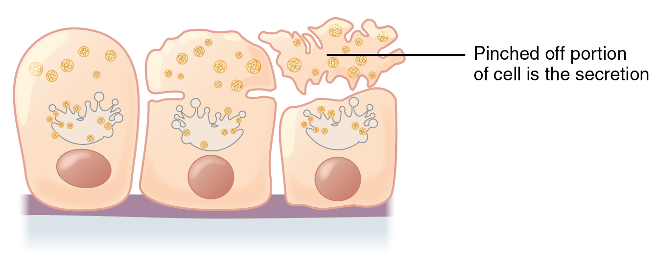
Apocrine () glands are a type of exocrine gland, which are themselves a type of gland, i.e. a group of cells specialized for the release of secretions. Exocrine glands secrete by one of three means: holocrine, merocrine and apocrine. In apocrine secretion, secretory cells accumulate material at their apical (anatomy), apical ends, and this material then buds off from the cells, forming extracellular vesicle (biology and chemistry), vesicles. The secretory cells therefore lose part of their cytoplasm in the process of secretion. An example of true apocrine glands is the mammary glands, responsible for secreting breast milk. Apocrine glands are also found in the anogenital region and axillae. Apocrine secretion is less damaging to the gland than holocrine secretion (which destroys a cell) but more damaging than merocrine secretion (exocytosis). Apocrine metaplasia Apocrine metaplasia is a reversible transformation of cells to an apocrine phenotype. It is common in the breast in ... [...More Info...] [...Related Items...] OR: [Wikipedia] [Google] [Baidu] |
Apical Membrane
The cell membrane (also known as the plasma membrane (PM) or cytoplasmic membrane, and historically referred to as the plasmalemma) is a biological membrane that separates and protects the interior of all cells from the outside environment (the extracellular space). The cell membrane consists of a lipid bilayer, made up of two layers of phospholipids with cholesterols (a lipid component) interspersed between them, maintaining appropriate membrane fluidity at various temperatures. The membrane also contains membrane proteins, including integral proteins that span the membrane and serve as membrane transporters, and peripheral proteins that loosely attach to the outer (peripheral) side of the cell membrane, acting as enzymes to facilitate interaction with the cell's environment. Glycolipids embedded in the outer lipid layer serve a similar purpose. The cell membrane controls the movement of substances in and out of cells and organelles, being selectively permeable to ions an ... [...More Info...] [...Related Items...] OR: [Wikipedia] [Google] [Baidu] |
Human Gastrointestinal Tract
The gastrointestinal tract (GI tract, digestive tract, alimentary canal) is the tract or passageway of the digestive system that leads from the mouth to the anus. The GI tract contains all the major organs of the digestive system, in humans and other animals, including the esophagus, stomach, and intestines. Food taken in through the mouth is digested to extract nutrients and absorb energy, and the waste expelled at the anus as feces. ''Gastrointestinal'' is an adjective meaning of or pertaining to the stomach and intestines. Most animals have a "through-gut" or complete digestive tract. Exceptions are more primitive ones: sponges have small pores ( ostia) throughout their body for digestion and a larger dorsal pore (osculum) for excretion, comb jellies have both a ventral mouth and dorsal anal pores, while cnidarians and acoels have a single pore for both digestion and excretion. The human gastrointestinal tract consists of the esophagus, stomach, and intestines, and is ... [...More Info...] [...Related Items...] OR: [Wikipedia] [Google] [Baidu] |
Duct (anatomy)
In anatomy and physiology, a duct is a circumscribed channel leading from an exocrine gland or organ. Types of ducts Examples include: Duct system As ducts travel from the acinus which generates the fluid to the target, the ducts become larger and the epithelium becomes thicker. The parts of the system are classified as follows: Some sources consider "lobar" ducts to be the same as "interlobar ducts", while others consider lobar ducts to be larger and more distal from the acinus. For sources that make the distinction, the interlobar ducts are more likely to classified with simple columnar epithelium (or pseudostratified epithelium), reserving the stratified columnar for the lobar ducts. File:Gray1025.png, Section of submaxillary gland of kitten. Duct semidiagrammatic. X 200. File:Gray1173.png, Section of portion of mamma. Intercalated duct The intercalated duct, also called intercalary duct (ducts of Boll), is the portion of an exocrine gland leading directly from th ... [...More Info...] [...Related Items...] OR: [Wikipedia] [Google] [Baidu] |
Exocrine Gland
Exocrine glands are glands that secrete substances on to an epithelial surface by way of a duct. Examples of exocrine glands include sweat, salivary, mammary, ceruminous, lacrimal, sebaceous, prostate and mucous. Exocrine glands are one of two types of glands in the human body, the other being endocrine glands, which secrete their products directly into the bloodstream. The liver and pancreas are both exocrine and endocrine glands; they are exocrine glands because they secrete products—bile and pancreatic juice—into the gastrointestinal tract through a series of ducts, and endocrine because they secrete other substances directly into the bloodstream. Exocrine sweat glands are part of the integumentary system; they have eccrine and apocrine types. Classification Structure Exocrine glands contain a glandular portion and a duct portion, the structures of which can be used to classify the gland. * The duct portion may be branched (called compound) or unbranched (called simple ... [...More Info...] [...Related Items...] OR: [Wikipedia] [Google] [Baidu] |


