|
Genicular Anastomosis
The patellar network (circulatory anastomosis around the knee-joint, patellar anastomosis, genicular anastomosis, articular vascular network of knee or rete articulare genushttp://medical-dictionary.thefreedictionary.com/rete+articulare+genus) is an intricate network of blood vessels around and above the patella, and on the contiguous ends of the femur and tibia, forming a superficial and a deep plexus. * The ''superficial plexus'' is situated between the fascia and skin around about the patella, and forms three well-defined arches: one, above the upper border of the patella, in the loose connective tissue over the Quadriceps femoris; the other two, below the level of the patella, are situated in the fat behind the ligamentum patellæ. * The ''deep plexus'', which forms a close net-work of vessels, lies on the lower end of the femur and upper end of the tibia around their articular surfaces, and sends numerous offsets into the interior of the joint. The arteries which form thi ... [...More Info...] [...Related Items...] OR: [Wikipedia] [Google] [Baidu] |
Circulatory Anastomosis
A circulatory anastomosis is a connection (an anastomosis) between two blood vessels, such as between arteries (arterio-arterial anastomosis), between veins (veno-venous anastomosis) or between an artery and a vein (arterio-venous anastomosis). Anastomoses between arteries and between veins result in a multitude of arteries and veins, respectively, serving the same volume of tissue. Such anastomoses occur normally in the body in the circulatory system, serving as backup routes for blood to flow if one link is blocked or otherwise compromised, but may also occur pathologically. Physiologic Arterio-arterial anastomoses include actual (e.g., palmar and plantar arches) and potential varieties (e.g., coronary arteries and cortical branch of cerebral arteries). There are many examples of normal arterio-arterial anastomoses in the body. Clinically important examples include: *Circle of Willis (in the brain) *Coronary: anterior interventricular artery and posterior interventricular art ... [...More Info...] [...Related Items...] OR: [Wikipedia] [Google] [Baidu] |
Descending Genicular Artery
The descending genicular artery (highest genicular artery) arises from the femoral artery just before it passes through the adductor hiatus. It immediately divides into two branches: * a saphenous branch, which classically joins with the medial inferior genicular artery. * muscular and articular branches. Structure Saphenous branch The saphenous branch pierces the aponeurotic covering of the adductor canal, and accompanies the saphenous nerve to the medial side of the knee. It passes between the sartorius muscle and the gracilis muscle, and, piercing the fascia lata, is distributed to the integument of the upper and medial part of the leg, anastomosing with the medial inferior genicular artery. Articular branches The articular branches descend within the vastus medialis muscle, and in front of the tendon of the adductor magnus muscle, to the medial side of the knee, where they join with the medial superior genicular and anterior recurrent tibial artery. A branch from this v ... [...More Info...] [...Related Items...] OR: [Wikipedia] [Google] [Baidu] |
X-ray Computed Tomography
An X-ray, or, much less commonly, X-radiation, is a penetrating form of high-energy electromagnetic radiation. Most X-rays have a wavelength ranging from 10 picometers to 10 nanometers, corresponding to frequencies in the range 30 petahertz to 30 exahertz ( to ) and energies in the range 145 eV to 124 keV. X-ray wavelengths are shorter than those of UV rays and typically longer than those of gamma rays. In many languages, X-radiation is referred to as Röntgen radiation, after the German scientist Wilhelm Conrad Röntgen, who discovered it on November 8, 1895. He named it ''X-radiation'' to signify an unknown type of radiation.Novelline, Robert (1997). ''Squire's Fundamentals of Radiology''. Harvard University Press. 5th edition. . Spellings of ''X-ray(s)'' in English include the variants ''x-ray(s)'', ''xray(s)'', and ''X ray(s)''. The most familiar use of X-rays is checking for fractures (broken bones), but X-rays are also used in other ways. Fo ... [...More Info...] [...Related Items...] OR: [Wikipedia] [Google] [Baidu] |
Astley Cooper
Sir Astley Paston Cooper, 1st Baronet (23 August 176812 February 1841) was a British surgeon and anatomist, who made contributions to otology, vascular surgery, the anatomy and pathology of the mammary glands and testicles, and the pathology and surgery of hernia. Life Cooper was born at Brooke Hall in Brooke, Norfolk on 23 August 1768 and baptised at the parish church on 9 September. His father, Dr Samuel Cooper, was a clergyman of the Church of England; his mother Maria Susanna Bransby was the author of several novels. At the age of sixteen he was sent to London and placed under Henry Cline (1750–1827), surgeon to St Thomas' Hospital. From the first he devoted himself to the study of anatomy, and had the privilege of attending the lectures of John Hunter. In 1789 he was appointed demonstrator of anatomy at St Thomas' Hospital, where in 1791 he became joint lecturer with Cline in anatomy and surgery, and in 1800 he was appointed surgeon to Guy's Hospital on the death of ... [...More Info...] [...Related Items...] OR: [Wikipedia] [Google] [Baidu] |
John Hunter (surgeon)
John Hunter (13 February 1728 – 16 October 1793) was a British surgeon, one of the most distinguished scientists and surgeons of his day. He was an early advocate of careful observation and scientific method in medicine. He was a teacher of, and collaborator with, Edward Jenner, pioneer of the smallpox vaccine. He is alleged to have paid for the stolen body of Charles Byrne, and proceeded to study and exhibit it against the deceased's explicit wishes. His wife, Anne Hunter (''née'' Home), was a poet, some of whose poems were set to music by Joseph Haydn. He learned anatomy by assisting his elder brother William with dissections in William's anatomy school in Central London, starting in 1748, and quickly became an expert in anatomy. He spent some years as an Army surgeon, worked with the dentist James Spence conducting tooth transplants, and in 1764 set up his own anatomy school in London. He built up a collection of living animals whose skeletons and other organs he prepa ... [...More Info...] [...Related Items...] OR: [Wikipedia] [Google] [Baidu] |
Gray's Anatomy
''Gray's Anatomy'' is a reference book of human anatomy written by Henry Gray, illustrated by Henry Vandyke Carter, and first published in London in 1858. It has gone through multiple revised editions and the current edition, the 42nd (October 2020), remains a standard reference, often considered "the doctors' bible". Earlier editions were called ''Anatomy: Descriptive and Surgical'', ''Anatomy of the Human Body'' and ''Gray's Anatomy: Descriptive and Applied'', but the book's name is commonly shortened to, and later editions are titled, ''Gray's Anatomy''. The book is widely regarded as an extremely influential work on the subject. Publication history Origins The English anatomist Henry Gray was born in 1827. He studied the development of the endocrine glands and spleen and in 1853 was appointed Lecturer on Anatomy at St George's Hospital Medical School in London. In 1855, he approached his colleague Henry Vandyke Carter with his idea to produce an inexpensive and ac ... [...More Info...] [...Related Items...] OR: [Wikipedia] [Google] [Baidu] |
Popliteal Artery
The popliteal artery is a deeply placed continuation of the femoral artery opening in the distal portion of the adductor magnus muscle. It courses through the popliteal fossa and ends at the lower border of the popliteus muscle, where it branches into the anterior tibial artery, anterior and Posterior tibial artery, posterior tibial arteries. The deepest (most anterior) structure in the fossa, the popliteal artery runs close to the joint capsule of the knee as it spans the Intercondylar fossa of femur, intercondylar fossa. Five genicular branches of the popliteal artery supply the capsule and ligaments of the knee joint. The genicular arteries are the superior lateral, superior medial, middle, inferior lateral, and inferior medial genicular arteries. They participate in the formation of the periarticular genicular anastomosis, a network of vessels surrounding the knee that provides collateral circulation capable of maintaining blood supply to the leg during full knee flexion, which ... [...More Info...] [...Related Items...] OR: [Wikipedia] [Google] [Baidu] |
Ligature (medicine)
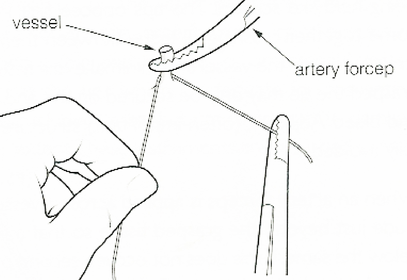
In surgery or medical procedure, a ligature consists of a piece of thread ( suture) tied around an anatomical structure, usually a blood vessel or another hollow structure (e.g. urethra) to shut it off. History The principle of ligation is attributed to Hippocrates and Galen. In ancient Rome, ligatures were used to treat hemorrhoids. The concept of a ligature was reintroduced some 1,500 years later by Ambroise Paré, and finally it found its modern use in 1870–80, made popular by Jules-Émile Péan. Procedure With a blood vessel the surgeon will clamp the vessel perpendicular to the axis of the artery or vein with a hemostat, then secure it by ligating it; i.e. using a piece of suture around it before dividing the structure and releasing the hemostat. It is different from a tourniquet in that the tourniquet will not be secured by knots and it can therefore be released/tightened at will. Ligature is one of the remedies to treat skin tag, or acrochorda. It is done by tying str ... [...More Info...] [...Related Items...] OR: [Wikipedia] [Google] [Baidu] |
Femoral Artery
The femoral artery is a large artery in the thigh and the main arterial supply to the thigh and leg. The femoral artery gives off the deep femoral artery or profunda femoris artery and descends along the anteromedial part of the thigh in the femoral triangle. It enters and passes through the adductor canal, and becomes the popliteal artery as it passes through the adductor hiatus in the adductor magnus near the junction of the middle and distal thirds of the thigh. Structure The femoral artery enters the thigh from behind the inguinal ligament as the continuation of the external iliac artery. Here, it lies midway between the anterior superior iliac spine and the symphysis pubis (Mid-inguinal point). Segments In clinical parlance, the femoral artery has the following segments: *The common femoral artery (CFA) is the segment of the femoral artery between the inferior margin of the inguinal ligament and the branching point of the deep femoral artery/profunda femoris artery. Its ... [...More Info...] [...Related Items...] OR: [Wikipedia] [Google] [Baidu] |
Aneurysm
An aneurysm is an outward bulging, likened to a bubble or balloon, caused by a localized, abnormal, weak spot on a blood vessel wall. Aneurysms may be a result of a hereditary condition or an acquired disease. Aneurysms can also be a nidus (starting point) for clot formation (thrombosis) and embolization. As an aneurysm increases in size, the risk of rupture, which leads to uncontrolled bleeding, increases. Although they may occur in any blood vessel, particularly lethal examples include aneurysms of the Circle of Willis in the brain, aortic aneurysms affecting the thoracic aorta, and abdominal aortic aneurysms. Aneurysms can arise in the heart itself following a heart attack, including both ventricular and atrial septal aneurysms. There are congenital atrial septal aneurysms, a rare heart defect. Etymology The word is from Greek: ἀνεύρυσμα, aneurysma, "dilation", from ἀνευρύνειν, aneurynein, "to dilate". Classification Aneurysms are classified by type, ... [...More Info...] [...Related Items...] OR: [Wikipedia] [Google] [Baidu] |
Popliteal Artery
The popliteal artery is a deeply placed continuation of the femoral artery opening in the distal portion of the adductor magnus muscle. It courses through the popliteal fossa and ends at the lower border of the popliteus muscle, where it branches into the anterior tibial artery, anterior and Posterior tibial artery, posterior tibial arteries. The deepest (most anterior) structure in the fossa, the popliteal artery runs close to the joint capsule of the knee as it spans the Intercondylar fossa of femur, intercondylar fossa. Five genicular branches of the popliteal artery supply the capsule and ligaments of the knee joint. The genicular arteries are the superior lateral, superior medial, middle, inferior lateral, and inferior medial genicular arteries. They participate in the formation of the periarticular genicular anastomosis, a network of vessels surrounding the knee that provides collateral circulation capable of maintaining blood supply to the leg during full knee flexion, which ... [...More Info...] [...Related Items...] OR: [Wikipedia] [Google] [Baidu] |
Anterior Tibial Recurrent Artery
The anterior tibial recurrent artery is a small artery in the leg. It arises from the anterior tibial artery, as soon as that vessel has passed through the interosseous space. It ascends in the tibialis anterior muscle, ramifies on the front and sides of the knee-joint, and assists in the formation of the patellar plexus by anastomosing An anastomosis (, plural anastomoses) is a connection or opening between two things (especially cavities or passages) that are normally diverging or branching, such as between blood vessels, leaf veins, or streams. Such a connection may be normal ... with the genicular branches of the popliteal, and with the highest genicular artery. References External links Arteries of the lower limb {{circulatory-stub ... [...More Info...] [...Related Items...] OR: [Wikipedia] [Google] [Baidu] |


