|
Fascia Of Scarpa
The fascia of Scarpa is the deep membranous layer ''(stratum membranosum)'' of the superficial fascia of the abdomen. It is a Abdominal wall#Layers, layer of the anterior abdominal wall. It is found ''deep'' to the fascia of Camper and ''superficial'' to the external oblique muscle. Structure It is thinner and more membranous in character than the superficial fascia of Camper, and contains a considerable quantity of orange elastic fibers. It is loosely connected by areolar tissue to the aponeurosis of the external oblique muscle, but in the midline it is more intimately adherent to the linea alba (abdomen), linea alba and the pubic symphysis, and is prolonged on to the dorsum of the penis, forming the fundiform ligament; above, it is continuous with the superficial fascia over the rest of the torso, trunk; inferiorly, it is continuous with the fascia of Colles of the perineum; however, it does not extend into the thigh as it just attaches to its fascia, which is known as fascia l ... [...More Info...] [...Related Items...] OR: [Wikipedia] [Google] [Baidu] |
Membranous Layer
The membranous layer or stratum membranosum is the deepest layer of subcutaneous tissue. It is a fusion of fibres into a homogeneous layer below the adipose tissue, for example, superficial to muscular fascias. It is considered a fascia by some sources, but not by others. However, prominent areas of the membranous layer are called fascias; these include the fascia of Scarpa in the abdomen and the fascia of Colles in the perineum. References Skin anatomy {{anatomy-stub ... [...More Info...] [...Related Items...] OR: [Wikipedia] [Google] [Baidu] |
Fascia Lata
The fascia lata is the deep fascia of the thigh. It encloses the thigh muscles and forms the outer limit of the fascial compartments of thigh, which are internally separated by the medial intermuscular septum and the lateral intermuscular septum. The fascia lata is thickened at its lateral side where it forms the iliotibial tract, a structure that runs to the tibia and serves as a site of muscle attachment. Structure The fascia lata is an investment for the whole of the thigh, but varies in thickness in different parts. It is thicker in the upper and lateral part of the thigh, where it receives a fibrous expansion from the gluteus maximus, and where the tensor fasciae latae is inserted between its layers; it is very thin behind and at the upper and medial part, where it covers the adductor muscles, and again becomes stronger around the knee, receiving fibrous expansions from the tendon of the biceps femoris laterally, from the sartorius medially, and from the quadriceps femoris ... [...More Info...] [...Related Items...] OR: [Wikipedia] [Google] [Baidu] |
Cullen's Sign
Cullen's sign is superficial edema and bruising in the subcutaneous fatty tissue around the umbilicus. It is named for gynecologist Thomas Stephen Cullen (1869–1953), who first described the sign in ruptured ectopic pregnancy in 1916.T.S. Cullen. Embryology, anatomy, and diseases of the umbilicus together with diseases of the urachus. Philadelphia, Saunders, and London, 1916. This sign takes 24–48 hours to appear and can predict acute pancreatitis, with mortality rising from 8–10% to 40%. It may be accompanied by Grey Turner's sign (bruising of the flank), which may then be indicative of pancreatic necrosis with retroperitoneal or intra-abdominal bleeding. Causes Causes include: * acute pancreatitis, where methemalbumin formed from digested blood tracks around the abdomen from the inflamed pancreas * bleeding from blunt abdominal trauma * bleeding from aortic rupture Aortic rupture is the rupture or breakage of the aorta, the largest artery in the body. Aortic rupture ... [...More Info...] [...Related Items...] OR: [Wikipedia] [Google] [Baidu] |
Clinical Signs
Signs and symptoms are the observed or detectable signs, and experienced symptoms of an illness, injury, or condition. A sign for example may be a higher or lower temperature than normal, raised or lowered blood pressure or an abnormality showing on a medical scan. A symptom is something out of the ordinary that is experienced by an individual such as feeling feverish, a headache or other pain or pains in the body. Signs and symptoms Signs A medical sign is an objective observable indication of a disease, injury, or abnormal physiological state that may be detected during a physical examination, examining the patient history, or diagnostic procedure. These signs are visible or otherwise detectable such as a rash or bruise. Medical signs, along with symptoms, assist in formulating diagnostic hypothesis. Examples of signs include elevated blood pressure, nail clubbing of the fingernails or toenails, staggering gait, and arcus senilis and arcus juvenilis of the eyes. Indications ... [...More Info...] [...Related Items...] OR: [Wikipedia] [Google] [Baidu] |
Abraham Colles
Abraham Colles (23 July 1773 – 16 November 1843) was Professor of Anatomy, Surgery and Physiology at the Royal College of Surgeons in Ireland, Royal College of Surgeons in Ireland (RCSI) and the President of RCSI in 1802 and 1830. A prestigious Colles Medal & Travelling Fellowship in Surgery is awarded competitively annually to an Irish surgical trainee embarking on higher specialist training abroad before returning to establish practice in Ireland. Life Descended from a Worcestershire family, some of whom had sat in Parliament, he was born to William Colles and Mary Anne Bates of Woodbroak, Co. Wexford. The family lived near Millmount, a townland near Kilkenny, Ireland, where his father owned and managed his inheritance which was the extensive Black Quarry that produced the famous Black Kilkenny Marble. His father died when Colles was 6, but his mother took over the management of the quarry and managed to give her children a good education. While at Kilkenny College, a flood ... [...More Info...] [...Related Items...] OR: [Wikipedia] [Google] [Baidu] |
Antonio Scarpa
Antonio Scarpa (9 May 1752 – 31 October 1832) was an Italian anatomist and professor. Biography Scarpa was born to an impoverished family in the frazione of Lorenzaga, Motta di Livenza, Veneto. An uncle, who was a member of the priesthood, gave him instruction until the age of 15, when he passed the entrance exam for the University of Padua. He was a pupil of Giovanni Battista Morgagni and Marc Antonio Caldani. Under the former, he became doctor of medicine on 19 May 1770; in 1772, he became professor at the University of Modena. For a time he chose to travel, visiting Holland, France and England. When he returned to Italy, he was made professor of anatomy at the University of Pavia in 1783, on the strong recommendation of Emperor Joseph II. His lectures were so popular with students that Emperor Joseph II commissioned Leopoldo Pollack to build a new anotamic theater, now called Aula Scarpa, inside the Old Campus of the University of Pavia. He remained in that post until 1804, ... [...More Info...] [...Related Items...] OR: [Wikipedia] [Google] [Baidu] |
Labia Majora
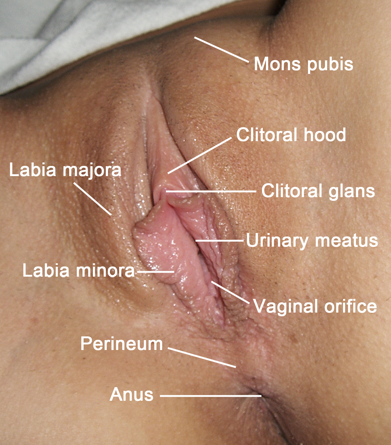
The labia majora (singular: ''labium majus'') are two prominent longitudinal cutaneous folds that extend downward and backward from the mons pubis to the perineum. Together with the labia minora they form the labia of the vulva. The labia majora are homologous to the male scrotum. Etymology ''Labia majora'' is the Latin plural for big ("major") lips; the singular is ''labium majus.'' The Latin term ''labium/labia'' is used in anatomy for a number of usually paired parallel structures, but in English it is mostly applied to two pairs of parts of female external genitals (vulva)—labia majora and labia minora. Labia majora are commonly known as the outer lips, while labia minora (Latin for ''small lips''), which run alongside between them, are referred to as the inner lips. Traditionally, to avoid confusion with other lip-like structures of the body, the labia of female genitals were termed by anatomists in Latin as ''labia majora (''or ''minora) pudendi.'' Embryology Embryolo ... [...More Info...] [...Related Items...] OR: [Wikipedia] [Google] [Baidu] |
Fascia Of The Perineum
The fascia of perineum (deep perineal fascia, superficial investing fascia of perineum or Gallaudet fascia) is the fascia which covers the muscles of the superficial perineal pouch. The muscles surrounded by the deep perineal fascia are the bulbospongiosus, ischiocavernosus, and superficial transverse perineal. The fascia is attached laterally to the ischiopubic rami and fused anteriorly with the suspensory ligament of the penis or clitoris. It is continuous anteriorly with the deep investing fascia of the abdominal wall muscles, and in males, it is continuous with Buck's fascia Buck's fascia (deep fascia of the penis, Gallaudet's fascia or fascia of the penis) is a layer of deep fascia covering the three erectile bodies of the penis. Structure Buck's fascia is continuous with the external spermatic fascia in the scrotu .... {{Authority control Fascia ... [...More Info...] [...Related Items...] OR: [Wikipedia] [Google] [Baidu] |
Dartos
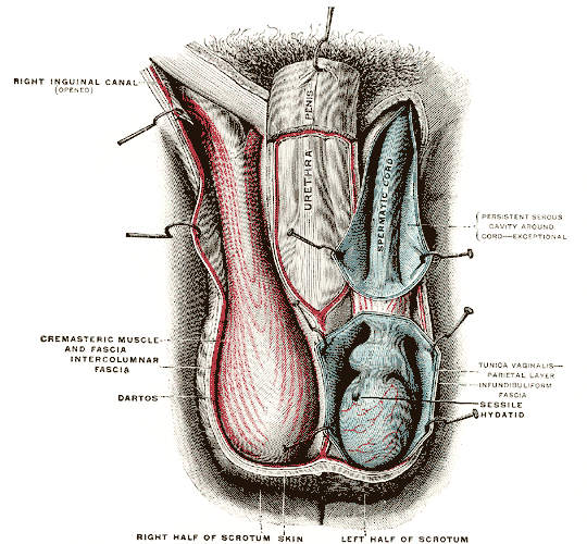
The dartos fascia or simply dartos is a layer of connective tissue found in the penile shaft, foreskin, scrotum and labia. The penile portion is referred to as the superficial fascia of penis or the subcutaneous tissue of penis, while the scrotal part is the dartos proper. In addition to being continuous with itself between the scrotum and the penis, it is also continuous with Colles fascia of the perineum and Scarpa's fascia of the abdomen. The dartos lies just below the skin, which places it just superficial to the external spermatic fascia in the scrotum and to Buck's fascia in the penile shaft. In the scrotum, it consists mostly of smooth muscle. The tone of this smooth muscle is responsible for the wrinkled (rugose) appearance of the scrotum. In females, the same muscle fibers are less well developed and termed ''dartos muliebris,'' lying beneath the skin of the labia majora. The dartos fascia receives innervation from postganglionic sympathetic nerve fibers arriving via the ... [...More Info...] [...Related Items...] OR: [Wikipedia] [Google] [Baidu] |
Scrotum
The scrotum or scrotal sac is an anatomical male reproductive structure located at the base of the penis that consists of a suspended dual-chambered sac of skin and smooth muscle. It is present in most terrestrial male mammals. The scrotum contains the external spermatic fascia, testes, epididymis, and ductus deferens. It is a distention of the perineum and carries some abdominal tissues into its cavity including the testicular artery, testicular vein, and pampiniform plexus. The perineal raphe is a small, vertical, slightly raised ridge of scrotal skin under which is found the scrotal septum. It appears as a thin longitudinal line that runs front to back over the entire scrotum. In humans and some other mammals the scrotum becomes covered with pubic hair at puberty. The scrotum will usually tighten during penile erection and when exposed to cold temperatures. One testis is typically lower than the other to avoid compression in the event of an impact. The scrotum is biologicall ... [...More Info...] [...Related Items...] OR: [Wikipedia] [Google] [Baidu] |
Spermatic Cord
The spermatic cord is the cord-like structure in males formed by the vas deferens (''ductus deferens'') and surrounding tissue that runs from the deep inguinal ring down to each testicle. Its serosal covering, the tunica vaginalis, is an extension of the peritoneum that passes through the transversalis fascia. Each testicle develops in the lower thoracic and upper lumbar region and migrates into the scrotum. During its descent it carries along with it the vas deferens, its vessels, nerves etc. There is one on each side. Structure The spermatic cord is ensheathed in three layers of tissue: * ''external spermatic fascia'', an extension of the innominate fascia that overlies the aponeurosis of the external oblique muscle. * ''cremasteric muscle and fascia'', formed from a continuation of the internal oblique muscle and its fascia. * ''internal spermatic fascia'', continuous with the transversalis fascia. The normal diameter of the spermatic cord is about 16 mm (range 11 to 22 mm). It ... [...More Info...] [...Related Items...] OR: [Wikipedia] [Google] [Baidu] |
Perineum
The perineum in humans is the space between the anus and scrotum in the male, or between the anus and the vulva in the female. The perineum is the region of the body between the pubic symphysis (pubic arch) and the coccyx (tail bone), including the perineal body and surrounding structures. There is some variability in how the boundaries are defined. The perineal raphe is visible and pronounced to varying degrees. The perineum is an erogenous zone. The word perineum entered English from late Latin via Greek περίναιος ~ περίνεος ''perinaios, perineos'', itself from περίνεος, περίνεοι 'male genitals' and earlier περίς ''perís'' 'penis' through influence from πηρίς ''pērís'' 'scrotum'. The term was originally understood as a purely male body-part with the perineal raphe seen as a continuation of the scrotal septum since masculinization causes the development of a large anogenital distance in men, in comparison to the corresponding lack ... [...More Info...] [...Related Items...] OR: [Wikipedia] [Google] [Baidu] |


