|
Fundus
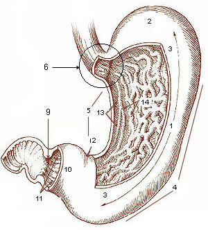
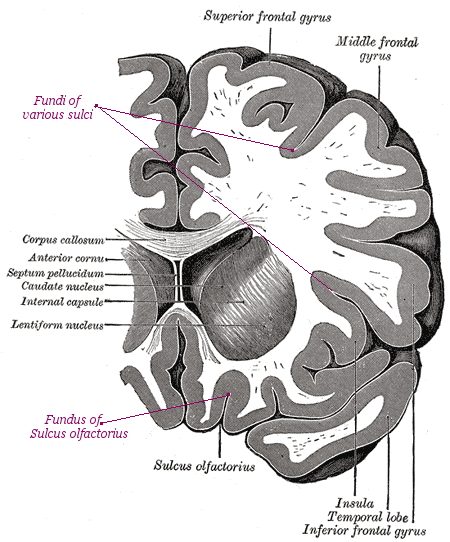
''Fundus'' (Latin for "bottom") is an anatomical term referring to that part of a concavity in any organ, which is at the far end from its opening. It may refer to: Anatomy * Fundus (brain), the deepest part of any sulcus of the cerebral cortex * Fundus (eye), the interior surface of the eye, opposite the lens, and including the retina, optic disc, macula and fovea, and posterior pole ** Fundus camera, equipment for photographing the interior of the eye ** Fundus photography * Fundus (stomach), the portion of the stomach which bulges up past the point of entry of the esophagus * Fundus (uterus), the top portion, opposite from the cervix * Fundus of gallbladder In vertebrates, the gallbladder, also known as the cholecyst, is a small hollow organ where bile is stored and concentrated before it is released into the small intestine. In humans, the pear-shaped gallbladder lies beneath the liver, althoug ..., the portion of the gallbladder which lies the farthest from the cystic ... [...More Info...] [...Related Items...] OR: [Wikipedia] [Google] [Baidu] |
Fundus Camera
Fundus photography involves photographing the rear of an eye, also known as the fundus (eye), fundus. Specialized fundus cameras consisting of an intricate microscope attached to a flash (photography), flash enabled camera are used in fundus photography. The main structures that can be visualized on a fundus photo are the central and peripheral retina, optic disc and Macula of retina, macula. Fundus photography can be performed with colored filters, or with specialized dyes including Fluorescein angiography, fluorescein and indocyanine green. The models and technology of fundus photography have advanced and evolved rapidly over the last century. Since the equipment is sophisticated and challenging to manufacture to clinical standards, only a few manufacturers/brands are available in the market: Welch Allyn, Digisight, Volk, Topcon, Carl Zeiss AG, Zeiss, Canon Inc., Canon, Nidek, Kowa (company), Kowa, CSO, CenterVue, Ezer and Optos are some example of fundus camera manufacturers. ... [...More Info...] [...Related Items...] OR: [Wikipedia] [Google] [Baidu] |
Fundus Photography
Fundus photography involves photographing the rear of an eye, also known as the fundus. Specialized fundus cameras consisting of an intricate microscope attached to a flash enabled camera are used in fundus photography. The main structures that can be visualized on a fundus photo are the central and peripheral retina, optic disc and macula. Fundus photography can be performed with colored filters, or with specialized dyes including fluorescein and indocyanine green. The models and technology of fundus photography have advanced and evolved rapidly over the last century. Since the equipment is sophisticated and challenging to manufacture to clinical standards, only a few manufacturers/brands are available in the market: Welch Allyn, Digisight, Volk, Topcon, Zeiss, Canon, Nidek, Kowa, CSO, CenterVue, Ezer and Optos are some example of fundus camera manufacturers. History The concept of fundus photography was first introduced in the mid 19th century, after the introduction of ... [...More Info...] [...Related Items...] OR: [Wikipedia] [Google] [Baidu] |
Fundus (eye)
The fundus of the eye is the interior surface of the eye opposite the lens and includes the retina, optic disc, macula, fovea, and posterior pole.Cassin, B. and Solomon, S. ''Dictionary of Eye Terminology''. Gainesville, Florida: Triad Publishing Company, 1990. The fundus can be examined by ophthalmoscopy and/or fundus photography. Variation The color of the fundus varies both between and within species. In one study of primates the retina is blue, green, yellow, orange, and red; only the human fundus (from a lightly pigmented blond person) is red. The major differences noted among the "higher" primate species were size and regularity of the border of macular area, size and shape of the optic disc, apparent 'texturing' of retina, and pigmentation of retina. Clinical significance Medical signs that can be detected from observation of eye fundus (generally by funduscopy) include hemorrhages, exudates, cotton wool spots, blood vessel abnormalities (tortuosity, pulsation and n ... [...More Info...] [...Related Items...] OR: [Wikipedia] [Google] [Baidu] |
Fundus (uterus)
The uterus (from Latin ''uterus'', plural ''uteri'') or womb () is the organ in the reproductive system of most female mammals, including humans that accommodates the embryonic and fetal development of one or more embryos until birth. The uterus is a hormone-responsive sex organ that contains glands in its lining that secrete uterine milk for embryonic nourishment. In the human, the lower end of the uterus, is a narrow part known as the isthmus that connects to the cervix, leading to the vagina. The upper end, the body of the uterus, is connected to the fallopian tubes, at the uterine horns, and the rounded part above the openings to the fallopian tubes is the fundus. The connection of the uterine cavity with a fallopian tube is called the uterotubal junction. The fertilized egg is carried to the uterus along the fallopian tube. It will have divided on its journey to form a blastocyst that will implant itself into the lining of the uterus – the endometrium, where it will re ... [...More Info...] [...Related Items...] OR: [Wikipedia] [Google] [Baidu] |
Fundus (stomach)
The stomach is a muscular, hollow organ in the gastrointestinal tract of humans and many other animals, including several invertebrates. The stomach has a dilated structure and functions as a vital organ in the digestive system. The stomach is involved in the gastric phase of digestion, following chewing. It performs a chemical breakdown by means of enzymes and hydrochloric acid. In humans and many other animals, the stomach is located between the oesophagus and the small intestine. The stomach secretes digestive enzymes and gastric acid to aid in food digestion. The pyloric sphincter controls the passage of partially digested food (chyme) from the stomach into the duodenum, where peristalsis takes over to move this through the rest of intestines. Structure In the human digestive system, the stomach lies between the oesophagus and the duodenum (the first part of the small intestine). It is in the left upper quadrant of the abdominal cavity. The top of the stomach lies against ... [...More Info...] [...Related Items...] OR: [Wikipedia] [Google] [Baidu] |
Fundus (brain)
The deepest part of a sulcus, such as the sulci in the human cerebral cortex The cerebral cortex, also known as the cerebral mantle, is the outer layer of neural tissue of the cerebrum of the brain in humans and other mammals. The cerebral cortex mostly consists of the six-layered neocortex, with just 10% consisting of .... {{Neuroanatomy-stub Neuroanatomy ... [...More Info...] [...Related Items...] OR: [Wikipedia] [Google] [Baidu] |
Fundus Of Gallbladder
In vertebrates, the gallbladder, also known as the cholecyst, is a small hollow organ where bile is stored and concentrated before it is released into the small intestine. In humans, the pear-shaped gallbladder lies beneath the liver, although the structure and position of the gallbladder can vary significantly among animal species. It receives and stores bile, produced by the liver, via the common hepatic duct, and releases it via the common bile duct into the duodenum, where the bile helps in the digestion of fats. The gallbladder can be affected by gallstones, formed by material that cannot be dissolved – usually cholesterol or bilirubin, a product of haemoglobin breakdown. These may cause significant pain, particularly in the upper-right corner of the abdomen, and are often treated with removal of the gallbladder (called a cholecystectomy). Cholecystitis, inflammation of the gallbladder, has a wide range of causes, including result from the impaction of gallstones, infec ... [...More Info...] [...Related Items...] OR: [Wikipedia] [Google] [Baidu] |
Fundus Of The Urinary Bladder
The urinary bladder, or simply bladder, is a hollow organ in humans and other vertebrates that stores urine from the kidneys before disposal by urination. In humans the bladder is a distensible organ that sits on the pelvic floor. Urine enters the bladder via the ureters and exits via the urethra. The typical adult human bladder will hold between 300 and (10.14 and ) before the urge to empty occurs, but can hold considerably more. The Latin phrase for "urinary bladder" is ''vesica urinaria'', and the term ''vesical'' or prefix ''vesico -'' appear in connection with associated structures such as vesical veins. The modern Latin word for "bladder" – ''cystis'' – appears in associated terms such as cystitis (inflammation of the bladder). Structure In humans, the bladder is a hollow muscular organ situated at the base of the pelvis. In gross anatomy, the bladder can be divided into a broad , a body, an apex, and a neck. The apex (also called the vertex) is directed forward ... [...More Info...] [...Related Items...] OR: [Wikipedia] [Google] [Baidu] |


