|
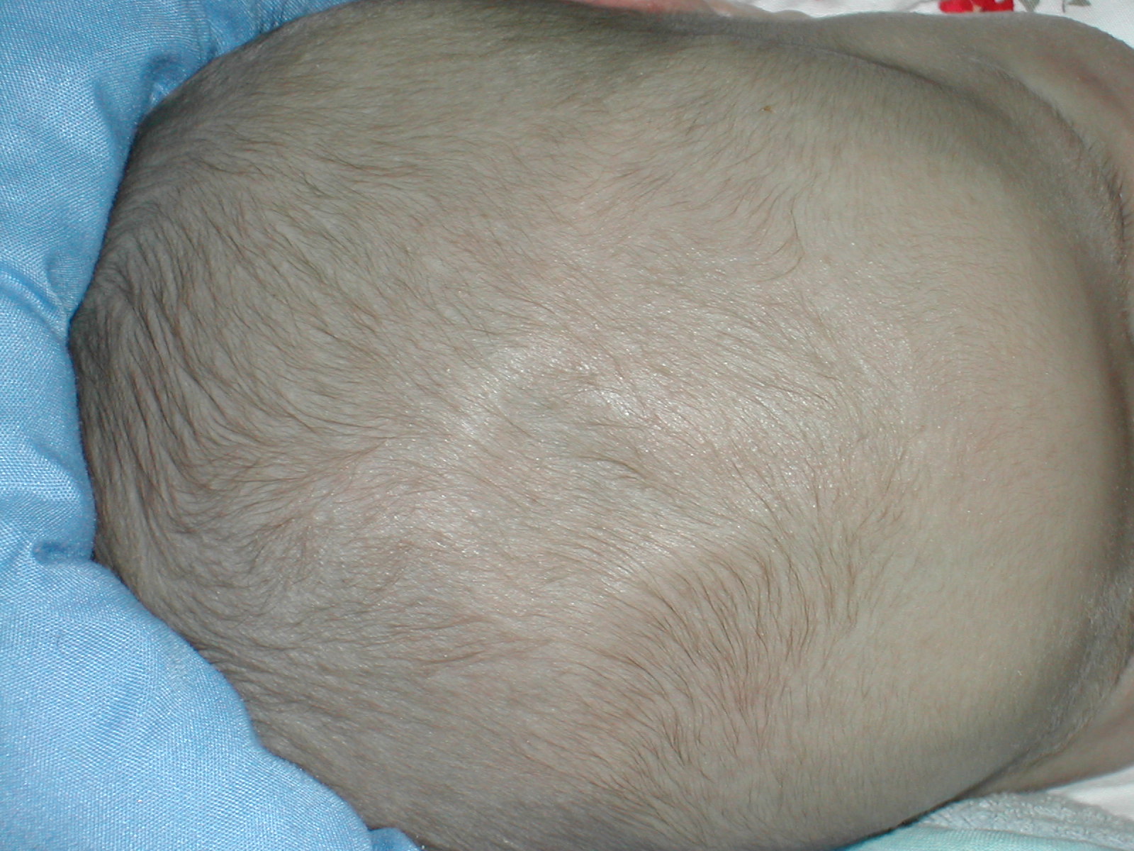
Fontanelle (other)
A fontanelle (or fontanel) (colloquially, soft spot) is an anatomical feature of the infant human skull comprising soft membranous gaps ( sutures) between the cranial bones that make up the calvaria of a fetus or an infant. Fontanelles allow for stretching and deformation of the neurocranium both during birth and later as the brain expands faster than the surrounding bone can grow. Premature complete ossification of the sutures is called craniosynostosis. After infancy, the anterior fontanelle is known as the bregma. Structure An infant's skull consists of five main bones: two frontal bones, two parietal bones, and one occipital bone. These are joined by fibrous sutures, which allow movement that facilitates childbirth and brain growth. * Posterior fontanelle is triangle-shaped. It lies at the junction between the sagittal suture and lambdoid suture. At birth, the skull features a small posterior fontanelle with an open area covered by a tough membrane, where ... [...More Info...] [...Related Items...] OR: [Wikipedia] [Google] [Baidu] |
Human Body
The human body is the structure of a Human, human being. It is composed of many different types of Cell (biology), cells that together create Tissue (biology), tissues and subsequently organ systems. They ensure homeostasis and the life, viability of the human body. It comprises a human head, head, hair, neck, Trunk (anatomy), trunk (which includes the thorax and abdomen), arms and hands, human leg, legs and feet. The study of the human body involves anatomy, physiology, histology and embryology. The body anatomical variability, varies anatomically in known ways. Physiology focuses on the systems and organs of the human body and their functions. Many systems and mechanisms interact in order to maintain homeostasis, with safe levels of substances such as sugar and oxygen in the blood. The body is studied by health professionals, physiologists, anatomists, and by artists to assist them in their work. Composition The composition of the human body, human body is composed of ... [...More Info...] [...Related Items...] OR: [Wikipedia] [Google] [Baidu] |
Childbirth
Childbirth, also known as labour and delivery, is the ending of pregnancy where one or more babies exits the internal environment of the mother via vaginal delivery or caesarean section. In 2019, there were about 140.11 million births globally. In the developed countries, most deliveries occur in hospitals, while in the developing countries most are home births. The most common childbirth method worldwide is vaginal delivery. It involves four stages of labour: the shortening and opening of the cervix during the first stage, descent and birth of the baby during the second, the delivery of the placenta during the third, and the recovery of the mother and infant during the fourth stage, which is referred to as the postpartum. The first stage is characterized by abdominal cramping or back pain that typically lasts half a minute and occurs every 10 to 30 minutes. Contractions gradually becomes stronger and closer together. Since the pain of childbirth correlates with contractions ... [...More Info...] [...Related Items...] OR: [Wikipedia] [Google] [Baidu] |
Meninges
In anatomy, the meninges (, ''singular:'' meninx ( or ), ) are the three membranes that envelop the brain and spinal cord. In mammals, the meninges are the dura mater, the arachnoid mater, and the pia mater. Cerebrospinal fluid is located in the subarachnoid space between the arachnoid mater and the pia mater. The primary function of the meninges is to protect the central nervous system. Structure Dura mater The dura mater ( la, tough mother) (also rarely called ''meninx fibrosa'' or ''pachymeninx'') is a thick, durable membrane, closest to the Human skull, skull and vertebrae. The dura mater, the outermost part, is a loosely arranged, fibroelastic layer of cells, characterized by multiple interdigitating cell processes, no extracellular collagen, and significant extracellular spaces. The middle region is a mostly fibrous portion. It consists of two layers: the endosteal layer, which lies closest to the Calvaria (skull), skull, and the inner meningeal layer, which lies closer ... [...More Info...] [...Related Items...] OR: [Wikipedia] [Google] [Baidu] |
Sphenoidal Fontanelle
The pterion is the region where the frontal, parietal, temporal, and sphenoid bones join. It is located on the side of the skull, just behind the temple. Structure The pterion is located in the temporal fossa, approximately 2.6 cm behind and 1.3 cm above the posterolateral margin of the frontozygomatic suture. It is the junction between four bones: * the parietal bone. * the squamous part of temporal bone. * the greater wing of sphenoid bone. * the frontal bone. These bones are typically joined by five cranial sutures: * the sphenoparietal suture joins the sphenoid and parietal bones. * the coronal suture joins the frontal bone to the sphenoid and parietal bones. * the squamous suture joins the temporal bone to the sphenoid and parietal bones. * the sphenofrontal suture joins the sphenoid and frontal bones. * the sphenosquamosal suture joins the sphenoid and temporal bones. Clinical significance Haematoma The pterion is known as the weakest part of the sk ... [...More Info...] [...Related Items...] OR: [Wikipedia] [Google] [Baidu] |
Mastoid Fontanelle
The asterion is a meeting point between three sutures between bones of the skull. It is an important surgical landmark. Structure In human anatomy, the asterion is a visible (craniometric) point on the exposed skull. It is just posterior to the ear. It is the point where three cranial sutures meet: * the lambdoid suture. * parietomastoid suture. * occipitomastoid suture. It is also the point where three cranial bones meet: * the parietal bone. * the occipital bone. * the mastoid portion of the temporal bone. In the adult, it lies 4 cm behind and 12 mm above the center of the entrance to the ear canal. Its relation to other anatomical structures is fairly variable. Clinical significance Neurosurgeons may use the asterion to orient themselves, in order to plan safe entry into the skull for some operations, such as when using a retro-sigmoid approach. Etymology The asterion receives its name from the Greek ἀστέριον (''astērion''), meaning "star" or "starry". T ... [...More Info...] [...Related Items...] OR: [Wikipedia] [Google] [Baidu] |
Cleidocranial Dysostosis
Cleidocranial dysostosis (CCD), also called cleidocranial dysplasia, is a birth defect that mostly affects the bones and teeth. The collarbones are typically either poorly developed or absent, which allows the shoulders to be brought close together. The front of the skull often does not close until later, and those affected are often shorter than average. Other symptoms may include a prominent forehead, wide set eyes, abnormal teeth, and a flat nose. Symptoms vary among people; however, intelligence is typically unaffected. The condition is either inherited from a person's parents or occurs as a new mutation. It is inherited in an autosomal dominant manner. It is due to a defect in the RUNX2 gene which is involved in bone formation. Diagnosis is suspected based on symptoms and X-rays with confirmation by genetic testing. Other conditions that can produce similar symptoms include mandibuloacral dysplasia, pyknodysostosis, osteogenesis imperfecta, and Hajdu-Cheney syndrome. Tre ... [...More Info...] [...Related Items...] OR: [Wikipedia] [Google] [Baidu] |
Coronal Suture
The coronal suture is a dense, fibrous connective tissue joint that separates the two parietal bones from the frontal bone of the skull. Structure The coronal suture lies between the paired parietal bones and the frontal bone of the skull. It runs from the pterion on each side. Nerve supply The coronal suture is likely supplied by a branch of the trigeminal nerve. Development The coronal suture is derived from the paraxial mesoderm. Clinical significance If certain bones of the skull grow too fast then premature fusion of the sutures may occur. This can result in skull deformities. There are two possible deformities that can be caused by the premature closure of the coronal suture: * a high, tower-like skull called "oxycephaly" or "turret skull". * a twisted and asymmetrical skull called "plagiocephaly". References * "Sagittal suture." ''Stedman's Medical Dictionary, 27th ed.'' (2000). * Moore, Keith L., and T.V.N. Persaud. ''The Developing Human: Clinically Orien ... [...More Info...] [...Related Items...] OR: [Wikipedia] [Google] [Baidu] |
Anterior Fontanelle
The anterior fontanelle (bregmatic fontanelle, frontal fontanelle) is the largest fontanelle, and is placed at the junction of the sagittal suture, coronal suture, and frontal suture; it is lozenge-shaped, and measures about 4 cm in its antero-posterior and 2.5 cm in its transverse diameter. The fontanelle allows the skull to deform during birth to ease its passage through the birth canal and for expansion of the brain after birth. The anterior fontanelle typically closes between the ages of 12 and 18 months. Clinical significance The anterior fontanelle is useful clinically. Examination of an infant includes palpating the anterior fontanelle. A sunken fontanelle indicates dehydration whereas a very tense or bulging anterior fontanelle indicates raised intracranial pressure. However, this is not a certain indicator for raised pressure as prolonged crying by the baby may produce the same effect. A full anterior fontanelle may also be indicative of neonatal meningitis, spe ... [...More Info...] [...Related Items...] OR: [Wikipedia] [Google] [Baidu] |
Lambda (anatomy)
The lambda is the meeting point of the sagittal suture and the lambdoid suture. This is also the point of the occipital angle. It is named after the Greek letter lambda. Structure The lambda is the meeting point of the sagittal suture and the lambdoid suture. It may be the exact midpoint of the lambdoid suture, but often deviates slightly from the midline. This is also the point of the occipital angle. Development In the foetus, the lambda is membranous, and is called the posterior fontanelle. Etymology The lambda is named after the Greek letter lambda Lambda (}, ''lám(b)da'') is the 11th letter of the Greek alphabet, representing the voiced alveolar lateral approximant . In the system of Greek numerals, lambda has a value of 30. Lambda is derived from the Phoenician Lamed . Lambda gave rise ..., whose lowercase form (λ) resembles the junction formed by the sutures. References External links * Skull {{musculoskeletal-stub ... [...More Info...] [...Related Items...] OR: [Wikipedia] [Google] [Baidu] |
Biological Membrane
A biological membrane, biomembrane or cell membrane is a selectively permeable membrane that separates the interior of a cell from the external environment or creates intracellular compartments by serving as a boundary between one part of the cell and another. Biological membranes, in the form of eukaryotic cell membranes, consist of a phospholipid bilayer with embedded, integral and peripheral proteins used in communication and transportation of chemicals and ions. The bulk of lipids in a cell membrane provides a fluid matrix for proteins to rotate and laterally diffuse for physiological functioning. Proteins are adapted to high membrane fluidity environment of the lipid bilayer with the presence of an annular lipid shell, consisting of lipid molecules bound tightly to the surface of integral membrane proteins. The cell membranes are different from the isolating tissues formed by layers of cells, such as mucous membranes, basement membranes, and serous membranes. Composition ... [...More Info...] [...Related Items...] OR: [Wikipedia] [Google] [Baidu] |
Lambdoid Suture
The lambdoid suture (or lambdoidal suture) is a dense, fibrous connective tissue joint on the posterior aspect of the skull that connects the parietal bones with the occipital bone. It is continuous with the occipitomastoid suture. Structure The lambdoid suture is between the paired parietal bones and the occipital bone of the skull. It runs from the asterion on each side. Nerve supply The lambdoid suture may be supplied by a branch of the supraorbital nerve, a branch of the frontal branch of the trigeminal nerve. Clinical significance At birth, the bones of the skull do not meet. If certain bones of the skull grow too fast, then craniosynostosis (premature closure of the sutures) may occur. This can result in skull deformities. If the lambdoid suture closes too soon on one side, the skull will appear twisted and asymmetrical, a condition called "plagiocephaly". Plagiocephaly refers to the shape and not the condition. The condition is craniosynostosis. The lambdoid sut ... [...More Info...] [...Related Items...] OR: [Wikipedia] [Google] [Baidu] |
Sagittal Suture
The sagittal suture, also known as the interparietal suture and the ''sutura interparietalis'', is a dense, fibrous connective tissue joint between the two parietal bones of the skull. The term is derived from the Latin word ''sagitta'', meaning arrow. Structure The sagittal suture is formed from the fibrous connective tissue joint between the two parietal bones of the skull. It has a varied and irregular shape which arises during development. The pattern is different between the inside and the outside. Two anatomical landmarks are found on the sagittal suture: the bregma, and the vertex of the skull. The bregma is formed by the intersection of the sagittal and coronal sutures. The vertex is the highest point on the skull and is often near the midpoint of the sagittal suture. Development At birth, the bones of the skull do not meet. The gap that remains, which is approximately 5 mm wide, allows for the brain to continue to grow normally after birth. The inner parts of ... [...More Info...] [...Related Items...] OR: [Wikipedia] [Google] [Baidu] |


