|
Fibrin Ring Granuloma
A fibrin ring granuloma, also known as doughnut granuloma, is a histopathological finding that is characteristic of Q fever. On hematoxylin-eosin staining, the fibrin ring granuloma consists of a central lipid vacuole (usually washed-out during fixing and staining, leaving only an empty hole) surrounded by a dense red fibrin ring and epithelioid macrophages. Fibrin ring granulomas may also be seen in Hodgkin's disease and infectious mononucleosis Infectious mononucleosis (IM, mono), also known as glandular fever, is an infection usually caused by the Epstein–Barr virus (EBV). Most people are infected by the virus as children, when the disease produces few or no symptoms. In young adult .... See also * Granuloma References Histopathology {{Pathology-stub ... [...More Info...] [...Related Items...] OR: [Wikipedia] [Google] [Baidu] |
Histopathology
Histopathology (compound of three Greek words: ''histos'' "tissue", πάθος ''pathos'' "suffering", and -λογία '' -logia'' "study of") refers to the microscopic examination of tissue in order to study the manifestations of disease. Specifically, in clinical medicine, histopathology refers to the examination of a biopsy or surgical specimen by a pathologist, after the specimen has been processed and histological sections have been placed onto glass slides. In contrast, cytopathology examines free cells or tissue micro-fragments (as "cell blocks"). Collection of tissues Histopathological examination of tissues starts with surgery, biopsy, or autopsy. The tissue is removed from the body or plant, and then, often following expert dissection in the fresh state, placed in a fixative which stabilizes the tissues to prevent decay. The most common fixative is 10% neutral buffered formalin (corresponding to 3.7% w/v formaldehyde in neutral buffered water, such as phosphate buf ... [...More Info...] [...Related Items...] OR: [Wikipedia] [Google] [Baidu] |
Q Fever
Q fever or query fever is a disease caused by infection with ''Coxiella burnetii'', a bacterium that affects humans and other animals. This organism is uncommon, but may be found in cattle, sheep, goats, and other domestic mammals, including cats and dogs. The infection results from inhalation of a spore-like small-cell variant, and from contact with the milk, urine, feces, vaginal mucus, or semen of infected animals. Rarely, the disease is tick-borne. The incubation period is 9–40 days. Humans are vulnerable to Q fever, and infection can result from even a few organisms. The bacterium is an obligate intracellular pathogenic parasite. Signs and symptoms Incubation period is usually two to three weeks. The most common manifestation is flu-like symptoms: abrupt onset of fever, malaise, profuse perspiration, severe headache, muscle pain, joint pain, loss of appetite, upper respiratory problems, dry cough, pleuritic pain, chills, confusion, and gastrointestinal s ... [...More Info...] [...Related Items...] OR: [Wikipedia] [Google] [Baidu] |
H&E Stain
Hematoxylin and eosin stain ( or haematoxylin and eosin stain or hematoxylin-eosin stain; often abbreviated as H&E stain or HE stain) is one of the principal tissue stains used in histology. It is the most widely used stain in medical diagnosis and is often the gold standard. For example, when a pathologist looks at a biopsy of a suspected cancer, the histological section is likely to be stained with H&E. H&E is the combination of two histological stains: hematoxylin and eosin. The hematoxylin stains cell nuclei a purplish blue, and eosin stains the extracellular matrix and cytoplasm pink, with other structures taking on different shades, hues, and combinations of these colors. Hence a pathologist can easily differentiate between the nuclear and cytoplasmic parts of a cell, and additionally, the overall patterns of coloration from the stain show the general layout and distribution of cells and provides a general overview of a tissue sample's structure. Thus, pattern recogniti ... [...More Info...] [...Related Items...] OR: [Wikipedia] [Google] [Baidu] |
Fibrin
Fibrin (also called Factor Ia) is a fibrous, non-globular protein involved in the clotting of blood. It is formed by the action of the protease thrombin on fibrinogen, which causes it to polymerize. The polymerized fibrin, together with platelets, forms a hemostatic plug or clot over a wound site. When the lining of a blood vessel is broken, platelets are attracted, forming a platelet plug. These platelets have thrombin receptors on their surfaces that bind serum thrombin molecules, which in turn convert soluble fibrinogen in the serum into fibrin at the wound site. Fibrin forms long strands of tough insoluble protein that are bound to the platelets. Factor XIII completes the cross-linking of fibrin so that it hardens and contracts. The cross-linked fibrin forms a mesh atop the platelet plug that completes the clot. Fibrin was discovered by Marcello Malpighi in 1666. Role in disease Excessive generation of fibrin due to activation of the coagulation cascade leads to thrombos ... [...More Info...] [...Related Items...] OR: [Wikipedia] [Google] [Baidu] |
Granuloma
A granuloma is an aggregation of macrophages that forms in response to chronic inflammation. This occurs when the immune system attempts to isolate foreign substances that it is otherwise unable to eliminate. Such substances include infectious organisms including bacteria and fungi, as well as other materials such as foreign objects, keratin, and suture fragments. Definition In pathology, a granuloma is an organized collection of macrophages. In medical practice, doctors occasionally use the term ''granuloma'' in its more literal meaning: "a small nodule". Since a small nodule can represent any tissue from a harmless nevus to a malignant tumor, this use of the term is not very specific. Examples of this use of the term ''granuloma'' are the lesions known as vocal cord granuloma (known as contact granuloma), pyogenic granuloma, and intubation granuloma, all of which are examples of granulation tissue, not granulomas. "Pulmonary hyalinizing granuloma" is a lesion characterized ... [...More Info...] [...Related Items...] OR: [Wikipedia] [Google] [Baidu] |
Lipid
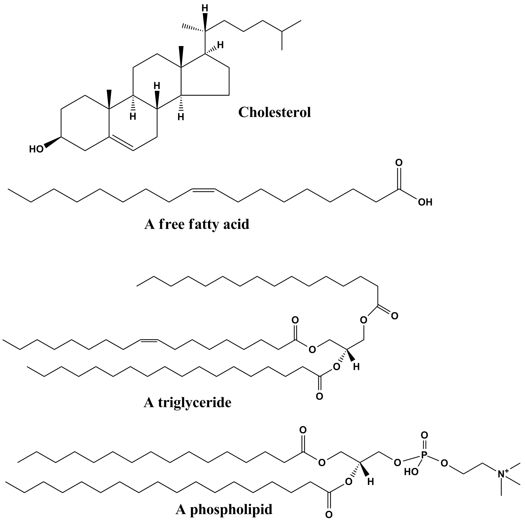
Lipids are a broad group of naturally-occurring molecules which includes fats, waxes, sterols, fat-soluble vitamins (such as vitamins A, D, E and K), monoglycerides, diglycerides, phospholipids, and others. The functions of lipids include storing energy, signaling, and acting as structural components of cell membranes. Lipids have applications in the cosmetic and food industries, and in nanotechnology. Lipids may be broadly defined as hydrophobic or amphiphilic small molecules; the amphiphilic nature of some lipids allows them to form structures such as vesicles, multilamellar/unilamellar liposomes, or membranes in an aqueous environment. Biological lipids originate entirely or in part from two distinct types of biochemical subunits or "building-blocks": ketoacyl and isoprene groups. Using this approach, lipids may be divided into eight categories: fatty acyls, glycerolipids, glycerophospholipids, sphingolipids, saccharolipids, and polyketides (derived from condensati ... [...More Info...] [...Related Items...] OR: [Wikipedia] [Google] [Baidu] |
Vacuole
A vacuole () is a membrane-bound organelle which is present in plant and fungal cells and some protist, animal, and bacterial cells. Vacuoles are essentially enclosed compartments which are filled with water containing inorganic and organic molecules including enzymes in solution, though in certain cases they may contain solids which have been engulfed. Vacuoles are formed by the fusion of multiple membrane vesicles and are effectively just larger forms of these. The organelle has no basic shape or size; its structure varies according to the requirements of the cell. Discovery Contractile vacuoles ("stars") were first observed by Spallanzani (1776) in protozoa, although mistaken for respiratory organs. Dujardin (1841) named these "stars" as ''vacuoles''. In 1842, Schleiden applied the term for plant cells, to distinguish the structure with cell sap from the rest of the protoplasm. In 1885, de Vries named the vacuole membrane as tonoplast. Function The function and signifi ... [...More Info...] [...Related Items...] OR: [Wikipedia] [Google] [Baidu] |
Epithelioid Macrophage
According to a common point of view epithelioid cells (also called epithelioid histiocytes) are derivatives of activated macrophages resembling epithelial cells. Structure and function Structurally, epithelioid cells (when examined by light microscopy after stained with hematoxylin and eosin), are elongated, with finely granular, pale eosinophilic (pink) cytoplasm, and central, ovoid nuclei (oval or elongate), which are less dense than that of a lymphocyte. They have indistinct shape and often appear to merge into one another, forming aggregates known as giant cells. When examined by transmission electron microscopy in epithelioid cells in the field of Golgi lamellar complex are taped not only zonated, but also sleek vesicles with dense center, and also great many (more than 100) large granulas with diameters up to 340 nm and with finegranular matrix more light than in macrophage granulas, sometimes with perigranular halo. “The most prominent feature of these cells is the ... [...More Info...] [...Related Items...] OR: [Wikipedia] [Google] [Baidu] |
Hodgkin's Disease
Hodgkin lymphoma (HL) is a type of lymphoma, in which cancer originates from a specific type of white blood cell called lymphocytes, where multinucleated Reed–Sternberg cells (RS cells) are present in the patient's lymph nodes. The condition was named after the English physician Thomas Hodgkin, who first described it in 1832. Symptoms may include fever, night sweats, and weight loss. Often, nonpainful enlarged lymph nodes occur in the neck, under the arm, or in the groin. Those affected may feel tired or be itchy. The two major types of Hodgkin lymphoma are classic Hodgkin lymphoma and nodular lymphocyte-predominant Hodgkin lymphoma. About half of cases of Hodgkin lymphoma are due to Epstein–Barr virus (EBV) and these are generally the classic form. Other risk factors include a family history of the condition and having HIV/AIDS. Diagnosis is conducted by confirming the presence of cancer and identifying RS cells in lymph node biopsies. The virus-positive cases are classified ... [...More Info...] [...Related Items...] OR: [Wikipedia] [Google] [Baidu] |
Infectious Mononucleosis
Infectious mononucleosis (IM, mono), also known as glandular fever, is an infection usually caused by the Epstein–Barr virus (EBV). Most people are infected by the virus as children, when the disease produces few or no symptoms. In young adults, the disease often results in fever, sore throat, enlarged lymph nodes in the neck, and tiredness. Most people recover in two to four weeks; however, feeling tired may last for months. The liver or spleen may also become swollen, and in less than one percent of cases splenic rupture may occur. While usually caused by the Epstein–Barr virus, also known as human herpesvirus 4, which is a member of the herpesvirus family, a few other viruses may also cause the disease. It is primarily spread through saliva but can rarely be spread through semen or blood. Spread may occur by objects such as drinking glasses or toothbrushes or through a cough or sneeze. Those who are infected can spread the disease weeks before symptoms develop. Mono is p ... [...More Info...] [...Related Items...] OR: [Wikipedia] [Google] [Baidu] |
Granuloma
A granuloma is an aggregation of macrophages that forms in response to chronic inflammation. This occurs when the immune system attempts to isolate foreign substances that it is otherwise unable to eliminate. Such substances include infectious organisms including bacteria and fungi, as well as other materials such as foreign objects, keratin, and suture fragments. Definition In pathology, a granuloma is an organized collection of macrophages. In medical practice, doctors occasionally use the term ''granuloma'' in its more literal meaning: "a small nodule". Since a small nodule can represent any tissue from a harmless nevus to a malignant tumor, this use of the term is not very specific. Examples of this use of the term ''granuloma'' are the lesions known as vocal cord granuloma (known as contact granuloma), pyogenic granuloma, and intubation granuloma, all of which are examples of granulation tissue, not granulomas. "Pulmonary hyalinizing granuloma" is a lesion characterized ... [...More Info...] [...Related Items...] OR: [Wikipedia] [Google] [Baidu] |


_mixed_cellulary_type.jpg)
