|
Feet
The foot ( : feet) is an anatomical structure found in many vertebrates. It is the terminal portion of a limb which bears weight and allows locomotion. In many animals with feet, the foot is a separate organ at the terminal part of the leg made up of one or more segments or bones, generally including claws or nails. Etymology The word "foot", in the sense of meaning the "terminal part of the leg of a vertebrate animal" comes from "Old English fot "foot," from Proto-Germanic *fot (source also of Old Frisian fot, Old Saxon fot, Old Norse fotr, Danish fod, Swedish fot, Dutch voet, Old High German fuoz, German Fuß, Gothic fotus "foot"), from PIE root *ped- "foot". The "plural form feet is an instance of i-mutation." Structure The human foot is a strong and complex mechanical structure containing 26 bones, 33 joints (20 of which are actively articulated), and more than a hundred muscles, tendons, and ligaments.Podiatry Channel, ''Anatomy of the foot and ankle'' The joints of the ... [...More Info...] [...Related Items...] OR: [Wikipedia] [Google] [Baidu] |
Arches Of The Foot
The arches of the foot, formed by the tarsus (skeleton), tarsal and metatarsal bones, strengthened by ligaments and tendons, allow the foot to support the weight of the body in the erect posture with the least weight. They are categorized as longitudinal and transverse arches. Structure Longitudinal arches The longitudinal arches of the foot can be divided into medial and lateral arches. Medial arch The medial arch is higher than the lateral longitudinal arch. It is made up by the calcaneus, the talus bone, talus, the navicular, the three Cuneiform (anatomy), cuneiforms (medial, intermediate, and lateral), and the first, second, and third metatarsals. Its summit is at the superior articular surface of the talus, and its two extremities or piers, on which it rests in standing, are the tuberosity on the plantar surface of the calcaneus posteriorly and the heads of the first, second, and third metatarsal bones anteriorly. The chief characteristic of this arch is its elasticity, ... [...More Info...] [...Related Items...] OR: [Wikipedia] [Google] [Baidu] |
Animal Locomotion
Animal locomotion, in ethology, is any of a variety of methods that animal (biology), animals use to move from one place to another. Some modes of locomotion are (initially) self-propelled, e.g., running, swimming, jumping, flying, hopping, soaring and gliding. There are also many animal species that depend on their environment for transportation, a type of mobility called Passive locomotion in animals, passive locomotion, e.g., sailing (some jellyfish), Ballooning (spider), kiting (spiders), rolling (some beetles and spiders) or riding other animals (phoresis (biology), phoresis). Animals move for a variety of reasons, such as to find Foraging, food, a Mating system, mate, a suitable microhabitat, or to Escape response, escape predators. For many animals, the ability to move is essential for survival and, as a result, natural selection has shaped the locomotion methods and mechanisms used by moving organisms. For example, Animal migration, migratory animals that travel vast dista ... [...More Info...] [...Related Items...] OR: [Wikipedia] [Google] [Baidu] |
Limb (anatomy)
A limb (from the Old English ''lim'', meaning "body part") or leg is a jointed, muscled appendage that tetrapod vertebrates use for weight-bearing and terrestrial locomotion such as walking, running and jumping, for paddle-swimming, or for grasping and climbing. The distalmost portion of a limb is known as its extremity. The limbs' bony endoskeleton, known as the appendicular skeleton, is homologous among all tetrapods. All tetrapods have four limbs that are organized into two bilaterally symmetrical pairs, with one pair at each end of the torso. The cranial pair are known as the forelimbs or ''front legs'', and the caudal pair the hindlimbs or ''back legs''. In animals with more upright posture (mainly hominid primates, particularly humans), the forelimbs and hindlimbs are often called upper and lower limbs, respectively. The fore-/upper limbs are connected to the thoracic cage via the shoulder girdles, and the hind-/lower limbs are connected to the pelvis via the hip ... [...More Info...] [...Related Items...] OR: [Wikipedia] [Google] [Baidu] |
Metti (cropped)
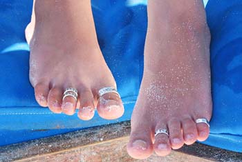
A toe ring is a ring made out of metals and non-metals worn on any of the toes. The second toe of either foot is where they are worn most commonly. This is because proportionately it is the longest toe and thus the easiest toe to put a ring on and stay without being connected to anything else. In most western countries they are a relatively new fashion accessory, and typically have no symbolic meaning. They are usually worn with barefoot sandals, anklets, bare feet or flip flops. Like finger rings, toe rings come in many shapes and forms, from intricately designed flowers embedded with jewels to simple bands. Fitted toe rings are rings that are of one size, whereas adjustable toe rings have a gap at the bottom so they can be easily made to fit snugly. Toe rings in India The wearing of toe rings has been practised in India since ancient times. In the Ramayana, there is a mention of Sita, on being abducted by Ravana, throwing her toe ring down so that lord Rama could find her. ... [...More Info...] [...Related Items...] OR: [Wikipedia] [Google] [Baidu] |
Anatomical
Anatomy () is the branch of biology concerned with the study of the structure of organisms and their parts. Anatomy is a branch of natural science that deals with the structural organization of living things. It is an old science, having its beginnings in prehistoric times. Anatomy is inherently tied to developmental biology, embryology, comparative anatomy, evolutionary biology, and phylogeny, as these are the processes by which anatomy is generated, both over immediate and long-term timescales. Anatomy and physiology, which study the structure and function of organisms and their parts respectively, make a natural pair of related disciplines, and are often studied together. Human anatomy is one of the essential basic sciences that are applied in medicine. The discipline of anatomy is divided into macroscopic and microscopic. Macroscopic anatomy, or gross anatomy, is the examination of an animal's body parts using unaided eyesight. Gross anatomy also includes the branch o ... [...More Info...] [...Related Items...] OR: [Wikipedia] [Google] [Baidu] |
Navicular Bone
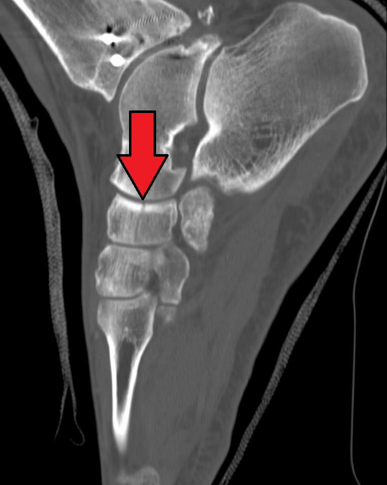
The navicular bone is a small bone found in the feet of most mammals. Human anatomy The navicular bone in humans is one of the tarsal bones, found in the foot. Its name derives from the human bone's resemblance to a small boat, caused by the strongly concave proximal articular surface. The term ''navicular bone'' or ''hand navicular bone'' was formerly used for the scaphoid bone, one of the carpal bones of the wrist. The navicular bone in humans is located on the medial side of the foot, and articulates proximally with the talus, distally with the three cuneiform bones, and laterally with the cuboid. It is the last of the foot bones to start ossification and does not tend to do so until the end of the third year in girls and the beginning of the fourth year in boys, although a large range of variation has been reported. The tibialis posterior is the only muscle that attaches to the navicular bone. The main portion of the muscle inserts into the tuberosity of the navi ... [...More Info...] [...Related Items...] OR: [Wikipedia] [Google] [Baidu] |
Talus Bone
The talus (; Latin for ankle or ankle bone), talus bone, astragalus (), or ankle bone is one of the group of foot bones known as the tarsus. The tarsus forms the lower part of the ankle joint. It transmits the entire weight of the body from the lower legs to the foot.Platzer (2004), p 216 The talus has joints with the two bones of the lower leg, the tibia and thinner fibula. These leg bones have two prominences (the lateral and medial malleoli) that articulate with the talus. At the foot end, within the tarsus, the talus articulates with the calcaneus (heel bone) below, and with the curved navicular bone in front; together, these foot articulations form the ball-and-socket-shaped talocalcaneonavicular joint. The talus is the second largest of the tarsal bones; it is also one of the bones in the human body with the highest percentage of its surface area covered by articular cartilage. It is also unusual in that it has a retrograde blood supply, i.e. arterial blood enters the ... [...More Info...] [...Related Items...] OR: [Wikipedia] [Google] [Baidu] |
Interphalangeal Joints Of The Foot
The interphalangeal joints of the foot are between the phalanx bones of the toes in the feet. Since the great toe only has two phalanx bones (proximal and distal phalanges), it only has one interphalangeal joint, which is often abbreviated as the "IP joint". The rest of the toes each have three phalanx bones (proximal, middle, and distal phalanges), so they have two interphalangeal joints: the proximal interphalangeal joint between the proximal and middle phalanges (abbreviated "PIP joint") and the distal interphalangeal joint between the middle and distal phalanges (abbreviated "DIP joint"). All interphalangeal joints are ginglymoid (hinge) joints, and each has a plantar (underside) and two collateral ligaments. In the arrangement of these ligaments, extensor tendons supply the places of dorsal ligaments, which is similar to that in the metatarsophalangeal articulations. Movements The only movements permitted in the joints of the digits are flexion and extension; these movem ... [...More Info...] [...Related Items...] OR: [Wikipedia] [Google] [Baidu] |
Subtalar Joint
In human anatomy, the subtalar joint, also known as the talocalcaneal joint, is a joint of the foot. It occurs at the meeting point of the Talus bone, talus and the calcaneus. The joint is classed structurally as a synovial joint, and functionally as a plane joint. Structure The talus is oriented slightly obliquely on the anterior surface of the calcaneus. There are three points of articulation between the two bones: two anteriorly and one posteriorly. The three articulations are known as facets, and they are the posterior, middle and anterior facets. * At the ''anterior and middle talocalcaneal articulation'', wikt:convex, convex areas of the talus fits on to wikt:concave, concave surfaces of the calcaneus. * The ''posterior talocalcaneal articulation'' is formed by a concave surface of the talus and a convex surface of the calcaneus. The sustentaculum tali forms the floor of middle facet, and the anterior facet articulates with the head of the talus, and sits lateral and co ... [...More Info...] [...Related Items...] OR: [Wikipedia] [Google] [Baidu] |
Ankle
The ankle, or the talocrural region, or the jumping bone (informal) is the area where the foot and the leg meet. The ankle includes three joints: the ankle joint proper or talocrural joint, the subtalar joint, and the inferior tibiofibular joint. The movements produced at this joint are dorsiflexion and plantarflexion of the foot. In common usage, the term ankle refers exclusively to the ankle region. In medical terminology, "ankle" (without qualifiers) can refer broadly to the region or specifically to the talocrural joint. The main bones of the ankle region are the talus (in the foot), and the tibia and fibula (in the leg). The talocrural joint is a synovial hinge joint that connects the distal ends of the tibia and fibula in the lower limb with the proximal end of the talus. The articulation between the tibia and the talus bears more weight than that between the smaller fibula and the talus. Structure Region The ankle region is found at the junction of the leg and the f ... [...More Info...] [...Related Items...] OR: [Wikipedia] [Google] [Baidu] |
Fibula
The fibula or calf bone is a leg bone on the lateral side of the tibia, to which it is connected above and below. It is the smaller of the two bones and, in proportion to its length, the most slender of all the long bones. Its upper extremity is small, placed toward the back of the head of the tibia, below the knee joint and excluded from the formation of this joint. Its lower extremity inclines a little forward, so as to be on a plane anterior to that of the upper end; it projects below the tibia and forms the lateral part of the ankle joint. Structure The bone has the following components: * Lateral malleolus * Interosseous membrane connecting the fibula to the tibia, forming a syndesmosis joint * The superior tibiofibular articulation is an arthrodial joint between the lateral condyle of the tibia and the head of the fibula. * The inferior tibiofibular articulation (tibiofibular syndesmosis) is formed by the rough, convex surface of the medial side of the lower end of the f ... [...More Info...] [...Related Items...] OR: [Wikipedia] [Google] [Baidu] |
Tibia
The tibia (; ), also known as the shinbone or shankbone, is the larger, stronger, and anterior (frontal) of the two bones in the leg below the knee in vertebrates (the other being the fibula, behind and to the outside of the tibia); it connects the knee with the ankle. The tibia is found on the medial side of the leg next to the fibula and closer to the median plane. The tibia is connected to the fibula by the interosseous membrane of leg, forming a type of fibrous joint called a syndesmosis with very little movement. The tibia is named for the flute ''tibia''. It is the second largest bone in the human body, after the femur. The leg bones are the strongest long bones as they support the rest of the body. Structure In human anatomy, the tibia is the second largest bone next to the femur. As in other vertebrates the tibia is one of two bones in the lower leg, the other being the fibula, and is a component of the knee and ankle joints. The ossification or formation of the bone ... [...More Info...] [...Related Items...] OR: [Wikipedia] [Google] [Baidu] |


.jpg)

