|
Fascin
Fascin is an actin bundling protein. Species and tissue distribution It is a 54-58 kilodalton monomeric actin filament bundling protein originally isolated from sea urchin egg but also found in ''Drosophila'' and vertebrates, including humans. Fascin (from the Latin for ''bundle'') is spaced at 11 nanometre intervals along the Protein filament, filament. The bundles in cross section are seen to be hexagonally packed, and the longitudinal spacing is compatible with a model where fascin cross-links at alternating 4 and 5 actins. It is calcium insensitive and monomeric. Three forms of fascin are found in vertebrates: Fascin1, widely found in the nervous system and elsewhere; fascin2 found in the retinal photoreceptor cells; fascin3, which is only found in the testes. Function Fascin binds beta-catenin, and colocalizes with it at the leading edges and borders of epithelial and endothelial cells. The role of Fascin in regulating cytoskeletal structures for the maint ... [...More Info...] [...Related Items...] OR: [Wikipedia] [Google] [Baidu] |
FSCN1
Fascin is a protein that in humans is encoded by the ''FSCN1'' gene. Interactions FSCN1 has been shown to Protein-protein interaction, interact with Low affinity nerve growth factor receptor and PKC alpha. References Further reading * * * * * * * * * * * * * * * * * {{gene-7-stub ... [...More Info...] [...Related Items...] OR: [Wikipedia] [Google] [Baidu] |
FSCN2
Fascin-2 is a protein that in humans is encoded by the ''FSCN2'' gene. This gene encodes a member of the fascin Fascin is an actin bundling protein. Species and tissue distribution It is a 54-58 kilodalton monomeric actin filament bundling protein originally isolated from sea urchin egg but also found in ''Drosophila'' and vertebrates, including ... protein family. Fascins crosslink actin into filamentous bundles within dynamic cell extensions. This family member is proposed to play a role in photoreceptor disk morphogenesis. A mutation in this gene results in one form of autosomal dominant retinitis pigmentosa and macular degeneration. Multiple transcript variants encoding different isoforms have been found for this gene. References Further reading * * * * * * * * * * External links GeneReviews/NCBI/NIH/UW entry on Retinitis Pigmentosa Overview {{gene-17-stub ... [...More Info...] [...Related Items...] OR: [Wikipedia] [Google] [Baidu] |
FSCN3
Fascin-3 also known as testis fascin is a protein that in humans is encoded by the ''FSCN3'' gene. See also * Fascin Fascin is an actin bundling protein. Species and tissue distribution It is a 54-58 kilodalton monomeric actin filament bundling protein originally isolated from sea urchin egg but also found in ''Drosophila'' and vertebrates, including ... References Further reading * * * * * * {{gene-7-stub ... [...More Info...] [...Related Items...] OR: [Wikipedia] [Google] [Baidu] |
Photoreceptor Cell
A photoreceptor cell is a specialized type of neuroepithelial cell found in the retina that is capable of visual phototransduction. The great biological importance of photoreceptors is that they convert light (visible electromagnetic radiation) into signals that can stimulate biological processes. To be more specific, photoreceptor proteins in the cell absorb photons, triggering a change in the cell's membrane potential. There are currently three known types of photoreceptor cells in mammalian eyes: rods, cones, and intrinsically photosensitive retinal ganglion cells. The two classic photoreceptor cells are rods and cones, each contributing information used by the visual system to form an image of the environment, sight. Rods primarily mediate scotopic vision (dim conditions) whereas cones primarily mediate to photopic vision (bright conditions), but the processes in each that supports phototransduction is similar. A third class of mammalian photoreceptor cell was discovered ... [...More Info...] [...Related Items...] OR: [Wikipedia] [Google] [Baidu] |
Signalling Pathway
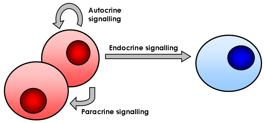
In biology, cell signaling (cell signalling in British English) or cell communication is the ability of a cell to receive, process, and transmit signals with its environment and with itself. Cell signaling is a fundamental property of all cellular life in prokaryotes and eukaryotes. Signals that originate from outside a cell (or extracellular signals) can be physical agents like mechanical pressure, voltage, temperature, light, or chemical signals (e.g., small molecules, peptides, or gas). Cell signaling can occur over short or long distances, and as a result can be classified as autocrine, juxtacrine, intracrine, paracrine, or endocrine. Signaling molecules can be synthesized from various biosynthetic pathways and released through passive or active transports, or even from cell damage. Receptors play a key role in cell signaling as they are able to detect chemical signals or physical stimuli. Receptors are generally proteins located on the cell surface or within the interior ... [...More Info...] [...Related Items...] OR: [Wikipedia] [Google] [Baidu] |
Cellular Infiltration
Infiltration is the diffusion or accumulation (in a tissue or cells) of foreign substances in amounts excess of the normal. The material collected in those tissues or cells is called infiltrate. Definitions of infiltration As part of a disease process, infiltration is sometimes used to define the invasion of cancer cells into the underlying matrix or the blood vessels. Similarly, the term may describe the deposition of amyloid protein. During leukocyte extravasation, white blood cells move in response to cytokines from within the blood, into the diseased or infected tissues, usually in the same direction as a chemical gradient,Kumar et al. 2014, p. 36 in a process called chemotaxis. The presence of lymphocytes in tissue in greater than normal numbers is likewise called infiltration. As part of medical intervention, local anaesthetics may be injected at more than one point so as to infiltrate an area prior to a surgical procedure. However, the term may also apply to unintended iatr ... [...More Info...] [...Related Items...] OR: [Wikipedia] [Google] [Baidu] |
Motility
Motility is the ability of an organism to move independently, using metabolic energy. Definitions Motility, the ability of an organism to move independently, using metabolic energy, can be contrasted with sessility, the state of organisms that do not possess a means of self-locomotion and are normally immobile. Motility differs from mobility, the ability of an object to be moved. The term vagility encompasses both motility and mobility; sessile organisms including plants and fungi often have vagile parts such as fruits, seeds, or spores which may be dispersed by other agents such as wind, water, or other organisms. Motility is genetically determined, but may be affected by environmental factors such as toxins. The nervous system and musculoskeletal system provide the majority of mammalian motility. In addition to animal locomotion, most animals are motile, though some are vagile, described as having passive locomotion. Many bacteria and other microorganisms, and multicellu ... [...More Info...] [...Related Items...] OR: [Wikipedia] [Google] [Baidu] |
Cell Adhesion
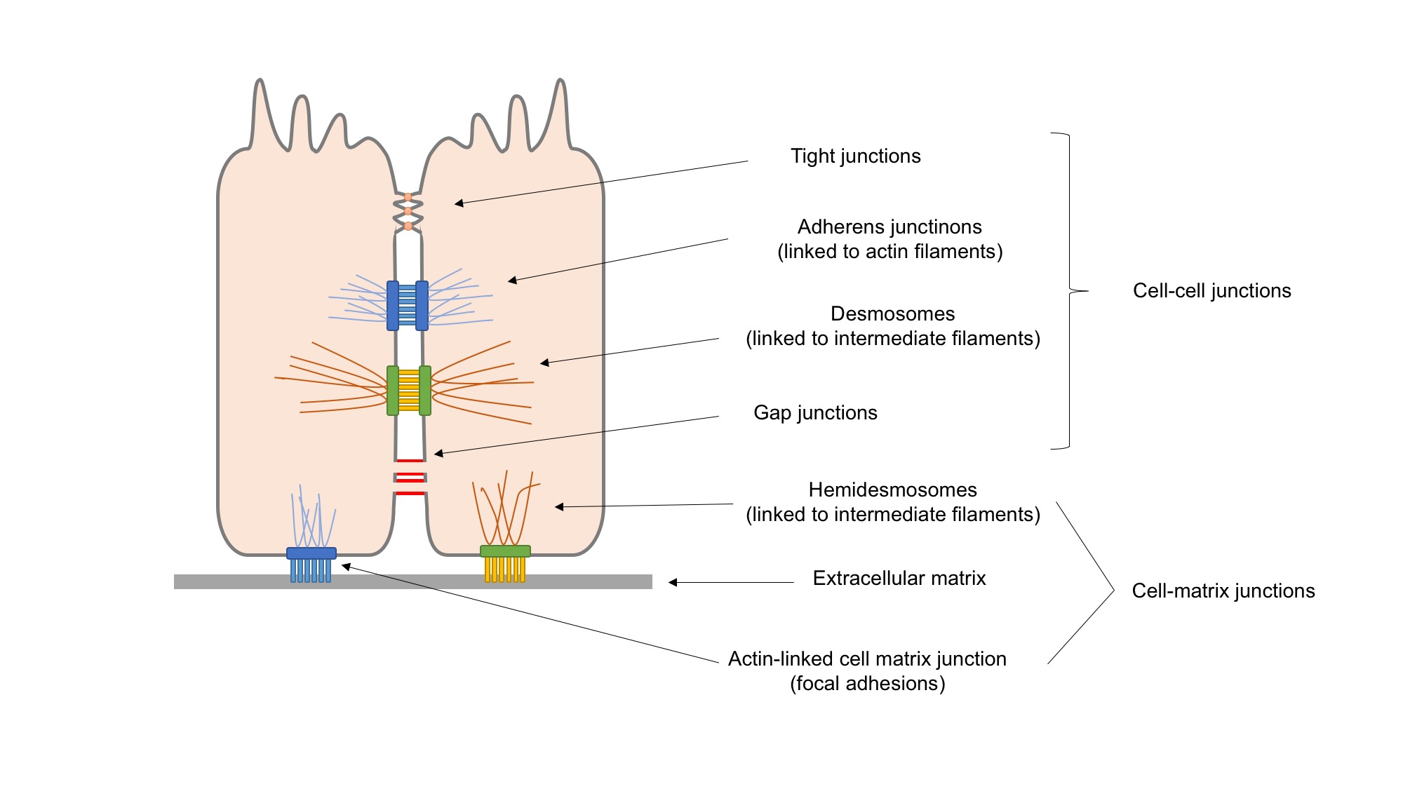
Cell adhesion is the process by which cells interact and attach to neighbouring cells through specialised molecules of the cell surface. This process can occur either through direct contact between cell surfaces such as cell junctions or indirect interaction, where cells attach to surrounding extracellular matrix, a gel-like structure containing molecules released by cells into spaces between them. Cells adhesion occurs from the interactions between cell-adhesion molecules (CAMs), transmembrane proteins located on the cell surface. Cell adhesion links cells in different ways and can be involved in signal transduction for cells to detect and respond to changes in the surroundings. Other cellular processes regulated by cell adhesion include cell migration and tissue development in multicellular organisms. Alterations in cell adhesion can disrupt important cellular processes and lead to a variety of diseases, including cancer and arthritis. Cell adhesion is also essential for in ... [...More Info...] [...Related Items...] OR: [Wikipedia] [Google] [Baidu] |
Cytoskeletal
The cytoskeleton is a complex, dynamic network of interlinking protein filaments present in the cytoplasm of all cells, including those of bacteria and archaea. In eukaryotes, it extends from the cell nucleus to the cell membrane and is composed of similar proteins in the various organisms. It is composed of three main components, microfilaments, intermediate filaments and microtubules, and these are all capable of rapid growth or disassembly dependent on the cell's requirements. A multitude of functions can be performed by the cytoskeleton. Its primary function is to give the cell its shape and mechanical resistance to deformation, and through association with extracellular connective tissue and other cells it stabilizes entire tissues. The cytoskeleton can also contract, thereby deforming the cell and the cell's environment and allowing cells to migrate. Moreover, it is involved in many cell signaling pathways and in the uptake of extracellular material ( endocytosis), the ... [...More Info...] [...Related Items...] OR: [Wikipedia] [Google] [Baidu] |
Endothelial
The endothelium is a single layer of squamous endothelial cells that line the interior surface of blood vessels and lymphatic vessels. The endothelium forms an interface between circulating blood or lymph in the lumen and the rest of the vessel wall. Endothelial cells form the barrier between vessels and tissue and control the flow of substances and fluid into and out of a tissue. Endothelial cells in direct contact with blood are called vascular endothelial cells whereas those in direct contact with lymph are known as lymphatic endothelial cells. Vascular endothelial cells line the entire circulatory system, from the heart to the smallest capillaries. These cells have unique functions that include fluid filtration, such as in the glomerulus of the kidney, blood vessel tone, hemostasis, neutrophil recruitment, and hormone trafficking. Endothelium of the interior surfaces of the heart chambers is called endocardium. An impaired function can lead to serious health issues throug ... [...More Info...] [...Related Items...] OR: [Wikipedia] [Google] [Baidu] |
Epithelial
Epithelium or epithelial tissue is one of the four basic types of animal tissue, along with connective tissue, muscle tissue and nervous tissue. It is a thin, continuous, protective layer of compactly packed cells with a little intercellular matrix. Epithelial tissues line the outer surfaces of organs and blood vessels throughout the body, as well as the inner surfaces of cavities in many internal organs. An example is the epidermis, the outermost layer of the skin. There are three principal shapes of epithelial cell: squamous (scaly), columnar, and cuboidal. These can be arranged in a singular layer of cells as simple epithelium, either squamous, columnar, or cuboidal, or in layers of two or more cells deep as stratified (layered), or ''compound'', either squamous, columnar or cuboidal. In some tissues, a layer of columnar cells may appear to be stratified due to the placement of the nuclei. This sort of tissue is called pseudostratified. All glands are made up of epitheli ... [...More Info...] [...Related Items...] OR: [Wikipedia] [Google] [Baidu] |
Rockefeller University Press
The Rockefeller University Press (RUP) is a department of The Rockefeller University. Journals Rockefeller University Press publishes three scientific journals: ''Journal of Experimental Medicine'', founded in 1896, '' Journal of General Physiology'', founded in 1918, and ''Journal of Cell Biology'', founded in 1955 under the title ''The Journal of Biophysical and Biochemical Cytology''. All editorial decisions on manuscripts submitted to the three journals are made by active scientists in conjunction with in-house scientific editors, and all peer-review operations and pre-press production functions are carried out at the Rockefeller University Press offices. In 2018, Rockefeller University Press partnered with EMBO Press and Cold Spring Harbor Laboratory Press to publish the "Life Science Alliance" journal. Focus Rockefeller University Press places a strong emphasis on preserving the integrity of primary research data, and it is a pioneer in the application of new technologies t ... [...More Info...] [...Related Items...] OR: [Wikipedia] [Google] [Baidu] |


