|
External Occipital Protuberance
Near the middle of the squamous part of occipital bone is the external occipital protuberance, the highest point of which is referred to as the inion. The inion is the most prominent projection of the protuberance which is located at the posterioinferior (rear lower) part of the human skull. The nuchal ligament and trapezius muscle attach to it. The inion (ἰνίον, iníon, Greek for the occipital bone) is used as a landmark in the 10-20 system in electroencephalography (EEG) recording. Extending laterally from it on either side is the superior nuchal line, and above it is the faintly marked highest nuchal line. A study of 16th-century Anatolian remains showed that the external occipital protuberance statistically tends to be less pronounced in female remains. Additional images See also * Internal occipital protuberance Along the internal surface of the occipital bone, at the point of intersection of the four divisions of the cruciform eminence, is the internal occipital ... [...More Info...] [...Related Items...] OR: [Wikipedia] [Google] [Baidu] |
Human Skull
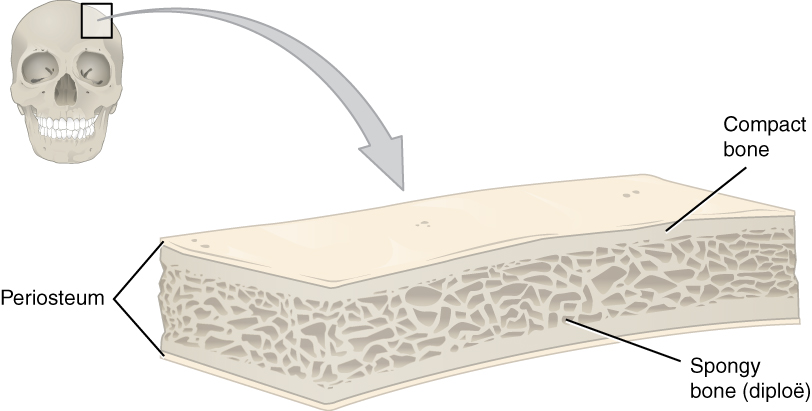
The skull is a bone protective cavity for the brain. The skull is composed of four types of bone i.e., cranial bones, facial bones, ear ossicles and hyoid bone. However two parts are more prominent: the cranium and the mandible. In humans, these two parts are the neurocranium and the viscerocranium ( facial skeleton) that includes the mandible as its largest bone. The skull forms the anterior-most portion of the skeleton and is a product of cephalisation—housing the brain, and several sensory structures such as the eyes, ears, nose, and mouth. In humans these sensory structures are part of the facial skeleton. Functions of the skull include protection of the brain, fixing the distance between the eyes to allow stereoscopic vision, and fixing the position of the ears to enable sound localisation of the direction and distance of sounds. In some animals, such as horned ungulates (mammals with hooves), the skull also has a defensive function by providing the mount (on the ... [...More Info...] [...Related Items...] OR: [Wikipedia] [Google] [Baidu] |
Occipital Bone
The occipital bone () is a cranial dermal bone and the main bone of the occiput (back and lower part of the skull). It is trapezoidal in shape and curved on itself like a shallow dish. The occipital bone overlies the occipital lobes of the cerebrum. At the base of skull in the occipital bone, there is a large oval opening called the foramen magnum, which allows the passage of the spinal cord. Like the other cranial bones, it is classed as a flat bone. Due to its many attachments and features, the occipital bone is described in terms of separate parts. From its front to the back is the basilar part, also called the basioccipital, at the sides of the foramen magnum are the lateral parts, also called the exoccipitals, and the back is named as the squamous part. The basilar part is a thick, somewhat quadrilateral piece in front of the foramen magnum and directed towards the pharynx. The squamous part is the curved, expanded plate behind the foramen magnum and is the largest par ... [...More Info...] [...Related Items...] OR: [Wikipedia] [Google] [Baidu] |
Squamous Part Of Occipital Bone
The squamous part of occipital bone is situated above and behind the foramen magnum, and is curved from above downward and from side to side. External surface The external surface is convex and presents midway between the summit of the bone and the foramen magnum a prominence, the external occipital protuberance and inion. Extending lateralward from this on either side are two curved lines, one a little above the other. The upper, often faintly marked, is named the highest nuchal line, and to it the epicranial aponeurosis is attached. The lower is termed the superior nuchal line. That area of the squamous part, which lies above the highest nuchal lines is named the occipital plane ''(planum occipitale)'' and is covered by the ''occipitalis muscle''. That below, termed the nuchal plane, is rough and irregular for the attachment of several muscles. From the external occipital protuberance, an often faintly marked ridge or crest, the median nuchal line, descends to the foram ... [...More Info...] [...Related Items...] OR: [Wikipedia] [Google] [Baidu] |
Skull
The skull is a bone protective cavity for the brain. The skull is composed of four types of bone i.e., cranial bones, facial bones, ear ossicles and hyoid bone. However two parts are more prominent: the cranium and the mandible. In humans, these two parts are the neurocranium and the viscerocranium ( facial skeleton) that includes the mandible as its largest bone. The skull forms the anterior-most portion of the skeleton and is a product of cephalisation—housing the brain, and several sensory structures such as the eyes, ears, nose, and mouth. In humans these sensory structures are part of the facial skeleton. Functions of the skull include protection of the brain, fixing the distance between the eyes to allow stereoscopic vision, and fixing the position of the ears to enable sound localisation of the direction and distance of sounds. In some animals, such as horned ungulates (mammals with hooves), the skull also has a defensive function by providing the mount (on the ... [...More Info...] [...Related Items...] OR: [Wikipedia] [Google] [Baidu] |
Nuchal Ligament
The nuchal ligament is a ligament at the back of the neck that is continuous with the supraspinous ligament. Structure The nuchal ligament extends from the external occipital protuberance on the skull and median nuchal line to the spinous process of the seventh cervical vertebra in the lower part of the neck. From the anterior border of the nuchal ligament, a fibrous lamina is given off. This is attached to the posterior tubercle of the atlas, and to the spinous processes of the cervical vertebrae, and forms a septum between the muscles on either side of the neck. The trapezius and splenius capitis muscle attach to the nuchal ligament. Function It is a tendon-like structure that has developed independently in humans and other animals well adapted for running. In some four-legged animals, particularly ungulates, the nuchal ligament serves to sustain the weight of the head. Clinical significance In Chiari malformation treatment, decompression and duraplasty with a harvest ... [...More Info...] [...Related Items...] OR: [Wikipedia] [Google] [Baidu] |
Trapezius Muscle
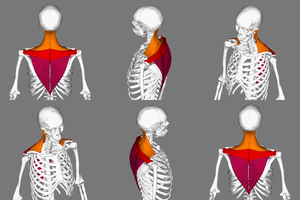
The trapezius is a large paired trapezoid-shaped surface muscle that extends longitudinally from the occipital bone to the lower thoracic vertebrae of the spine and laterally to the spine of the scapula. It moves the scapula and supports the arm. The trapezius has three functional parts: an upper (descending) part which supports the weight of the arm; a middle region (transverse), which retracts the scapula; and a lower (ascending) part which medially rotates and depresses the scapula. Name and history The trapezius muscle resembles a trapezium, also known as a trapezoid, or diamond-shaped quadrilateral. The word "spinotrapezius" refers to the human trapezius, although it is not commonly used in modern texts. In other mammals, it refers to a portion of the analogous muscle. Similarly, the term "tri-axle back plate" was historically used to describe the trapezius muscle. Structure The ''superior'' or ''upper'' (or descending) fibers of the trapezius originate from the s ... [...More Info...] [...Related Items...] OR: [Wikipedia] [Google] [Baidu] |
10-20 System (EEG)
10-20 system may refer to: * 10-20 system (swimming safety) A lifeguard is a rescuer who supervises the safety and rescue of swimmers, surfers, and other water sports participants such as in a swimming pool, water park, beach, spa, river and lake. Lifeguards are trained in swimming and CPR/ AED fi ..., a technique used by lifeguards * 10-20 system (EEG), an electrode placement method used in electroencephalography systems {{Disambiguation ... [...More Info...] [...Related Items...] OR: [Wikipedia] [Google] [Baidu] |
Electroencephalography
Electroencephalography (EEG) is a method to record an electrogram of the spontaneous electrical activity of the brain. The biosignals detected by EEG have been shown to represent the postsynaptic potentials of pyramidal neurons in the neocortex and allocortex. It is typically non-invasive, with the EEG electrodes placed along the scalp (commonly called "scalp EEG") using the International 10-20 system, or variations of it. Electrocorticography, involving surgical placement of electrodes, is sometimes called "intracranial EEG". Clinical interpretation of EEG recordings is most often performed by visual inspection of the tracing or quantitative EEG analysis. Voltage fluctuations measured by the EEG bioamplifier and electrodes allow the evaluation of normal brain activity. As the electrical activity monitored by EEG originates in neurons in the underlying brain tissue, the recordings made by the electrodes on the surface of the scalp vary in accordance with their orientation ... [...More Info...] [...Related Items...] OR: [Wikipedia] [Google] [Baidu] |
Nuchal Lines
The nuchal lines are four curved lines on the external surface of the occipital bone: * The upper, often faintly marked, is named the highest nuchal line, but is sometimes referred to as the Mempin line or linea suprema, and it attaches to the epicranial aponeurosis. * Below the highest nuchal line is the superior nuchal line. To it is attached, the splenius capitis muscle, the trapezius muscle, and the occipitalis. * From the external occipital protuberance a ridge or crest, the external occipital crest also called the median nuchal line, often faintly marked, descends to the foramen magnum, and affords attachment to the nuchal ligament. * Running from the middle of this line is the inferior nuchal line. Attached are the obliquus capitis superior muscle, rectus capitis posterior major muscle, and rectus capitis posterior minor muscle The rectus capitis posterior minor (or rectus capitis posticus minor, both being Latin for ''lesser posterior straight muscle of the head'') ari ... [...More Info...] [...Related Items...] OR: [Wikipedia] [Google] [Baidu] |
Anatolia
Anatolia, tr, Anadolu Yarımadası), and the Anatolian plateau, also known as Asia Minor, is a large peninsula in Western Asia and the westernmost protrusion of the Asian continent. It constitutes the major part of modern-day Turkey. The region is bounded by the Turkish Straits to the northwest, the Black Sea to the north, the Armenian Highlands to the east, the Mediterranean Sea to the south, and the Aegean Sea to the west. The Sea of Marmara forms a connection between the Black and Aegean seas through the Bosporus and Dardanelles straits and separates Anatolia from Thrace on the Balkan peninsula of Southeast Europe. The eastern border of Anatolia has been held to be a line between the Gulf of Alexandretta and the Black Sea, bounded by the Armenian Highlands to the east and Mesopotamia to the southeast. By this definition Anatolia comprises approximately the western two-thirds of the Asian part of Turkey. Today, Anatolia is sometimes considered to be synonymous w ... [...More Info...] [...Related Items...] OR: [Wikipedia] [Google] [Baidu] |
Internal Occipital Protuberance
Along the internal surface of the occipital bone, at the point of intersection of the four divisions of the cruciform eminence, is the internal occipital protuberance. Running transversely on either side is a groove for the transverse sinus The transverse sinuses (left and right lateral sinuses), within the human head, are two areas beneath the brain which allow blood to drain from the back of the head. They run laterally in a groove along the interior surface of the occipital bone. .... Additional images See also * External occipital protuberance References External links * Diagram at uni-mainz.de Bones of the head and neck {{musculoskeletal-stub ... [...More Info...] [...Related Items...] OR: [Wikipedia] [Google] [Baidu] |
Occipital Bun
The occipital bone () is a cranial dermal bone and the main bone of the occiput (back and lower part of the skull). It is trapezoidal in shape and curved on itself like a shallow dish. The occipital bone overlies the occipital lobes of the cerebrum. At the base of skull in the occipital bone, there is a large oval opening called the foramen magnum, which allows the passage of the spinal cord. Like the other cranial bones, it is classed as a flat bone. Due to its many attachments and features, the occipital bone is described in terms of separate parts. From its front to the back is the basilar part, also called the basioccipital, at the sides of the foramen magnum are the lateral parts, also called the exoccipitals, and the back is named as the squamous part. The basilar part is a thick, somewhat quadrilateral piece in front of the foramen magnum and directed towards the pharynx. The squamous part is the curved, expanded plate behind the foramen magnum and is the largest part o ... [...More Info...] [...Related Items...] OR: [Wikipedia] [Google] [Baidu] |


