|
Extensor Carpi Radialis Brevis
In human anatomy, extensor carpi radialis brevis is a muscle in the forearm that acts to extend and abduct the wrist. It is shorter and thicker than its namesake extensor carpi radialis longus which can be found above the proximal end of the extensor carpi radialis brevis. Origin and insertion It arises from the lateral epicondyle of the humerus, by the common extensor tendon; from the radial collateral ligament of the elbow-joint; from a strong aponeurosis which covers its surface; and from the intermuscular septa between it and the adjacent muscles.''Gray's Anatomy'' 1918, see infobox The fibres end approximately at the middle of the forearm in the form of a flat tendon, which is closely connected with that of the extensor carpi radialis longus, and accompanies it to the wrist; it passes beneath the abductor pollicis longus and extensor pollicis brevis, beneath the extensor retinaculum, and inserts into the lateral dorsal surface of the base of the third metacarpal bone, with ... [...More Info...] [...Related Items...] OR: [Wikipedia] [Google] [Baidu] |
Humerus
The humerus (; ) is a long bone in the arm that runs from the shoulder to the elbow. It connects the scapula and the two bones of the lower arm, the radius and ulna, and consists of three sections. The humeral upper extremity consists of a rounded head, a narrow neck, and two short processes (tubercles, sometimes called tuberosities). The body is cylindrical in its upper portion, and more prismatic below. The lower extremity consists of 2 epicondyles, 2 processes (trochlea & capitulum), and 3 fossae (radial fossa, coronoid fossa, and olecranon fossa). As well as its true anatomical neck, the constriction below the greater and lesser tubercles of the humerus is referred to as its surgical neck due to its tendency to fracture, thus often becoming the focus of surgeons. Etymology The word "humerus" is derived from la, humerus, umerus meaning upper arm, shoulder, and is linguistically related to Gothic ''ams'' shoulder and Greek ''ōmos''. Structure Upper extremity The upper or pr ... [...More Info...] [...Related Items...] OR: [Wikipedia] [Google] [Baidu] |
Aponeurosis
An aponeurosis (; plural: ''aponeuroses'') is a type or a variant of the deep fascia, in the form of a sheet of pearly-white fibrous tissue that attaches sheet-like muscles needing a wide area of attachment. Their primary function is to join muscles and the body parts they act upon, whether bone or other muscles. They have a shiny, whitish-silvery color, are histologically similar to tendons, and are very sparingly supplied with blood vessels and nerves. When dissected, aponeuroses are papery and peel off by sections. The primary regions with thick aponeuroses are in the ventral abdominal region, the dorsal lumbar region, the ventriculus in birds, and the palmar (palms) and plantar (soles) regions. Anatomy Anterior abdominal aponeuroses The anterior abdominal aponeuroses are located just superficial to the rectus abdominis muscle. It has for its borders the external oblique, pectoralis muscles, and the latissimus dorsi. Posterior lumbar aponeuroses The posterior lumbar apo ... [...More Info...] [...Related Items...] OR: [Wikipedia] [Google] [Baidu] |
Wrist Curl
The wrist curl is a weight training exercise for developing just the wrist flexion, flexor muscles of the forearm. It is therefore an isolation exercise. Ideally, it should be done in combination with the "reverse wrist curl" (also called wrist extension) to ensure equal development of the wrist flexor and wrist extension (kinesiology), extensor muscles. Wrist curls can be performed with a dumbbell or with both hands holding a barbell. To perform a seated wrist curl, the lifter should be seated on a Bench (weight training), bench with knees bent and the forearm(s) resting on the thigh, or with forearms on a bench and hands hanging off the edge. The hand, palm should be facing up and the hand should be free to move completely up and down. At the starting point, the wrist should be bent back so that the fingers are almost pointing down at the floor. In a steady motion, the lifter should raise the weight by using the forearm muscles to bring the hand up as far as possible. The forearm ... [...More Info...] [...Related Items...] OR: [Wikipedia] [Google] [Baidu] |
Strength Training
Strength training or resistance training involves the performance of physical exercises that are designed to improve strength and endurance. It is often associated with the lifting of weights. It can also incorporate a variety of training techniques such as bodyweight exercises, isometrics, and plyometrics. Training works by progressively increasing the force output of the muscles and uses a variety of exercises and types of equipment. Strength training is primarily an anaerobic activity, although circuit training also is a form of aerobic exercise. Strength training can increase muscle, tendon, and ligament strength as well as bone density, metabolism, and the lactate threshold; improve joint and cardiac function; and reduce the risk of injury in athletes and the elderly. For many sports and physical activities, strength training is central or is used as part of their training regimen. Principles and training methods The basic principles of strength training involve ... [...More Info...] [...Related Items...] OR: [Wikipedia] [Google] [Baidu] |
Joint
A joint or articulation (or articular surface) is the connection made between bones, ossicles, or other hard structures in the body which link an animal's skeletal system into a functional whole.Saladin, Ken. Anatomy & Physiology. 7th ed. McGraw-Hill Connect. Webp.274/ref> They are constructed to allow for different degrees and types of movement. Some joints, such as the knee, elbow, and shoulder, are self-lubricating, almost frictionless, and are able to withstand compression and maintain heavy loads while still executing smooth and precise movements. Other joints such as sutures between the bones of the skull permit very little movement (only during birth) in order to protect the brain and the sense organs. The connection between a tooth and the jawbone is also called a joint, and is described as a fibrous joint known as a gomphosis. Joints are classified both structurally and functionally. Classification The number of joints depends on if sesamoids are included, age of the ... [...More Info...] [...Related Items...] OR: [Wikipedia] [Google] [Baidu] |
Wrist
In human anatomy, the wrist is variously defined as (1) the Carpal bones, carpus or carpal bones, the complex of eight bones forming the proximal skeletal segment of the hand; "The wrist contains eight bones, roughly aligned in two rows, known as the carpal bones." (2) the wrist joint or radiocarpal joint, the joint between the radius (bone), radius and the Carpal bones, carpus and; (3) the anatomical region surrounding the carpus including the distal parts of the bones of the forearm and the proximal parts of the metacarpus or five metacarpal bones and the series of joints between these bones, thus referred to as ''wrist joints''. "With the large number of bones composing the wrist (ulna, radius, eight carpas, and five metacarpals), it makes sense that there are many, many joints that make up the structure known as the wrist." This region also includes the carpal tunnel, the anatomical snuff box, bracelet lines, the Flexor retinaculum of the hand, flexor retinaculum, and the ex ... [...More Info...] [...Related Items...] OR: [Wikipedia] [Google] [Baidu] |
Radial Nerve
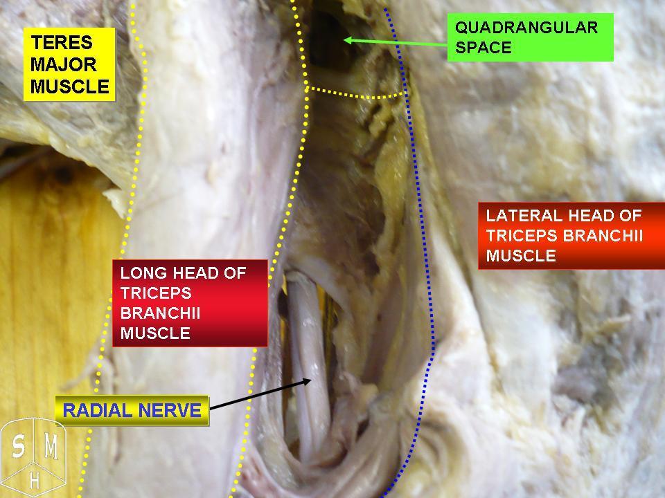
The radial nerve is a nerve in the human body that supplies the posterior portion of the upper limb. It innervates the medial and lateral heads of the triceps brachii muscle of the arm, as well as all 12 muscles in the posterior osteofascial compartment of the forearm and the associated joints and overlying skin. It originates from the brachial plexus, carrying fibers from the ventral roots of spinal nerves C5, C6, C7, C8 & T1. The radial nerve and its branches provide motor innervation to the dorsal arm muscles (the triceps brachii and the anconeus) and the extrinsic extensors of the wrists and hands; it also provides cutaneous sensory innervation to most of the back of the hand, except for the back of the little finger and adjacent half of the ring finger (which are innervated by the ulnar nerve). The radial nerve divides into a deep branch, which becomes the posterior interosseous nerve, and a superficial branch, which goes on to innervate the dorsum (back) of the hand. Th ... [...More Info...] [...Related Items...] OR: [Wikipedia] [Google] [Baidu] |
Forearm
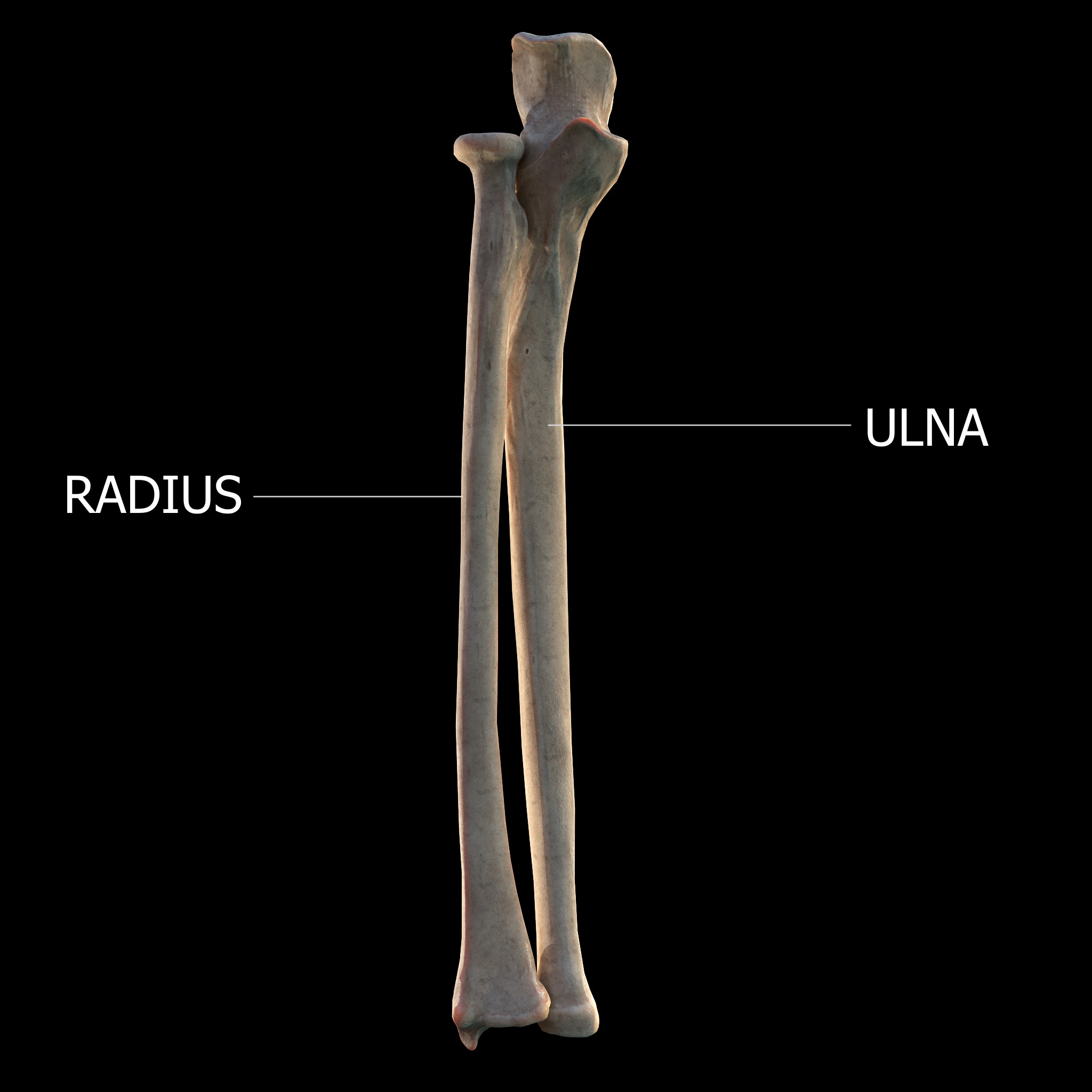
The forearm is the region of the upper limb between the elbow and the wrist. The term forearm is used in anatomy to distinguish it from the arm, a word which is most often used to describe the entire appendage of the upper limb, but which in anatomy, technically, means only the region of the upper arm, whereas the lower "arm" is called the forearm. It is homologous to the region of the leg that lies between the knee and the ankle joints, the crus. The forearm contains two long bones, the radius and the ulna, forming the two radioulnar joints. The interosseous membrane connects these bones. Ultimately, the forearm is covered by skin, the anterior surface usually being less hairy than the posterior surface. The forearm contains many muscles, including the flexors and extensors of the wrist, flexors and extensors of the digits, a flexor of the elbow (brachioradialis), and pronators and supinators that turn the hand to face down or upwards, respectively. In cross-section, the for ... [...More Info...] [...Related Items...] OR: [Wikipedia] [Google] [Baidu] |
Second Metacarpal Bone
The second metacarpal bone (metacarpal bone of the index finger) is the longest, and its base the largest, of all the metacarpal bones.''Gray's Anatomy'' (1918). See infobox. Human anatomy Its base is prolonged upward and medialward, forming a prominent ridge. It presents four articular facets, three on the upper surface and one on the ulnar side: * Of the facets on the upper surface: ** the ''intermediate'' is the largest and is concave from side to side, convex from before backward for articulation with the lesser multangular; ** the ''lateral'' is small, flat and oval for articulation with the greater multangular; ** the ''medial'', on the summit of the ridge, is long and narrow for articulation with the capitate. * The facet on the ulnar side articulates with the third metacarpal. The extensor carpi radialis longus muscle is inserted on the dorsal surface and the flexor carpi radialis muscle on the volar surface of the base. The shaft gives origin to the first palmar in ... [...More Info...] [...Related Items...] OR: [Wikipedia] [Google] [Baidu] |
Third Metacarpal Bone
The third metacarpal bone (metacarpal bone of the middle finger) is a little smaller than the second. The dorsal aspect of its base presents on its radial side a pyramidal eminence, the styloid process, which extends upward behind the capitate; immediately distal to this is a rough surface for the attachment of the extensor carpi radialis brevis muscle. The carpal articular facet is concave behind, flat in front, and articulates with the capitate. On the radial side is a smooth, concave facet for articulation with the second metacarpal, and on the ulnar side two small oval facets for the fourth metacarpal. Ossification The ossification process begins in the shaft during prenatal life, and in the head between 11th and 27th months. Additional images File:Third metacarpal bone (left hand) - animation01.gif, Third metacarpal bone of the left hand (shown in red). Animation. File:Third metacarpal bone (left hand) - animation02.gif, Third metacarpal bone of the left hand. Close ... [...More Info...] [...Related Items...] OR: [Wikipedia] [Google] [Baidu] |
Extensor Retinaculum Of The Hand
The extensor retinaculum (dorsal carpal ligament, or posterior annular ligament) is an anatomical term for the thickened part of the antebrachial fascia that holds the tendons of the extensor muscles in place. It is located on the back of the forearm, just proximal to the hand. It is continuous with the palmar carpal ligament, which is located on the anterior side of the forearm. Structure The extensor retinaculum is a strong, fibrous band, extending obliquely downward and medialward across the back of the wrist. It consists of part of the deep fascia of the back of the forearm, strengthened by the addition of some transverse fibers. The extensor retinaculum is attached laterally to the lateral margin of the radius. However, it is not attached to the ulna, as the distance between these two bones varies with supination and pronation of the forearm. Instead the medial attachment is to the pisiform bone and triquetral bone. Other authors may state the medial attachment of extensor ... [...More Info...] [...Related Items...] OR: [Wikipedia] [Google] [Baidu] |
Extensor Pollicis Brevis
In human anatomy, the extensor pollicis brevis is a skeletal muscle on the dorsal side of the forearm. It lies on the medial side of, and is closely connected with, the abductor pollicis longus. The extensor pollicis brevis (EPB) belongs to the deep group of the posterior fascial compartment of the forearm. It is a part of the lateral border of the anatomical snuffbox. Structure The extensor pollicis brevis arises from the ulna distal to the abductor pollicis longus, from the interosseous membrane, and from the dorsal surface of the radius. Its direction is similar to that of the abductor pollicis longus, its tendon passing the same groove on the lateral side of the lower end of the radius, to be inserted into the base of the first phalanx of the thumb. Variation Absence; fusion of tendon with that of the extensor pollicis longus or abductor pollicis longus muscle. Function In a close relationship to the abductor pollicis longus, the extensor pollicis brevis both exten ... [...More Info...] [...Related Items...] OR: [Wikipedia] [Google] [Baidu] |


