|
Erythrocyte Rosetting
Erythrocyte rosetting or E-rosetting is a phenomenon seen through a microscope where red blood cells ''(erythrocytes)'' are arranged around a central cell to form a cluster that looks like a flower. The red blood cells surrounding the cell form the ''petal,'' while the central cell forms the ''stigma'' of the flower shape. This formation occurs due to an immunological reaction between an epitope on the central cell's surface and a receptor or antibody on a red cell. The presence of E-rosetting can be used as a test for T cells although more modern tests such as immunohistochemistry are available. Rosetting is caused by parasites in the genus ''Plasmodium'' and is a cause of some malaria-associated symptoms. Rosetting techniques Three types of rosette techniques have been developed and used experimentally. Rosette test for Rh factor The ''Rosette test'' is performed on postpartum maternal blood to estimate the volume of fetal-maternal hemorrhage in case of an Rh negative mot ... [...More Info...] [...Related Items...] OR: [Wikipedia] [Google] [Baidu] |
Microscope
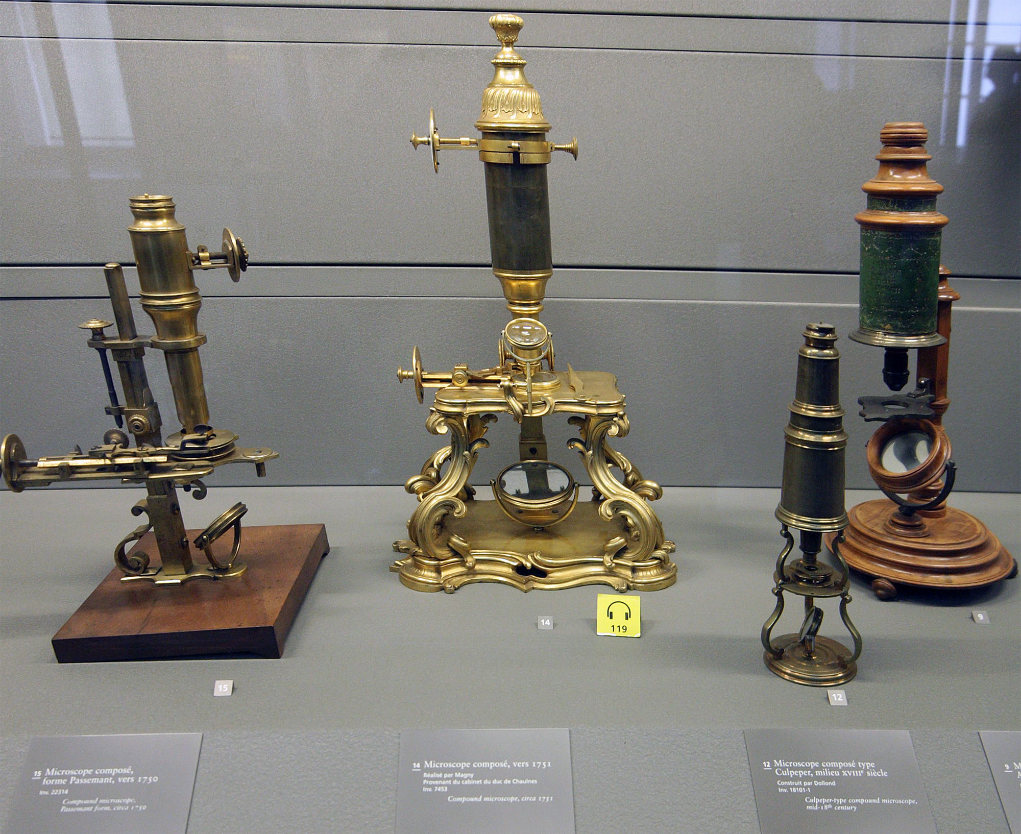
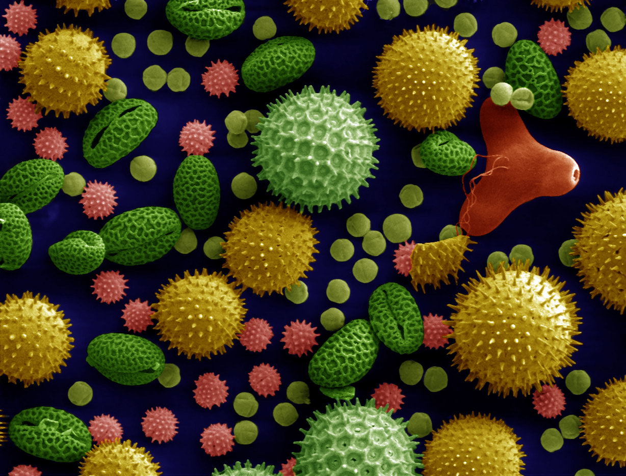
A microscope () is a laboratory instrument used to examine objects that are too small to be seen by the naked eye. Microscopy is the science of investigating small objects and structures using a microscope. Microscopic means being invisible to the eye unless aided by a microscope. There are many types of microscopes, and they may be grouped in different ways. One way is to describe the method an instrument uses to interact with a sample and produce images, either by sending a beam of light or electrons through a sample in its optical path, by detecting photon emissions from a sample, or by scanning across and a short distance from the surface of a sample using a probe. The most common microscope (and the first to be invented) is the optical microscope, which uses lenses to refract visible light that passed through a thinly sectioned sample to produce an observable image. Other major types of microscopes are the fluorescence microscope, electron microscope (both the transmi ... [...More Info...] [...Related Items...] OR: [Wikipedia] [Google] [Baidu] |
Red Blood Cell
Red blood cells (RBCs), also referred to as red cells, red blood corpuscles (in humans or other animals not having nucleus in red blood cells), haematids, erythroid cells or erythrocytes (from Greek ''erythros'' for "red" and ''kytos'' for "hollow vessel", with ''-cyte'' translated as "cell" in modern usage), are the most common type of blood cell and the vertebrate's principal means of delivering oxygen (O2) to the body tissues—via blood flow through the circulatory system. RBCs take up oxygen in the lungs, or in fish the gills, and release it into tissues while squeezing through the body's capillaries. The cytoplasm of a red blood cell is rich in hemoglobin, an iron-containing biomolecule that can bind oxygen and is responsible for the red color of the cells and the blood. Each human red blood cell contains approximately 270 million hemoglobin molecules. The cell membrane is composed of proteins and lipids, and this structure provides properties essential for physiolo ... [...More Info...] [...Related Items...] OR: [Wikipedia] [Google] [Baidu] |
Immunology
Immunology is a branch of medicineImmunology for Medical Students, Roderick Nairn, Matthew Helbert, Mosby, 2007 and biology that covers the medical study of immune systems in humans, animals, plants and sapient species. In such we can see there is a difference of human immunology and comparative immunology in veterinary medicine and animal biosciences. Immunology measures, uses charts and differentiate in context in medicine the studies of immunity on cell and molecular level, and the immune system as part of the physiological level as its functioning is of major importance. In the different states of both health, occurring symptoms and diseases; the functioning of the immune system and immunological responses such as autoimmune diseases, allergic hypersensitivities, or in some cases malfunctioning of immune system as for example in immunological disorders or in immune deficiency, and the specific transplant rejection) Immunology has applications in numerous disciplines of ... [...More Info...] [...Related Items...] OR: [Wikipedia] [Google] [Baidu] |
T Cell
A T cell is a type of lymphocyte. T cells are one of the important white blood cells of the immune system and play a central role in the adaptive immune response. T cells can be distinguished from other lymphocytes by the presence of a T-cell receptor (TCR) on their cell surface. T cells are born from hematopoietic stem cells, found in the bone marrow. Developing T cells then migrate to the thymus gland to develop (or mature). T cells derive their name from the thymus. After migration to the thymus, the precursor cells mature into several distinct types of T cells. T cell differentiation also continues after they have left the thymus. Groups of specific, differentiated T cell subtypes have a variety of important functions in controlling and shaping the immune response. One of these functions is immune-mediated cell death, and it is carried out by two major subtypes: CD8+ "killer" and CD4+ "helper" T cells. (These are named for the presence of the cell surface proteins CD8 or ... [...More Info...] [...Related Items...] OR: [Wikipedia] [Google] [Baidu] |
Immunohistochemistry
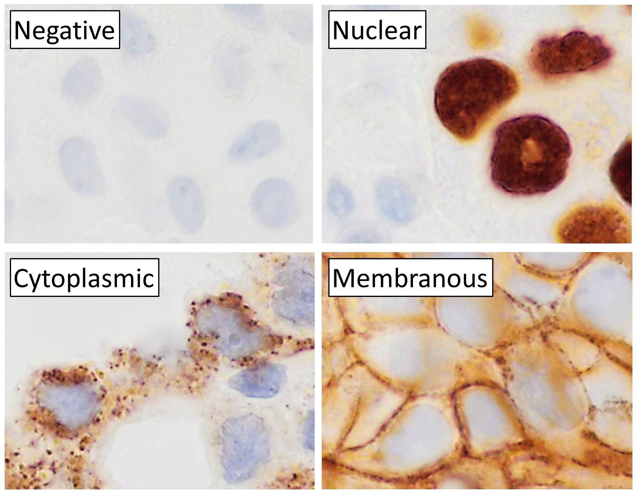
Immunohistochemistry (IHC) is the most common application of immunostaining. It involves the process of selectively identifying antigens (proteins) in cells of a tissue section by exploiting the principle of antibodies binding specifically to antigens in biological tissues. IHC takes its name from the roots "immuno", in reference to antibodies used in the procedure, and "histo", meaning tissue (compare to immunocytochemistry). Albert Coons conceptualized and first implemented the procedure in 1941. Visualising an antibody-antigen interaction can be accomplished in a number of ways, mainly either of the following: * ''Chromogenic immunohistochemistry'' (CIH), wherein an antibody is conjugated to an enzyme, such as peroxidase (the combination being termed immunoperoxidase), that can catalyse a colour-producing reaction. * '' Immunofluorescence'', where the antibody is tagged to a fluorophore, such as fluorescein or rhodamine. Immunohistochemical staining is widely used in the dia ... [...More Info...] [...Related Items...] OR: [Wikipedia] [Google] [Baidu] |
Fetal-maternal Hemorrhage
Fetal-maternal haemorrhage is the loss of fetal blood cells into the maternal circulation. It takes place in normal pregnancies as well as when there are obstetric or trauma related complications to pregnancy. Normally the maternal circulation and the fetal circulation are kept from direct contact with each other, with gas and nutrient exchange taking place across a membrane in the placenta made of two layers, the syncytiotrophoblast and the cytotrophoblast. Fetal-maternal haemorrhage occurs when this membrane ceases to function as a barrier and fetal cells may come in contact with and enter the maternal vessels in the decidua/endometrium. Description Normal pregnancy It is estimated that less than 1ml of fetal blood is lost to the maternal circulation during normal labour in around 96% of normal deliveries. The loss of this small amount of blood may however be a sensitising event and stimulate antibody production to the foetal red blood cells, an example of which is Rhesus diseas ... [...More Info...] [...Related Items...] OR: [Wikipedia] [Google] [Baidu] |
Rh Negative
The Rh blood group system is a human blood group system. It contains proteins on the surface of red blood cells. After the ABO blood group system, it is the most likely to be involved in transfusion reactions. The Rh blood group system consists of 49 defined blood group antigens, among which the five antigens D, C, c, E, and e are the most important. There is no d antigen. Rh(D) status of an individual is normally described with a ''positive'' (+) or ''negative'' (−) suffix after the ABO type (e.g., someone who is A+ has the A antigen and Rh(D) antigen, whereas someone who is A− has the A antigen but lacks the Rh(D) antigen). The terms ''Rh factor'', ''Rh positive'', and ''Rh negative'' refer to the Rh(D) antigen only. Antibodies to Rh antigens can be involved in hemolytic transfusion reactions and antibodies to the Rh(D) and Rh antigens confer significant risk of hemolytic disease of the fetus and newborn. Nomenclature The Rh blood group system has two sets of nomenclatur ... [...More Info...] [...Related Items...] OR: [Wikipedia] [Google] [Baidu] |
Rho(D) Immune Globulin
Rho(D) immune globulin (RhIG) is a medication used to prevent RhD isoimmunization in mothers who are RhD negative and to treat idiopathic thrombocytopenic purpura (ITP) in people who are Rh positive. It is often given both during and following pregnancy. It may also be used when RhD-negative people are given RhD-positive blood. It is given by injection into muscle or a vein. A single dose lasts 12 weeks. It is made from human blood plasma. Common side effects include fever, headache, pain at the site of injection, and red blood cell breakdown. Other side effects include allergic reactions, kidney problems, and a very small risk of viral infections. In those with ITP, the amount of red blood cell breakdown may be significant. Use is safe with breastfeeding. Rho(D) immune globulin is made up of antibodies to the antigen Rho(D) present on some red blood cells. It is believed to work by blocking a person's immune system from recognizing this antigen. Rho(D) immune globulin ... [...More Info...] [...Related Items...] OR: [Wikipedia] [Google] [Baidu] |
Light Microscopy
Microscopy is the technical field of using microscopes to view objects and areas of objects that cannot be seen with the naked eye (objects that are not within the resolution range of the normal eye). There are three well-known branches of microscopy: optical microscope, optical, electron microscope, electron, and scanning probe microscopy, along with the emerging field of X-ray microscopy. Optical microscopy and electron microscopy involve the diffraction, reflection (physics), reflection, or refraction of electromagnetic radiation/electron beams interacting with the Laboratory specimen, specimen, and the collection of the scattered radiation or another signal in order to create an image. This process may be carried out by wide-field irradiation of the sample (for example standard light microscopy and transmission electron microscope, transmission electron microscopy) or by scanning a fine beam over the sample (for example confocal laser scanning microscopy and scanning electro ... [...More Info...] [...Related Items...] OR: [Wikipedia] [Google] [Baidu] |
Kleihauer–Betke Test
The Kleihauer–Betke ("KB") test, Kleihauer–Betke ("KB") stain, Kleihauer test or acid elution test is a blood test used to measure the amount of fetal hemoglobin transferred from a fetus to a mother's bloodstream. It is usually performed on Rh-negative mothers to determine the required dose of Rho(D) immune globulin (RhIg) to inhibit formation of Rh antibodies in the mother and prevent Rh disease in future Rh-positive children. It is named after Enno Kleihauer and Klaus Betke who described it in 1957. Test details The KB test is the standard method of quantitating fetal–maternal hemorrhage (FMH). It takes advantage of the differential resistance of fetal hemoglobin to acid. A standard blood smear is prepared from the mother's blood and exposed to an acid bath. This removes adult hemoglobin, but not fetal hemoglobin, from the red blood cells. Subsequent staining, using Shepard's method, makes fetal cells (containing fetal hemoglobin) appear rose-pink in color, while adult ... [...More Info...] [...Related Items...] OR: [Wikipedia] [Google] [Baidu] |
LFA-3
CD58, or lymphocyte function-associated antigen 3 (LFA-3), is a cell adhesion molecule expressed on Antigen Presenting Cells (APC), particularly macrophages. It binds to CD2 (LFA-2) on T cells and is important in strengthening the adhesion between the T cells and Professional Antigen Presenting Cells. This adhesion occurs as part of the transitory initial encounters between T cells and Antigen Presenting Cells before T cell activation, when T cells are roaming the lymph nodes looking at the surface of APCs for peptide:MHC complexes the T-cell receptors are reactive to. Polymorphisms in the CD58 gene are associated with increased risk for multiple sclerosis. Genomic region containing the single-nucleotide polymorphism rs1335532, associated with high risk of multiple sclerosis, has enhancer properties and can significantly boost the CD58 promoter activity in lymphoblast cells. The protective (C) rs1335532 allele creates functional binding site for ASCL2 transcription factor, a ta ... [...More Info...] [...Related Items...] OR: [Wikipedia] [Google] [Baidu] |


