|
Electrophysiology Study
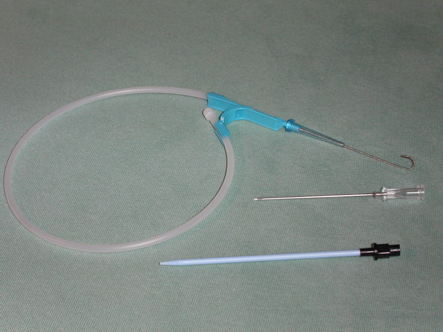
A cardiac electrophysiology study (EP test or EP study) is a minimally invasive procedure using catheters introduced through a vein or artery to record electrical activity from within the heart. This electrical activity is recorded when the heart is in a normal rhythm (sinus rhythm) to assess the conduction system of the heart and to look for additional electrical connections (accessory pathways), and during any abnormal heart rhythms that can be induced. EP studies are used to investigate the cause, location of origin, and best treatment for various abnormal heart rhythms, and are often followed by a catheter ablation during the same procedure. Preparation It is important for patients not to eat or drink for up to 12 hours before the procedure. This is to prevent vomiting, which can result in aspiration, and also cause severe bleeding from the insertion site of the catheter. Failure to follow this simple preparation may result in dangerous consequences. In general, small amoun ... [...More Info...] [...Related Items...] OR: [Wikipedia] [Google] [Baidu] |
Cardiac Electrophysiology
Cardiac electrophysiology is a branch of cardiology and basic science focusing on the electrical activities of the heart. The term is usually used in clinical context, to describe studies of such phenomena by invasive (intracardiac) catheter recording of spontaneous activity as well as of cardiac responses to programmed electrical stimulation - clinical cardiac electrophysiology. However, cardiac electrophysiology also encompasses basic research and translational research components. Specialists studying cardiac electrophysiology, either clinically or solely through research, are known as cardiac electrophysiologists. Description Electrophysiological (EP) studies are performed to assess complex arrhythmias, elucidate symptoms, evaluate abnormal electrocardiograms, assess risk of developing arrhythmias in the future, and design treatment. These procedures include therapeutic methods (typically radiofrequency ablation, or cryoablation) in addition to diagnostic and prognostic proc ... [...More Info...] [...Related Items...] OR: [Wikipedia] [Google] [Baidu] |
Cannula
A cannula (; Latin meaning 'little reed'; plural or ) is a tube that can be inserted into the body, often for the delivery or removal of fluid or for the gathering of samples. In simple terms, a cannula can surround the inner or outer surfaces of a trocar needle thus extending the effective needle length by at least half the length of the original needle. Its size mainly ranges from 14 to 24 gauge. Different-sized cannula have different colours as coded. Decannulation is the permanent removal of a cannula ( extubation), especially of a tracheostomy cannula, once a physician determines it is no longer needed for breathing. Medicine Cannulas normally come with a trocar inside. The trocar is a needle, which punctures the body in order to get into the intended space. Many types of cannulas exist: Intravenous cannulas are the most common in hospital use. A variety of cannulas are used to establish cardiopulmonary bypass in cardiac surgery. A nasal cannula is a piece of pla ... [...More Info...] [...Related Items...] OR: [Wikipedia] [Google] [Baidu] |
Microwave
Microwave is a form of electromagnetic radiation with wavelengths ranging from about one meter to one millimeter corresponding to frequency, frequencies between 300 MHz and 300 GHz respectively. Different sources define different frequency ranges as microwaves; the above broad definition includes both Ultra high frequency, UHF and Extremely high frequency, EHF (millimeter wave) bands. A more common definition in radio-frequency engineering is the range between 1 and 100 GHz (wavelengths between 0.3 m and 3 mm). In all cases, microwaves include the entire Super high frequency, SHF band (3 to 30 GHz, or 10 to 1 cm) at minimum. Frequencies in the microwave range are often referred to by their Radio band#IEEE, IEEE radar band designations: S band, S, C band (IEEE), C, X band, X, Ku band, Ku, K band (IEEE), K, or Ka band, Ka band, or by similar NATO or EU designations. The prefix ' in ''microwave'' is not meant to suggest a wavelength in the M ... [...More Info...] [...Related Items...] OR: [Wikipedia] [Google] [Baidu] |
Radio Frequency
Radio frequency (RF) is the oscillation rate of an alternating electric current or voltage or of a magnetic, electric or electromagnetic field or mechanical system in the frequency range from around to around . This is roughly between the upper limit of audio frequencies and the lower limit of infrared frequencies; these are the frequencies at which energy from an oscillating current can radiate off a conductor into space as radio waves. Different sources specify different upper and lower bounds for the frequency range. Electric current Electric currents that oscillate at radio frequencies (RF currents) have special properties not shared by direct current or lower audio frequency alternating current, such as the 50 or 60 Hz current used in electrical power distribution. * Energy from RF currents in conductors can radiate into space as electromagnetic waves ( radio waves). This is the basis of radio technology. * RF current does not penetrate deeply into electrica ... [...More Info...] [...Related Items...] OR: [Wikipedia] [Google] [Baidu] |
Radiofrequency Ablation
Radiofrequency ablation (RFA), also called fulguration, is a medical procedure in which part of the electrical conduction system of the heart, tumor or other dysfunctional tissue is ablated using the heat generated from medium frequency alternating current (in the range of 350–500 kHz). RFA is generally conducted in the outpatient setting, using either local anesthetics or conscious sedation anesthesia. When it is delivered via catheter, it is called radiofrequency catheter ablation. Two important advantages of radio frequency current (over previously used low frequency AC or pulses of DC) are that it does not directly stimulate nerves or heart muscle and therefore can often be used without the need for general anesthesia, and that it is very specific for treating the desired tissue without significant collateral damage; due to this, it is gaining in popularity as an alternative for eligible patients who do not want to undergo surgery. Documented benefits have led to RFA ... [...More Info...] [...Related Items...] OR: [Wikipedia] [Google] [Baidu] |
Proarrhythmic Agent
A Pro-arrhythmic agent is a chemical, drug, or food that promotes cardiac arrhythmias. Substances Supplements Omega 3 fatty acids. Foods Chocolate, Coffee, Tea Drugs Caffeine, cocaine, beta-adrenergic agonists Encainide, Lorcainide See also *Antiarrhythmic agent *Electrophysiology study A cardiac electrophysiology study (EP test or EP study) is a minimally invasive procedure using catheters introduced through a vein or artery to record electrical activity from within the heart. This electrical activity is recorded when the hea ... References {{Reflist, 1 Antiarrhythmic agents Cardiac electrophysiology Medical emergencies ... [...More Info...] [...Related Items...] OR: [Wikipedia] [Google] [Baidu] |
Endocardium
The endocardium is the innermost layer of tissue that lines the chambers of the heart. Its cells are embryologically and biologically similar to the endothelial cells that line blood vessels. The endocardium also provides protection to the valves and heart chambers. The endocardium underlies the much more voluminous myocardium, the muscular tissue responsible for the contraction of the heart. The outer layer of the heart is termed epicardium and the heart is surrounded by a small amount of fluid enclosed by a fibrous sac called the pericardium. Function The endocardium, which is primarily made up of endothelial cells, controls myocardial function. This modulating role is separate from the homeometric and heterometric regulatory mechanisms that control myocardial contractility. Moreover, the endothelium of the myocardial (heart muscle) capillaries, which is also closely appositioned to the cardiomyocytes (heart muscle cells), is involved in this modulatory role. Thus, th ... [...More Info...] [...Related Items...] OR: [Wikipedia] [Google] [Baidu] |
Seldinger Technique
The Seldinger technique, also known as Seldinger wire technique, is a medical procedure to obtain safe access to blood vessels and other hollow organs. It is named after Sven Ivar Seldinger (1921–1998), a Swedish radiologist who introduced the procedure in 1953. Uses The Seldinger technique is used for angiography, insertion of chest drains and central venous catheters, insertion of PEG tubes using the push technique, insertion of the leads for an artificial pacemaker or implantable cardioverter-defibrillator, and numerous other interventional medical procedures. Complications The initial puncture is with a sharp instrument, and this may lead to hemorrhage or perforation of the organ in question. Infection is a possible complication, and hence asepsis is practiced during most Seldinger procedures. Loss of the guidewire into the cavity or blood vessel is a significant and generally preventable complication. Description The desired vessel or cavity is punctured with a s ... [...More Info...] [...Related Items...] OR: [Wikipedia] [Google] [Baidu] |
Femoral Artery
The femoral artery is a large artery in the thigh and the main arterial supply to the thigh and leg. The femoral artery gives off the deep femoral artery or profunda femoris artery and descends along the anteromedial part of the thigh in the femoral triangle. It enters and passes through the adductor canal, and becomes the popliteal artery as it passes through the adductor hiatus in the adductor magnus near the junction of the middle and distal thirds of the thigh. Structure The femoral artery enters the thigh from behind the inguinal ligament as the continuation of the external iliac artery. Here, it lies midway between the anterior superior iliac spine and the symphysis pubis (Mid-inguinal point). Segments In clinical parlance, the femoral artery has the following segments: *The common femoral artery (CFA) is the segment of the femoral artery between the inferior margin of the inguinal ligament and the branching point of the deep femoral artery/profunda femoris arte ... [...More Info...] [...Related Items...] OR: [Wikipedia] [Google] [Baidu] |
Subclavian Vein
The subclavian vein is a paired large vein, one on either side of the body, that is responsible for draining blood from the upper extremities, allowing this blood to return to the heart. The left subclavian vein plays a key role in the absorption of lipids, by allowing products that have been carried by lymph in the thoracic duct to enter the bloodstream. The diameter of the subclavian veins is approximately 1–2 cm, depending on the individual. Structure Each subclavian vein is a continuation of the axillary vein and runs from the outer border of the first rib to the medial border of anterior scalene muscle. From here it joins with the internal jugular vein to form the brachiocephalic vein (also known as "innominate vein"). The angle of union is termed the venous angle. The subclavian vein follows the subclavian artery and is separated from the subclavian artery by the insertion of anterior scalene. Thus, the subclavian vein lies anterior to the anterior scalene while the ... [...More Info...] [...Related Items...] OR: [Wikipedia] [Google] [Baidu] |
Femoral Vein
In the human body, the femoral vein is a blood vessel that accompanies the femoral artery in the femoral sheath. It begins at the adductor hiatus (an opening in the adductor magnus muscle) as the continuation of the popliteal vein. It ends at the inferior margin of the inguinal ligament where it becomes the external iliac vein. The femoral vein bears valves which are mostly bicuspid and whose number is variable between individuals and often between left and right leg. Structure Segments *The common femoral vein is the segment of the femoral vein between the branching point of the deep femoral vein and the inferior margin of the inguinal ligament.Page 590 in: *The subsartorial vein or superficial femoral vein are designat ... [...More Info...] [...Related Items...] OR: [Wikipedia] [Google] [Baidu] |


