|
Eye Development
Eye formation in the human embryo begins at approximately three weeks into embryonic development and continues through the tenth week.Ort, D., David, H."Development of the Eye" Retrieved 22 April 2015. Cells from both the mesodermal and the ectodermal tissues contribute to the formation of the eye. Specifically, the eye is derived from the neuroepithelium, surface ectoderm, and the extracellular mesenchyme which consists of both the neural crest and mesoderm. Neuroepithelium forms the retina, ciliary body, iris, and optic nerves. Surface ectoderm forms the lens, corneal epithelium and eyelid. The extracellular mesenchyme forms the sclera, the corneal endothelium and stroma, blood vessels, muscles, and vitreous. The eye begins to develop as a pair of optic vesicles on each side of the forebrain at the end of the 4th week of pregnancy. Optic vesicles are outgrowings of the brain which make contact with the surface ectoderm and this contact induces changes necessary for further ... [...More Info...] [...Related Items...] OR: [Wikipedia] [Google] [Baidu] |
Stroma Of Cornea
The stroma of the cornea (or substantia propria) is a fibrous, tough, unyielding, perfectly transparent and the thickest layer of the cornea of the eye. It is between Bowman's membrane anteriorly, and Descemet's membrane posteriorly. At its centre, human corneal stroma is composed of about 200 flattened ''lamellæ'' (layers of collagen fibrils), superimposed one on another. They are each about 1.5-2.5 μm in thickness. The anterior lamellæ interweave more than posterior lamellæ. The fibrils of each lamella are parallel with one another, but at different angles to those of adjacent lamellæ. The lamellæ are produced by keratocytes (corneal connective tissue cells), which occupy about 10% of the substantia propria. Apart from the cells, the major non-aqueous constituents of the stroma are collagen fibrils and proteoglycans. The collagen fibrils are made of a mixture of type I and type V collagens. These molecules are tilted by about 15 degrees to the fibril axis, and because ... [...More Info...] [...Related Items...] OR: [Wikipedia] [Google] [Baidu] |
Invagination
Invagination is the process of a surface folding in on itself to form a cavity, pouch or tube. In developmental biology, invagination is a mechanism that takes place during gastrulation. This mechanism or cell movement happens mostly in the vegetal pole. Invagination consists of the folding of an area of the exterior sheet of cells towards the inside of the blastula. In each organism, the complexity will be different depending on the number of cells. Invagination can be referenced as one of the steps of the establishment of the body plan. The term, originally used in embryology, has been adopted in other disciplines as well. There is more than one type of movement for invagination. Two common types are axial and orthogonal. The difference between the production of the tube formed in the cytoskeleton and extracellular matrix. Axial can be formed at a single point along the axis of a surface. Orthogonal is linear and trough. Biology * Invagination is the morphogenetic processes by ... [...More Info...] [...Related Items...] OR: [Wikipedia] [Google] [Baidu] |
Optic Cup (embryology)
During embryonic development of the eye, the outer wall of the bulb of the optic vesicles becomes thickened and invaginated, and the bulb is thus converted into a cup, the optic cup (or ophthalmic cup), consisting of two strata of cells. These two strata are continuous with each other at the cup margin, which ultimately overlaps the front of the lens and reaches as far forward as the future aperture of the pupil. The optic cup is part of the diencephalon and gives rise to the retina The retina (from la, rete "net") is the innermost, light-sensitive layer of tissue of the eye of most vertebrates and some molluscs. The optics of the eye create a focused two-dimensional image of the visual world on the retina, which then ... of the eye. References External links Overview at temple.edu Embryology of nervous system Eye {{eye-stub ... [...More Info...] [...Related Items...] OR: [Wikipedia] [Google] [Baidu] |
Optic Vesicles
The eyes begin to develop as a pair of diverticula (pouches) from the lateral aspects of the forebrain. These diverticula make their appearance before the closure of the anterior end of the neural tube; after the closure of the tube around the 4th week of development, they are known as the optic vesicles. Previous studies of optic vesicles suggest that the surrounding extraocular tissues – the surface ectoderm and extraocular mesenchyme – are necessary for normal eye growth and differentiation. They project toward the sides of the head, and the peripheral part of each expands to form a hollow bulb, while the proximal part remains narrow and constitutes the optic stalk, which goes on to form the optic nerve In neuroanatomy, the optic nerve, also known as the second cranial nerve, cranial nerve II, or simply CN II, is a paired cranial nerve that transmits visual information from the retina to the brain. In humans, the optic nerve is derived fro .... Additional images ... [...More Info...] [...Related Items...] OR: [Wikipedia] [Google] [Baidu] |
Evagination
Endodermic evagination relates to the inner germ layers of cells of the very early embryo, from which is formed the lining of the digestive tract, of other internal organs, and of certain glands, implies the extension of a layer of body tissue to form a pouch, or the turning inside out (protrusion) of some body part or organ from its basic position, for example the para-nasal sinuses are believed to be formed in the fetus by 'ballooning' of the developing nasal canal, and the prostate or Skene's gland formed out of evaginations of the urethra. See also *List of human cell types derived from the germ layers This is a list of cells in humans derived from the three embryonic germ layers – ectoderm, mesoderm, and endoderm. Cells derived from ectoderm Surface ectoderm Skin * Trichocyte * Keratinocyte Anterior pituitary * Gonadotrope * Corticot ... References Embryology Developmental biology {{developmental-biology-stub ... [...More Info...] [...Related Items...] OR: [Wikipedia] [Google] [Baidu] |
Drosophila
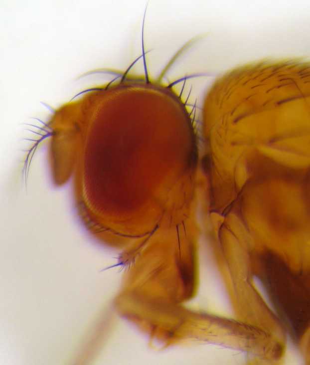
''Drosophila'' () is a genus of flies, belonging to the family Drosophilidae, whose members are often called "small fruit flies" or (less frequently) pomace flies, vinegar flies, or wine flies, a reference to the characteristic of many species to linger around overripe or rotting fruit. They should not be confused with the Tephritidae, a related family, which are also called fruit flies (sometimes referred to as "true fruit flies"); tephritids feed primarily on unripe or ripe fruit, with many species being regarded as destructive agricultural pests, especially the Mediterranean fruit fly. One species of ''Drosophila'' in particular, '' D. melanogaster'', has been heavily used in research in genetics and is a common model organism in developmental biology. The terms "fruit fly" and "''Drosophila''" are often used synonymously with ''D. melanogaster'' in modern biological literature. The entire genus, however, contains more than 1,500 species and is very diverse in appea ... [...More Info...] [...Related Items...] OR: [Wikipedia] [Google] [Baidu] |
PAX6
Paired box protein Pax-6, also known as aniridia type II protein (AN2) or oculorhombin, is a protein that in humans is encoded by the ''PAX6'' gene. Function PAX6 is a member of the Pax gene family which is responsible for carrying the genetic information that will encode the Pax-6 protein. It acts as a "master control" gene for the development of eyes and other sensory organs, certain neural and epidermal tissues as well as other homologous structures, usually derived from ectodermal tissues. However, it has been recognized that a suite of genes is necessary for eye development, and therefore the term of "master control" gene may be inaccurate. Pax-6 is expressed as a transcription factor when neural ectoderm receives a combination of weak Sonic hedgehog (SHH) and strong TGF-Beta signaling gradients. Expression is first seen in the forebrain, hindbrain, head ectoderm and spinal cord followed by later expression in midbrain. This transcription factor is most noted for its ... [...More Info...] [...Related Items...] OR: [Wikipedia] [Google] [Baidu] |
Surface Ectoderm
The surface ectoderm (or external ectoderm) forms the following structures: *Skin (only epidermis; dermis is derived from mesoderm) (along with glands, hair, and nails) * Epithelium of the mouth and nasal cavity and glands of the mouth and nasal cavity *Tooth enamel (as a side note, dentin and dental pulp are formed from ectomesenchyme which is derived from ectoderm (specifically neural crest cells and travels with mesenchmyal cells) *Epithelium of anterior pituitary *Lens, cornea, lacrimal gland, tarsal glands and the conjunctiva of the eye *Apical ectodermal ridge inducing development of the limb buds of the embryo. *Sensory receptors in epidermis See also *List of human cell types derived from the germ layers This is a list of cells in humans derived from the three embryonic germ layers – ectoderm, mesoderm, and endoderm. Cells derived from ectoderm Surface ectoderm Skin * Trichocyte * Keratinocyte Anterior pituitary * Gonadotrope * Corticot ... References ... [...More Info...] [...Related Items...] OR: [Wikipedia] [Google] [Baidu] |
Neural Tube
In the developing chordate (including vertebrates), the neural tube is the embryonic precursor to the central nervous system, which is made up of the brain and spinal cord. The neural groove gradually deepens as the neural fold become elevated, and ultimately the folds meet and coalesce in the middle line and convert the groove into the closed neural tube. In humans, neural tube closure usually occurs by the fourth week of pregnancy (the 28th day after conception). The ectodermal wall of the tube forms the rudiment of the nervous system. The centre of the tube is the ''neural canal''.It is an important structure for the development of fetus's brain and spine Development The neural tube develops in two ways: primary neurulation and secondary neurulation. Primary neurulation divides the ectoderm into three cell types: * The internally located neural tube * The externally located epidermis * The neural crest cells, which develop in the region between the neural tube and e ... [...More Info...] [...Related Items...] OR: [Wikipedia] [Google] [Baidu] |
Choroid Fissure
The tela choroidea (or tela chorioidea) is a region of meningeal pia mater that adheres to the underlying ependyma, and gives rise to the choroid plexus in each of the brain’s four ventricles. ''Tela'' is Latin for ''woven'' and is used to describe a web-like membrane or layer. The tela choroidea is a very thin part of the loose connective tissue of pia mater overlying and closely adhering to the ependyma. It has a rich blood supply. The ependyma and vascular pia mater – the tela choroidea, form regions of minute projections known as a choroid plexus that projects into each ventricle. The choroid plexus produces most of the cerebrospinal fluid of the central nervous system that circulates through the ventricles of the brain, the central canal of the spinal cord, and the subarachnoid space. The tela choroidea in the ventricles forms from different parts of the roof plate in the development of the embryo. Structure In the lateral ventricles the tela choroidea–a double-layere ... [...More Info...] [...Related Items...] OR: [Wikipedia] [Google] [Baidu] |
Optic Vesicle
The eyes begin to develop as a pair of diverticula (pouches) from the lateral aspects of the forebrain. These diverticula make their appearance before the closure of the anterior end of the neural tube; after the closure of the tube around the 4th week of development, they are known as the optic vesicles. Previous studies of optic vesicles suggest that the surrounding extraocular tissues – the surface ectoderm and extraocular mesenchyme – are necessary for normal eye growth and differentiation. They project toward the sides of the head, and the peripheral part of each expands to form a hollow bulb, while the proximal part remains narrow and constitutes the optic stalk, which goes on to form the optic nerve In neuroanatomy, the optic nerve, also known as the second cranial nerve, cranial nerve II, or simply CN II, is a paired cranial nerve that transmits visual information from the retina to the brain. In humans, the optic nerve is derived fro .... Additional images ... [...More Info...] [...Related Items...] OR: [Wikipedia] [Google] [Baidu] |


-_Drosophila_Model.jpg)
