|
External Iliac Lymph Nodes
The external iliac lymph nodes are lymph nodes, from eight to ten in number, that lie along the external iliac vessels. They are arranged in three groups, one on the lateral, another on the medial, and a third on the anterior aspect of the vessels; the third group is, however, sometimes absent. Their principal afferents are derived from the inguinal lymph nodes, the deep lymphatics of the abdominal wall below the umbilicus and of the adductor region of the thigh, and the lymphatics from the glans penis, glans clitoridis, the membranous urethra, the prostate, the fundus of the urinary bladder, the cervix uteri, and upper part of the vagina. Additional images File:Lymph_node_regions.svg, Regional lymph tissue File:Gray611.png , The parietal lymph glands of the pelvis. File:Gray612.png , Iliopelvic glands (lateral view). File:Lymphatics of the prostate-Gray619.png , Lymphatics of the prostate. File:Gray621.png, Deep lymph nodes and vessels of the thorax and abdomen. ... [...More Info...] [...Related Items...] OR: [Wikipedia] [Google] [Baidu] |
Inguinal Lymph Node
Inguinal lymph nodes are lymph nodes in the human groin. Located in the femoral triangle of the inguinal region, they are grouped into superficial and deep lymph nodes. The superficial have three divisions: the superomedial, superolateral, and inferior superficial. Superficial inguinal lymph nodes * The superficial inguinal lymph nodes are the inguinal lymph nodes that form a chain immediately below the inguinal ligament. They lie deep to the fascia of Camper that overlies the femoral vessels at the medial aspect of the thigh. They are bounded superiorly by the inguinal ligament in the femoral triangle; laterally by the border of the sartorius muscle, and medially by the adductor longus muscle. They are divided into three groups: * inferior – inferior of the saphenous opening of the leg, receive drainage from lower legs * superolateral – on the side of the saphenous opening, receive drainage from the side buttocks and the lower abdominal wall. * superomedial – located at t ... [...More Info...] [...Related Items...] OR: [Wikipedia] [Google] [Baidu] |
Common Iliac Lymph Nodes
The common iliac lymph nodes, four to six in number, are grouped behind and on the sides of the common iliac artery, one or two being placed below the bifurcation of the aorta, in front of the fifth lumbar vertebra. They drain chiefly the hypogastric and external iliac glands, and their efferents pass to the lateral aortic glands The periaortic lymph nodes (also known as lumbar) are a group of lymph nodes that lie in front of the lumbar vertebrae near the aorta. These lymph nodes receive drainage from the gastrointestinal tract and the abdominal organs. The periaortic ly .... References Lymphatics of the torso {{Portal bar, Anatomy ... [...More Info...] [...Related Items...] OR: [Wikipedia] [Google] [Baidu] |
Lymph Nodes
A lymph node, or lymph gland, is a kidney-shaped organ of the lymphatic system and the adaptive immune system. A large number of lymph nodes are linked throughout the body by the lymphatic vessels. They are major sites of lymphocytes that include B and T cells. Lymph nodes are important for the proper functioning of the immune system, acting as filters for foreign particles including cancer cells, but have no detoxification function. In the lymphatic system a lymph node is a secondary lymphoid organ. A lymph node is enclosed in a fibrous capsule and is made up of an outer cortex and an inner medulla. Lymph nodes become inflamed or enlarged in various diseases, which may range from trivial throat infections to life-threatening cancers. The condition of lymph nodes is very important in cancer staging, which decides the treatment to be used and determines the prognosis. Lymphadenopathy refers to glands that are enlarged or swollen. When inflamed or enlarged, lymph nodes can be fi ... [...More Info...] [...Related Items...] OR: [Wikipedia] [Google] [Baidu] |
External Iliac Vessels (other)
The external iliac vessels are: * External iliac artery The external iliac arteries are two major Artery, arteries which bifurcate off the common iliac arteries anterior to the sacroiliac joint of the pelvis. Structure The external iliac artery arises from the bifurcation of the common iliac arter ... * External iliac vein {{disambig ... [...More Info...] [...Related Items...] OR: [Wikipedia] [Google] [Baidu] |
Navel
The navel (clinically known as the umbilicus, commonly known as the belly button or tummy button) is a protruding, flat, or hollowed area on the abdomen at the attachment site of the umbilical cord. All placental mammals have a navel, although it is generally more conspicuous in humans. Structure The umbilicus is used to visually separate the abdomen into quadrants. The umbilicus is a prominent scar on the abdomen, with its position being relatively consistent among humans. The skin around the waist at the level of the umbilicus is supplied by the tenth thoracic spinal nerve (T10 dermatome). The umbilicus itself typically lies at a vertical level corresponding to the junction between the L3 and L4 vertebrae, with a normal variation among people between the L3 and L5 vertebrae. Parts of the adult navel include the "umbilical cord remnant" or "umbilical tip", which is the often protruding scar left by the detachment of the umbilical cord. This is located in the center of the ... [...More Info...] [...Related Items...] OR: [Wikipedia] [Google] [Baidu] |
Glans Penis
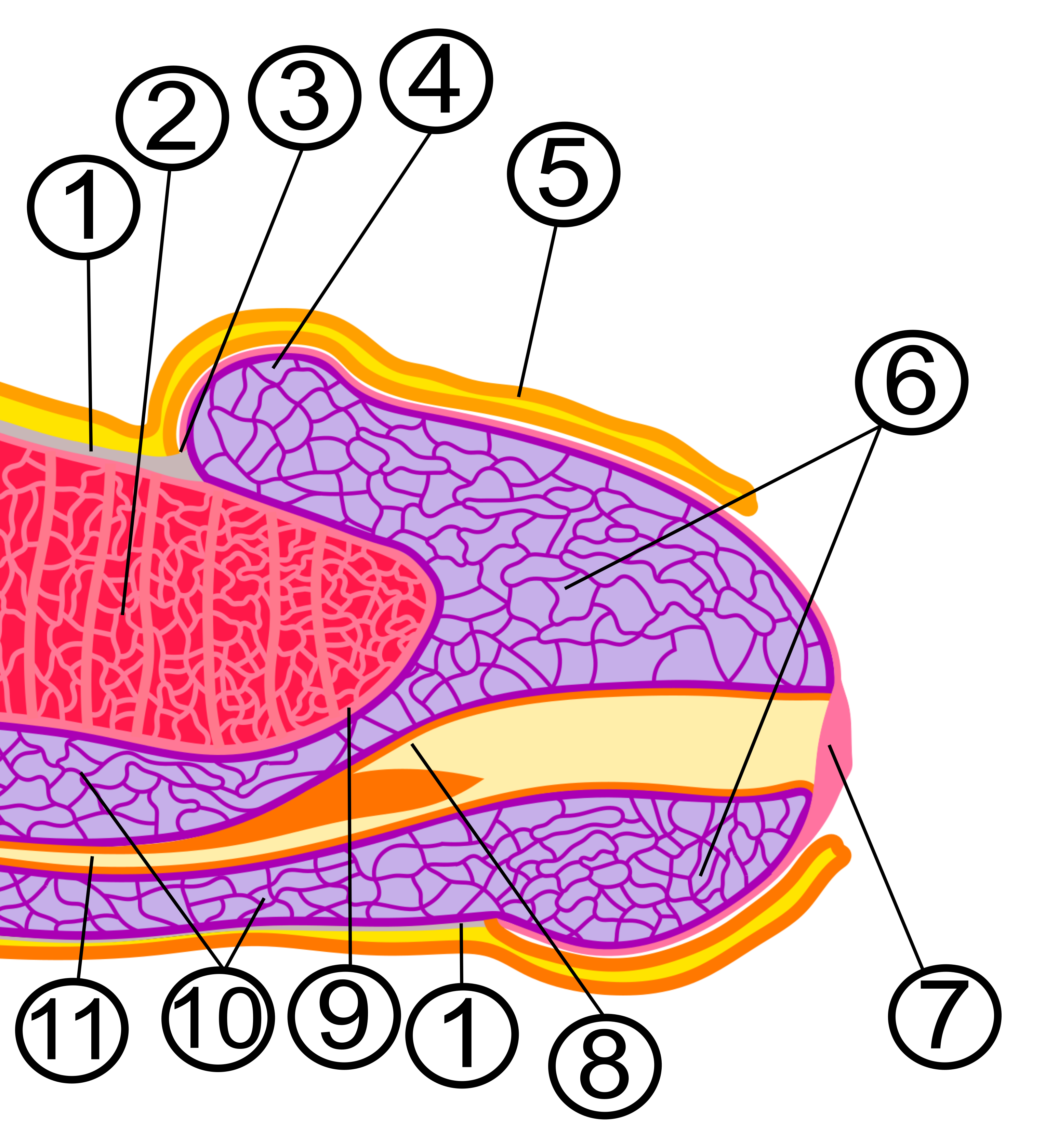
In male human anatomy, the glans penis, commonly referred to as the glans, is the bulbous structure at the distal end of the human penis that is the human male's most sensitive erogenous zone and their primary anatomical source of sexual pleasure. It is anatomically homologous to the clitoral glans. The glans penis is part of the male reproductive organs in humans and other mammals where it may appear smooth, spiny, elongated or divided. It is externally lined with mucosal tissue, which creates a smooth texture and glossy appearance. In humans, the glans is a continuation of the corpus spongiosum of the penis. At the summit appears the urinary meatus and at the base forms the corona glandis. An elastic band of tissue, known as the frenulum, runs on its ventral surface. In men who are not circumcised, it is completely or partially covered by the foreskin. In adults, the foreskin can generally be retracted over and past the glans manually or sometimes automatically during an ... [...More Info...] [...Related Items...] OR: [Wikipedia] [Google] [Baidu] |
Clitoris
The clitoris ( or ) is a female sex organ present in mammals, ostriches and a limited number of other animals. In humans, the visible portion – the glans – is at the front junction of the labia minora (inner lips), above the opening of the urethra. Unlike the penis, the male homologue (equivalent) to the clitoris, it usually does not contain the distal portion (or opening) of the urethra and is therefore not used for urination. In most species, the clitoris lacks any reproductive function. While few animals urinate through the clitoris or use it reproductively, the spotted hyena, which has an especially large clitoris, urinates, mates, and gives birth via the organ. Some other mammals, such as lemurs and spider monkeys, also have a large clitoris. The clitoris is the human female's most sensitive erogenous zone and generally the primary anatomical source of human female sexual pleasure. In humans and other mammals, it develops from an outgrowth in the embry ... [...More Info...] [...Related Items...] OR: [Wikipedia] [Google] [Baidu] |
Membranous Urethra
The membranous urethra or intermediate part of male urethra is the shortest, least dilatable, and, with the exception of the urinary meatus, the narrowest part of the urethra. It extends downward and forward, with a slight anterior concavity, between the apex of the prostate and the bulb of the urethra, perforating the urogenital diaphragm about 2.5 cm below and behind the pubic symphysis. The hinder part of the urethral bulb lies in apposition with the inferior fascia of the urogenital diaphragm, but its upper portion diverges somewhat from this fascia: the anterior wall of the membranous urethra is thus prolonged for a short distance in front of the urogenital diaphragm; it measures about 2 cm in length, while the posterior wall which is between the two fasciæ of the diaphragm is only 1.25 cm long. The anatomical variation in membranous urethral length measurements in men have been reported to range from 0.5 cm to 3.4 cm. The membranous portion of th ... [...More Info...] [...Related Items...] OR: [Wikipedia] [Google] [Baidu] |
Prostate
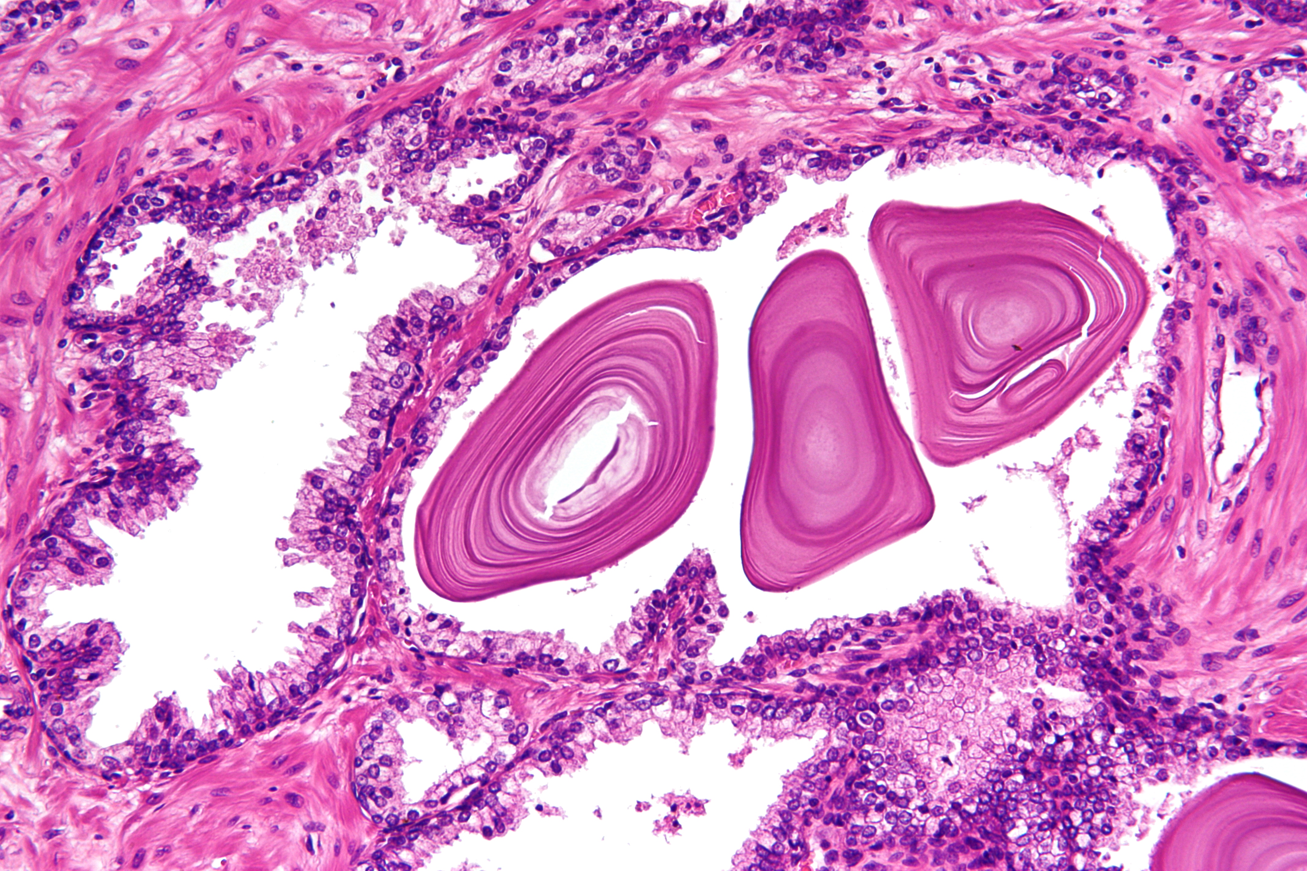
The prostate is both an Male accessory gland, accessory gland of the male reproductive system and a muscle-driven mechanical switch between urination and ejaculation. It is found only in some mammals. It differs between species anatomically, chemically, and physiologically. Anatomically, the prostate is found below the Urinary bladder, bladder, with the urethra passing through it. It is described in gross anatomy as consisting of lobes and in microanatomy by zone. It is surrounded by an elastic, fibromuscular capsule and contains glandular tissue as well as connective tissue. The prostate glands produce and contain fluid that forms part of semen, the substance emitted during ejaculation as part of the male Human sexual response cycle, sexual response. This prostatic fluid is slightly alkaline, milky or white in appearance. The alkalinity of semen helps neutralize the acidity of the vagina, vaginal tract, prolonging the lifespan of sperm. The prostatic fluid is expelled in the ... [...More Info...] [...Related Items...] OR: [Wikipedia] [Google] [Baidu] |
Fundus Of The Urinary Bladder
The urinary bladder, or simply bladder, is a hollow organ in humans and other vertebrates that stores urine from the kidneys before disposal by urination. In humans the bladder is a distensible organ that sits on the pelvic floor. Urine enters the bladder via the ureters and exits via the urethra. The typical adult human bladder will hold between 300 and (10.14 and ) before the urge to empty occurs, but can hold considerably more. The Latin phrase for "urinary bladder" is ''vesica urinaria'', and the term ''vesical'' or prefix ''vesico -'' appear in connection with associated structures such as vesical veins. The modern Latin word for "bladder" – ''cystis'' – appears in associated terms such as cystitis (inflammation of the bladder). Structure In humans, the bladder is a hollow muscular organ situated at the base of the pelvis. In gross anatomy, the bladder can be divided into a broad , a body, an apex, and a neck. The apex (also called the vertex) is directed forward ... [...More Info...] [...Related Items...] OR: [Wikipedia] [Google] [Baidu] |
Cervix Uteri
The cervix or cervix uteri (Latin, 'neck of the uterus') is the lower part of the uterus (womb) in the human female reproductive system. The cervix is usually 2 to 3 cm long (~1 inch) and roughly cylindrical in shape, which changes during pregnancy. The narrow, central cervical canal runs along its entire length, connecting the uterine cavity and the lumen of the vagina. The opening into the uterus is called the internal os, and the opening into the vagina is called the external os. The lower part of the cervix, known as the vaginal portion of the cervix (or ectocervix), bulges into the top of the vagina. The cervix has been documented anatomically since at least the time of Hippocrates, over 2,000 years ago. The cervical canal is a passage through which sperm must travel to fertilize an egg cell after sexual intercourse. Several methods of contraception, including cervical caps and cervical diaphragms, aim to block or prevent the passage of sperm through the cervical ca ... [...More Info...] [...Related Items...] OR: [Wikipedia] [Google] [Baidu] |
Vagina
In mammals, the vagina is the elastic, muscular part of the female genital tract. In humans, it extends from the vestibule to the cervix. The outer vaginal opening is normally partly covered by a thin layer of mucosal tissue called the hymen. At the deep end, the cervix (neck of the uterus) bulges into the vagina. The vagina allows for sexual intercourse and birth. It also channels menstrual flow, which occurs in humans and closely related primates as part of the menstrual cycle. Although research on the vagina is especially lacking for different animals, its location, structure and size are documented as varying among species. Female mammals usually have two external openings in the vulva; these are the urethral opening for the urinary tract and the vaginal opening for the genital tract. This is different from male mammals, who usually have a single urethral opening for both urination and reproduction. The vaginal opening is much larger than the nearby urethral opening, an ... [...More Info...] [...Related Items...] OR: [Wikipedia] [Google] [Baidu] |
.jpg)


.png)

