|
Extensor Hood
An extensor expansion (extensor hood, dorsal expansion, dorsal hood, dorsal aponeurosis) is the special connective attachments by which the extensor tendons insert into the phalanges. These flattened tendons (aponeurosis) of extensor muscles span the proximal and middle phalanges. At the distal end of the metacarpal, the extensor tendon will expand to form a hood, which covers the back and sides of the head of the metacarpal and the proximal phalanx. Bands The expansion soon divides into three bands: * lateral bands pass on either side of the proximal phalanx and stretch all the way to the distal phalanx. The lumbricals of the hand, extensor indicis muscle, dorsal interossei of the hand, and palmar interossei In human anatomy, the palmar or volar interossei (interossei volares in older literature) are three small, unipennate muscles in the hand that lie between the metacarpal bones and are attached to the index, ring, and little fingers. They are sm ... insert on these ba ... [...More Info...] [...Related Items...] OR: [Wikipedia] [Google] [Baidu] |
Extensor Tendon Compartments Of The Wrist
Extensor tendon compartments of the wrist are anatomical tunnels on the back of the wrist that contain tendons of muscles that extend (as opposed to flex) the wrist and the digits (fingers and thumb). The extensor tendons are held in place by the extensor retinaculum. As the tendons travel over the posterior (back) aspect of the wrist they are enclosed within synovial tendon sheaths. These sheaths reduce the friction to the extensor tendons as they traverse the compartments that are formed by the attachments of the extensor retinaculum to the distal (far end) of the radius and ulna. Structure The compartments are numbered with each compartment containing specific extensor tendons. Clinical significance Any of the dorsal compartments of the wrist can develop tenosynovial inflammation. The first compartment is the most frequently affected site, called De Quervain's disease (syndrome or tenosynovitis). The other two most commonly injured are the sixth (extensor carpi ulnaris ... [...More Info...] [...Related Items...] OR: [Wikipedia] [Google] [Baidu] |
Phalanx Bone
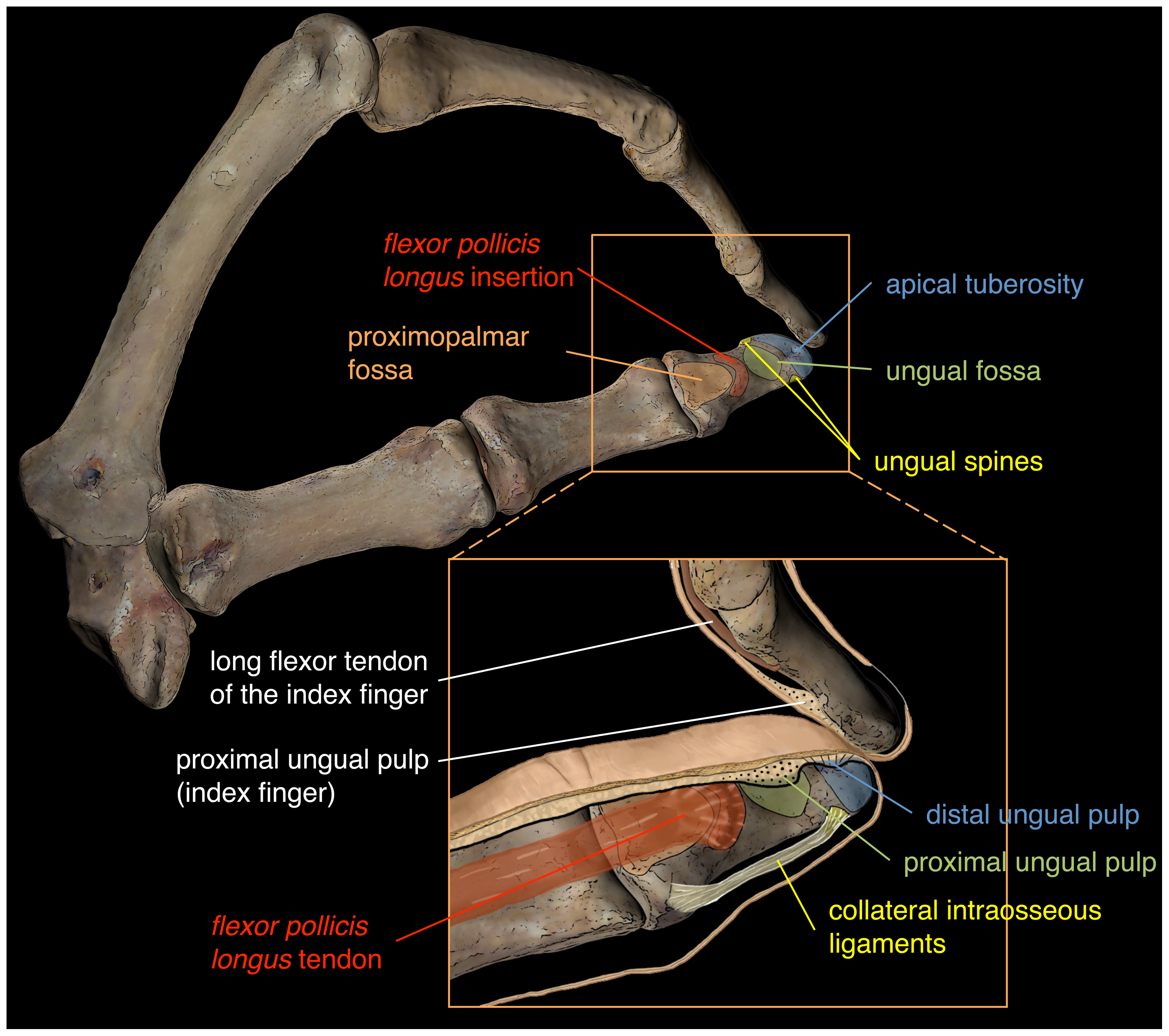
The phalanges (singular: ''phalanx'' ) are digital bones in the hands and feet of most vertebrates. In primates, the thumbs and big toes have two phalanges while the other digits have three phalanges. The phalanges are classed as long bones. Structure The phalanges are the bones that make up the fingers of the hand and the toes of the foot. There are 56 phalanges in the human body, with fourteen on each hand and foot. Three phalanges are present on each finger and toe, with the exception of the thumb and large toe, which possess only two. The middle and far phalanges of the fifth toes are often fused together (symphalangism). The phalanges of the hand are commonly known as the finger bones. The phalanges of the foot differ from the hand in that they are often shorter and more compressed, especially in the proximal phalanges, those closest to the torso. A phalanx is named according to whether it is proximal, middle, or distal and its associated finger or toe. The proxi ... [...More Info...] [...Related Items...] OR: [Wikipedia] [Google] [Baidu] |
Aponeurosis
An aponeurosis (; plural: ''aponeuroses'') is a type or a variant of the deep fascia, in the form of a sheet of pearly-white fibrous tissue that attaches sheet-like muscles needing a wide area of attachment. Their primary function is to join muscles and the body parts they act upon, whether bone or other muscles. They have a shiny, whitish-silvery color, are histologically similar to tendons, and are very sparingly supplied with blood vessels and nerves. When dissected, aponeuroses are papery and peel off by sections. The primary regions with thick aponeuroses are in the ventral abdominal region, the dorsal lumbar region, the ventriculus in birds, and the palmar (palms) and plantar (soles) regions. Anatomy Anterior abdominal aponeuroses The anterior abdominal aponeuroses are located just superficial to the rectus abdominis muscle. It has for its borders the external oblique, pectoralis muscles, and the latissimus dorsi. Posterior lumbar aponeuroses The posterior lumbar a ... [...More Info...] [...Related Items...] OR: [Wikipedia] [Google] [Baidu] |
Metacarpal
In human anatomy, the metacarpal bones or metacarpus form the intermediate part of the skeletal hand located between the phalanges of the fingers and the carpal bones of the wrist, which forms the connection to the forearm. The metacarpal bones are analogous to the metatarsal bones in the foot. Structure The metacarpals form a transverse arch to which the rigid row of distal carpal bones are fixed. The peripheral metacarpals (those of the thumb and little finger) form the sides of the cup of the palmar gutter and as they are brought together they deepen this concavity. The index metacarpal is the most firmly fixed, while the thumb metacarpal articulates with the trapezium and acts independently from the others. The middle metacarpals are tightly united to the carpus by intrinsic interlocking bone elements at their bases. The ring metacarpal is somewhat more mobile while the fifth metacarpal is semi-independent.Tubiana ''et al'' 1998, p 11 Each metacarpal bone consists of a bod ... [...More Info...] [...Related Items...] OR: [Wikipedia] [Google] [Baidu] |
Phalanx Bones
The phalanges (singular: ''phalanx'' ) are digital bones in the hands and feet of most vertebrates. In primates, the thumbs and big toes have two phalanges while the other digits have three phalanges. The phalanges are classed as long bones. Structure The phalanges are the bones that make up the fingers of the hand and the toes of the foot. There are 56 phalanges in the human body, with fourteen on each hand and foot. Three phalanges are present on each finger and toe, with the exception of the thumb and large toe, which possess only two. The middle and far phalanges of the fifth toes are often fused together (symphalangism). The phalanges of the hand are commonly known as the finger bones. The phalanges of the foot differ from the hand in that they are often shorter and more compressed, especially in the proximal phalanges, those closest to the torso. A phalanx is named according to whether it is proximal, middle, or distal and its associated finger or toe. The proxi ... [...More Info...] [...Related Items...] OR: [Wikipedia] [Google] [Baidu] |
Lumbricals Of The Hand
The lumbricals are intrinsic muscles of the hand that flex the metacarpophalangeal joints, and extend the interphalangeal joints. p. 97 The lumbrical muscles of the foot also have a similar action, though they are of less clinical concern. Structure The lumbricals are four, small, worm-like muscles on each hand. These muscles are unusual in that they do not attach to bone. Instead, they attach proximally to the tendons of flexor digitorum profundus, and distally to the extensor expansions. The first and second lumbricals are unipennate, while the third and fourth lumbricals are bipennate. Nerve supply The first and second lumbricals (the most radial two) are innervated by the median nerve. The third and fourth lumbricals (most ulnar two) are innervated by the deep branch of ulnar nerve. This is the usual innervation of the lumbricals (occurring in 60% of individuals). However 1:3 (median:ulnar - 20% of individuals) and 3:1 (median:ulnar - 20% of individuals) also ... [...More Info...] [...Related Items...] OR: [Wikipedia] [Google] [Baidu] |
Extensor Indicis Muscle
In human anatomy, the extensor indicis roprius'' is a narrow, elongated skeletal muscle in the deep layer of the dorsal forearm, placed medial to, and parallel with, the extensor pollicis longus In human anatomy, the extensor pollicis longus muscle (EPL) is a skeletal muscle located dorsally on the forearm. It is much larger than the extensor pollicis brevis, the origin of which it partly covers and acts to stretch the thumb together wi .... Its tendon goes to the index finger, which it extends. Structure It arises from the distal third of the dorsal part of the body of ulna and from the interosseous membrane. It runs through the fourth tendon compartment together with the extensor digitorum, from where it projects into the dorsal aponeurosis of the index finger. Opposite the head of the second metacarpal bone, it joins the ulnar side of the tendon of the extensor digitorum which belongs to the index finger. Like the extensor digiti minimi (i.e. the extensor of the ... [...More Info...] [...Related Items...] OR: [Wikipedia] [Google] [Baidu] |
Dorsal Interossei Of The Hand
In human anatomy, the dorsal interossei (DI) are four muscles in the back of the hand that act to abduct (spread) the index, middle, and ring fingers away from hand's midline (ray of middle finger) and assist in flexion at the metacarpophalangeal joints and extension at the interphalangeal joints of the index, middle and ring fingers. Structure There are four dorsal interossei in each hand. They are specified as 'dorsal' to contrast them with the palmar interossei, which are located on the anterior side of the metacarpals. The dorsal interosseous muscles are bipennate, with each muscle arising by two heads from the adjacent sides of the metacarpal bones, but more extensively from the metacarpal bone of the finger into which the muscle is inserted. They are inserted into the bases of the proximal phalanges and into the extensor expansion of the corresponding extensor digitorum tendon. The middle digit has two dorsal interossei insert onto it while the first digit (thumb) and ... [...More Info...] [...Related Items...] OR: [Wikipedia] [Google] [Baidu] |
Palmar Interossei
In human anatomy, the palmar or volar interossei (interossei volares in older literature) are three small, unipennate muscles in the hand that lie between the metacarpal bones and are attached to the index, ring, and little fingers. They are smaller than the dorsal interossei of the hand. Structure All palmar interossei originate along the shaft of the metacarpal bone of the digit on which they act. They are inserted into the base of the proximal phalanx and the extensor expansion of the extensor digitorum of the same digit. Pollical palmar interosseous The first palmar interosseous is located at the thumb's medial side. Passing between the first dorsal interosseous and the oblique head of adductor pollicis, it is inserted on the base of the thumb's proximal phalanx together with adductor pollicis. The "pollical" palmar interosseous muscle (PPIM), is present in more than 80% of individuals and was first described by . Its presence has been verified by numerous anatomists ... [...More Info...] [...Related Items...] OR: [Wikipedia] [Google] [Baidu] |
Extensor Compartment Of The Forearm
The posterior compartment of the forearm (or extensor compartment) contains twelve muscles which primarily extend the wrist and digits. It is separated from the anterior compartment by the interosseous membrane between the radius and ulna. Structure Muscles There are generally twelve muscles in the posterior compartment of the forearm, which can be further divided into superficial, intermediate, and deep. Most of the muscles in the superficial and the intermediate layers share a common origin which is the outer part of the elbow, the lateral epicondyle of humerus. The deep muscles arise from the distal part of the ulna and the surrounding interosseous membrane. The brachioradialis, flexor of the elbow, is unusual in that it is located in the posterior compartment, but it is actually a muscle of flexor / anterior compartment of the forearm. The anconeus, assisting in extension of the elbow joint, is by some considered part of the posterior compartment of the arm. The majority o ... [...More Info...] [...Related Items...] OR: [Wikipedia] [Google] [Baidu] |
Muscular System
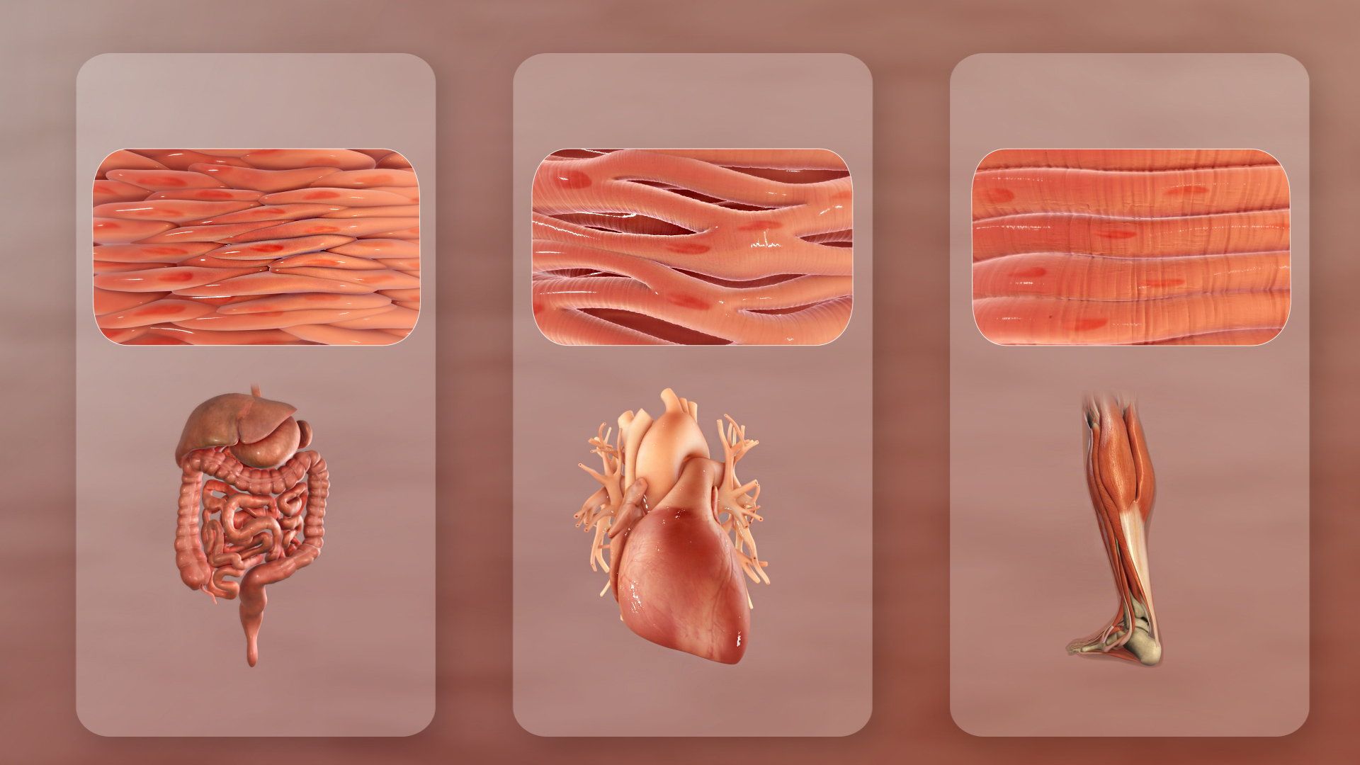
The muscular system is an organ (anatomy), organ system consisting of skeletal muscle, skeletal, smooth muscle, smooth, and cardiac muscle, cardiac muscle. It permits movement of the body, maintains posture, and circulates blood throughout the body. The muscular systems in vertebrates are controlled through the nervous system although some muscles (such as the cardiac muscle) can be completely autonomous. Together with the skeletal system in the human, it forms the musculoskeletal system, which is responsible for the movement of the human body, body. Types There are three distinct types of muscle: skeletal muscle, cardiac muscle, cardiac or heart muscle, and smooth muscle, smooth (non-striated) muscle. Muscles provide strength, balance, posture, movement, and heat for the body to keep warm. There are over 650 muscles in the human body. A kind of elastic tissue makes up each muscle, which consists of thousands, or tens of thousands, of small muscle fibers. Each fiber comprises ... [...More Info...] [...Related Items...] OR: [Wikipedia] [Google] [Baidu] |

_dorsal_view.png)


