|
Ethmoidal Nerve
The ethmoidal nerves, which arise from the nasociliary nerve, supply the ethmoidal cells; the posterior branch leaves the orbital cavity through the posterior ethmoidal foramen and gives some filaments to the sphenoidal sinus The sphenoid sinus is a paired paranasal sinus occurring within the within the body of the sphenoid bone. It represents one pair of the four paired paranasal sinuses.Illustrated Anatomy of the Head and Neck, Fehrenbach and Herring, Elsevier, 201 .... There are two ethmoidal nerves on each side of the face: * posterior ethmoidal nerve * anterior ethmoidal nerve References External links ufl.edu Trigeminal nerve {{neuroanatomy-stub ... [...More Info...] [...Related Items...] OR: [Wikipedia] [Google] [Baidu] |
Ciliary Ganglion
The ciliary ganglion is a bundle of nerve parasympathetic ganglion located just behind the eye in the posterior orbit. It is 1–2 mm in diameter and in humans contains approximately 2,500 neurons. The ganglion contains postganglionic parasympathetic neurons. These neurons supply the pupillary sphincter muscle, which constricts the pupil, and the ciliary muscle which contracts to make the lens more convex. Both of these muscles are involuntary since they are controlled by the parasympathetic division of the autonomic nervous system. The ciliary ganglion is one of four parasympathetic ganglia of the head. The others are the submandibular ganglion, pterygopalatine ganglion, and otic ganglion. Structure The ciliary ganglion contains postganglionic parasympathetic neurons that supply the ciliary muscle and the pupillary sphincter muscle. Because of the much larger size of the ciliary muscle, 95% of the neurons in the ciliary ganglion innervate it compared to the pupillary sphin ... [...More Info...] [...Related Items...] OR: [Wikipedia] [Google] [Baidu] |
Nasociliary Nerve
The nasociliary nerve is a branch of the ophthalmic nerve, itself a branch of the trigeminal nerve (CN V). It is intermediate in size between the other two branches of the ophthalmic nerve, the frontal nerve and lacrimal nerve. Structure The nasociliary nerve enters the orbit via the superior orbital fissure, between the two heads of the lateral rectus muscle and between the superior and inferior rami of the oculomotor nerve. It passes across the optic nerve (CN II) and runs obliquely beneath the superior rectus muscle and superior oblique muscle to the medial wall of the orbital cavity. It passes through the anterior ethmoidal opening as the anterior ethmoidal nerve and enters the cranial cavity just below the cribriform plate of the ethmoid bone. It supplies branches to the mucous membrane of the nasal cavity and finally emerges between the inferior border of the nasal bone and the side nasal cartilages as the external nasal branch. Branches * posterior ethmoidal nerve ... [...More Info...] [...Related Items...] OR: [Wikipedia] [Google] [Baidu] |
Ethmoidal Cell
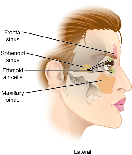
The ethmoid sinuses or ethmoid air cells of the ethmoid bone are one of the four paired paranasal sinuses. The cells are variable in both size and number in the lateral mass of each of the ethmoid bones and cannot be palpated during an extraoral examination. They are divided into anterior and posterior groups. The ethmoid air cells are numerous thin-walled cavities situated in the ethmoidal labyrinth and completed by the frontal, maxilla, lacrimal, sphenoidal, and palatine bones. They lie between the upper parts of the nasal cavities and the orbits, and are separated from these cavities by thin bony lamellae. Groups of sinuses The groups of the ethmoidal air cells drain into the nasal meatuses.Otorhinolaryngology, Head and Neck Surgery, Anniko, Springer, 2010, page 188 * The posterior group the ''posterior ethmoidal sinus'' drains into the superior meatus above the middle nasal concha; sometimes one or more opens into the sphenoidal sinus. * The anterior group the ''anterior ethm ... [...More Info...] [...Related Items...] OR: [Wikipedia] [Google] [Baidu] |
Orbital Cavity
In anatomy, the orbit is the cavity or socket of the skull in which the eye and its appendages are situated. "Orbit" can refer to the bony socket, or it can also be used to imply the contents. In the adult human, the volume of the orbit is , of which the eye occupies . The orbital contents comprise the eye, the orbital and retrobulbar fascia, extraocular muscles, cranial nerves II, III, IV, V, and VI, blood vessels, fat, the lacrimal gland with its sac and duct, the eyelids, medial and lateral palpebral ligaments, cheek ligaments, the suspensory ligament, septum, ciliary ganglion and short ciliary nerves. Structure The orbits are conical or four-sided pyramidal cavities, which open into the midline of the face and point back into the head. Each consists of a base, an apex and four walls."eye, human."Encyclopædia Britannica from Encyclopædia Britannica 2006 Ultimate Reference Suite DVD 2009 Openings There are two important foramina, or windows, two important f ... [...More Info...] [...Related Items...] OR: [Wikipedia] [Google] [Baidu] |
Posterior Ethmoidal Foramen
Lateral to either olfactory groove are the internal openings of the anterior and posterior ethmoidal foramina (or canals). The posterior ethmoidal foramen opens at the back part of this margin under cover of the projecting lamina of the sphenoid, and transmits the posterior ethmoidal vessels and nerve A nerve is an enclosed, cable-like bundle of nerve fibers (called axons) in the peripheral nervous system. A nerve transmits electrical impulses. It is the basic unit of the peripheral nervous system. A nerve provides a common pathway for the .... External links * () (#4) Foramina of the skull {{musculoskeletal-stub ... [...More Info...] [...Related Items...] OR: [Wikipedia] [Google] [Baidu] |
Sphenoidal Sinus
The sphenoid sinus is a paired paranasal sinus occurring within the within the body of the sphenoid bone. It represents one pair of the four paired paranasal sinuses.Illustrated Anatomy of the Head and Neck, Fehrenbach and Herring, Elsevier, 2012, page 64 The pair of sphenoid sinuses are separated in the middle by a septum of sphenoid sinuses. Each sphenoid sinus communicates with the nasal cavity via the opening of sphenoidal sinus. The two sphenoid sinuses vary in size and shape, and are usually asymmetrical. Anatomy On average, a sphenoid sinus measures 2.2 cm vertical height, 2 cm in transverse breadth; and 2.2 cm antero-posterior depth. Each spehoid sinus is contained within the body of sphenoid bone, being situated just inferior to the sella turcica. The two sphenoid sinuses are separated medially by the septum of sphenoidal sinuses (which is usually asymmetrical). An opening of sphenoidal sinus forms a passage between each sphenoidal sinus, and the nasal ... [...More Info...] [...Related Items...] OR: [Wikipedia] [Google] [Baidu] |
Posterior Ethmoidal Nerve
The posterior ethmoidal nerve is a nerve of the orbit around the eye. It is a branch of the nasociliary nerve from the ophthalmic nerve (CN V1). It supplies sensation to the sphenoid sinus, the ethmoid sinus, and part of the dura mater in the anterior cranial fossa. Structure The posterior ethmoidal nerve is a branch of the nasociliary nerve, itself a branch of the ophthalmic nerve (CN V1), itself a branch of the trigeminal nerve (CN V). It passes through the posterior ethmoidal foramen, with the posterior ethmoidal artery. It gives branches to the sphenoid sinus and the ethmoid sinus. It also gives a branch to supply part of the dura mater in the anterior cranial fossa. Variation The posterior ethmoidal nerve is absent in a significant proportion of people. This may be around 30%. Function The posterior ethmoidal nerve supplies sensation to the sphenoid sinus and the ethmoid sinus. It also supplies sensation to part of the dura mater in the anterior cranial fossa. Othe ... [...More Info...] [...Related Items...] OR: [Wikipedia] [Google] [Baidu] |
Anterior Ethmoidal Nerve
The anterior ethmoidal nerve is a nerve of the nose. It is a branch of the nasociliary nerve, itself a branch of the ophthalmic nerve (V1). It provides sensory innervation to some structures around the nasal cavity. Structure The anterior ethmoidal nerve is a terminal branch of the nasociliary nerve, a branch of the ophthalmic nerve (CN V1), itself a branch of the trigeminal nerve (CN V). It branches near the medial wall of the orbit. The anterior ethmoidal nerve arises only after the nasociliary has given off its four branches (the ramus communicans to the ciliary ganglion, the long ciliary nerves, the infratrochlear nerve, and the posterior ethmoidal nerve). It travels through the anterior ethmoidal foramen to reach the anterior cranial fossa. It then moves forward and passes through the cribriform plate to enter the nasal cavity. It gives off branches to the roof of the nasal cavity, and bifurcates into a lateral internal nasal branch and medial internal nasal branch. ... [...More Info...] [...Related Items...] OR: [Wikipedia] [Google] [Baidu] |

