|
Erb's Point (neurology)
The nerve point of the neck, also known as Erb's point is a site at the upper trunk of the brachial plexus located 2–3 cm above the clavicle. It is named for Wilhelm Heinrich Erb. Taken together, there are six types of nerves that meet at this point. "Erb's point" is also a term used in head and neck surgery to describe the point on the posterior border of the sternocleidomastoid muscle where the four superficial branches of the cervical plexus—the greater auricular, lesser occipital, transverse cervical, and supraclavicular nerves—emerge from behind the muscle. This point is located approximately at the junction of the upper and middle thirds of this muscle. From here, the accessory nerve courses through the posterior triangle of the neck to enter the anterior border of the trapezius muscle at a point located approximately at the junction of the middle and lower thirds of the anterior border of this muscle. The spinal accessory nerve can often be found 1 cm abov ... [...More Info...] [...Related Items...] OR: [Wikipedia] [Google] [Baidu] |
Brachial Plexus
The brachial plexus is a network () of nerves formed by the anterior rami of the lower four cervical nerves and first thoracic nerve ( C5, C6, C7, C8, and T1). This plexus extends from the spinal cord, through the cervicoaxillary canal in the neck, over the first rib, and into the armpit, it supplies afferent and efferent nerve fibers the to chest, shoulder, arm, forearm, and hand. Structure The brachial plexus is divided into five ''roots'', three ''trunks'', six ''divisions'' (three anterior and three posterior), three ''cords'', and five ''branches''. There are five "terminal" branches and numerous other "pre-terminal" or "collateral" branches, such as the subscapular nerve, the thoracodorsal nerve, and the long thoracic nerve, that leave the plexus at various points along its length. A common structure used to identify part of the brachial plexus in cadaver dissections is the M or W shape made by the musculocutaneous nerve, lateral cord, median nerve, medial cord, and ... [...More Info...] [...Related Items...] OR: [Wikipedia] [Google] [Baidu] |
Anterior
Standard anatomical terms of location are used to unambiguously describe the anatomy of animals, including humans. The terms, typically derived from Latin or Greek roots, describe something in its standard anatomical position. This position provides a definition of what is at the front ("anterior"), behind ("posterior") and so on. As part of defining and describing terms, the body is described through the use of anatomical planes and anatomical axes. The meaning of terms that are used can change depending on whether an organism is bipedal or quadrupedal. Additionally, for some animals such as invertebrates, some terms may not have any meaning at all; for example, an animal that is radially symmetrical will have no anterior surface, but can still have a description that a part is close to the middle ("proximal") or further from the middle ("distal"). International organisations have determined vocabularies that are often used as standard vocabularies for subdisciplines of anatomy ... [...More Info...] [...Related Items...] OR: [Wikipedia] [Google] [Baidu] |
Porter (doorkeeper)
An ostiarius, a Latin word sometimes anglicized as ostiary but often literally translated as porter or Doorman (profession), doorman, originally was a servant or guard posted at the entrance of a building. See also gatekeeper. In the Roman Catholic Church, this "porter" became the lowest of the four minor orders prescribed by the Council of Trent. This was the first order a seminarian was admitted to after receiving the tonsure. The porter had in ancient times the duty of opening and closing the church-door and of guarding the church, especially to ensure no unbaptised persons would enter during the Eucharist. Later on, the porter would also guard, open and close the doors of the sacristy, baptistry and elsewhere in the church. The porter was not a part of holy orders administering sacraments but simply a preparatory job on the way to the major orders: subdiaconate (until its suppression, after the Second Vatican Council by Pope Paul VI), deacon, diaconate and the priesthood. L ... [...More Info...] [...Related Items...] OR: [Wikipedia] [Google] [Baidu] |
Erb's Palsy
Erb's palsy is a paralysis of the arm caused by injury to the upper group of the arm's main nerves, specifically the severing of the upper trunk C5–C6 nerves. These form part of the brachial plexus, comprising the ventral rami of spinal nerves C5–C8 and thoracic nerve T1. pp.1037–1047 pp.370–374 pp.76–77 These injuries arise most commonly, but not exclusively, from shoulder dystocia during a difficult birth.A.D.A.M Healthcare center Depending on the nature of the damage, the paralysis can either resolve on its own over a period of months, necessitate rehabilitative therapy, or require surgery. Presentation The paralysis can be partial or complete; the damage to each nerve can range from bruising to tearing. The most commonly involved root is C5 (aka[...More Info...] [...Related Items...] OR: [Wikipedia] [Google] [Baidu] |
Axillary Nerve
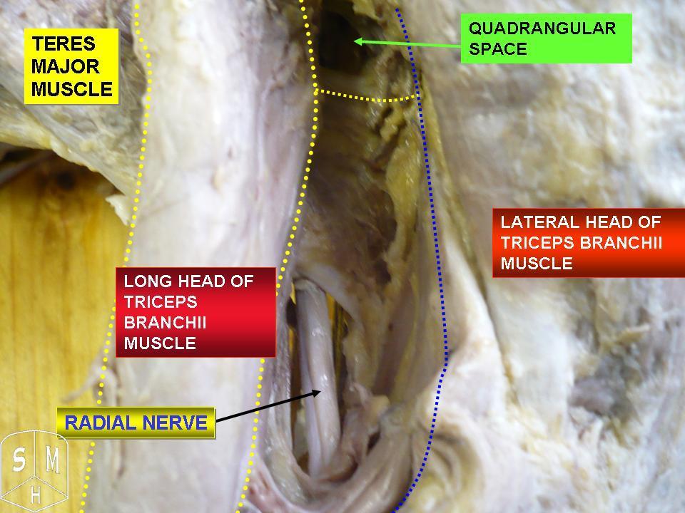
The axillary nerve or the circumflex nerve is a nerve of the human body, that originates from the brachial plexus (upper trunk, posterior division, posterior cord) at the level of the axilla (armpit) and carries nerve fibers from C5 and C6. The axillary nerve travels through the quadrangular space with the posterior circumflex humeral artery and vein to innervate the deltoid and teres minor. Structure The nerve lies at first behind the axillary artery, and in front of the subscapularis, and passes downward to the lower border of that muscle. It then winds from anterior to posterior around the neck of the humerus, in company with the posterior humeral circumflex artery, through the quadrangular space (bounded above by the teres minor, below by the teres major, medially by the long head of the triceps brachii, and laterally by the surgical neck of the humerus), and divides into an anterior, a posterior, and a collateral branch to the long head of the triceps brachii branch. * The ... [...More Info...] [...Related Items...] OR: [Wikipedia] [Google] [Baidu] |
Deltoid Muscle
The deltoid muscle is the muscle forming the rounded contour of the human shoulder. It is also known as the 'common shoulder muscle', particularly in other animals such as the domestic cat. Anatomically, the deltoid muscle appears to be made up of three distinct sets of muscle fibers, namely the # anterior or clavicular part (pars clavicularis) # posterior or scapular part (pars scapularis) # intermediate or acromial part (pars acromialis) However, electromyography suggests that it consists of at least seven groups that can be independently coordinated by the nervous system. It was previously called the deltoideus (plural ''deltoidei'') and the name is still used by some anatomists. It is called so because it is in the shape of the Greek capital letter delta (Δ). Deltoid is also further shortened in slang as "delt". A study of 30 shoulders revealed an average mass of in humans, ranging from to . Structure Previous studies showed that the insertions of the tendons of the delto ... [...More Info...] [...Related Items...] OR: [Wikipedia] [Google] [Baidu] |
Radial Nerve
The radial nerve is a nerve in the human body that supplies the posterior portion of the upper limb. It innervates the medial and lateral heads of the triceps brachii muscle of the arm, as well as all 12 muscles in the posterior osteofascial compartment of the forearm and the associated joints and overlying skin. It originates from the brachial plexus, carrying fibers from the ventral roots of spinal nerves C5, C6, C7, C8 & T1. The radial nerve and its branches provide motor innervation to the dorsal arm muscles (the triceps brachii and the anconeus) and the extrinsic extensors of the wrists and hands; it also provides cutaneous sensory innervation to most of the back of the hand, except for the back of the little finger and adjacent half of the ring finger (which are innervated by the ulnar nerve). The radial nerve divides into a deep branch, which becomes the posterior interosseous nerve, and a superficial branch, which goes on to innervate the dorsum (back) of the hand. Th ... [...More Info...] [...Related Items...] OR: [Wikipedia] [Google] [Baidu] |
Brachioradialis
The brachioradialis is a muscle of the forearm that flexes the forearm at the elbow. It is also capable of both pronation and supination, depending on the position of the forearm. It is attached to the distal styloid process of the radius by way of the brachioradialis tendon, and to the lateral supracondylar ridge of the humerus. Structure The brachioradialis is a superficial, fusiform muscle on the lateral side of the forearm. It originates proximally on the lateral supracondylar ridge of the humerus. It inserts distally on the radius, at the base of its styloid process. Near the elbow, it forms the lateral limit of the cubital fossa, or elbow pit. Nerve supply Despite the bulk of the muscle body being visible from the anterior aspect of the forearm, the brachioradialis is a posterior compartment muscle and consequently is innervated by the radial nerve. Of the muscles that receive innervation from the radial nerve, it is one of only four that receive input directly from the ra ... [...More Info...] [...Related Items...] OR: [Wikipedia] [Google] [Baidu] |
Musculocutaneous Nerve
The musculocutaneous nerve arises from the lateral cord of the brachial plexus, opposite the lower border of the pectoralis major, its fibers being derived from C5, C6 and C7. Structure The musculocutaneous nerve arises from the lateral cord of the brachial plexus, courses through the anterior part of the arm, and terminates at 2 cm above elbow as the lateral cutaneous nerve of the forearm. Musculocutaneous nerve arises from the lateral cord of the brachial plexus with root value of C5 to C7 of the spinal cord. It follows the course of the third part of the axillary artery (part of the axillary artery distal to the pectoralis minor) laterally and enters the frontal aspect of the arm where it penetrates the coracobrachialis muscle. It then passes downwards and laterally between the biceps brachii (above) and the brachialis muscles (below), to the lateral side of the arm; at 2 cm above the elbow it pierces the deep fascia lateral to the tendon of the biceps brachii ... [...More Info...] [...Related Items...] OR: [Wikipedia] [Google] [Baidu] |
Coracobrachialis
The coracobrachialis muscle is the smallest of the three muscles that attach to the coracoid process of the scapula. (The other two muscles are pectoralis minor and the short head of the biceps brachii.) It is situated at the upper and medial part of the arm. Structure Coracobrachialis muscle arises from the apex of the coracoid process, in common with the short head of the biceps brachii, and from the intermuscular septum between the two muscles. It is inserted by means of a flat tendon into an impression at the middle of the medial surface and border of the body of the humerus (shaft of the humerus) between the origins of the triceps brachii and brachialis. Innervation Coracobrachialis muscle is perforated by and innervated by the musculocutaneous nerve, which arises from the anterior division of the upper trunk ( C5, C6) and middle trunk ( C7) of the brachial plexus. Development Variation Function The action of the coracobrachialis is to flex and adduct the arm at the ... [...More Info...] [...Related Items...] OR: [Wikipedia] [Google] [Baidu] |
Brachialis
The brachialis (brachialis anticus), also known as the Teichmann muscle, is a muscle in the upper arm that flexes the elbow. It lies deeper than the biceps brachii, and makes up part of the floor of the region known as the cubital fossa (elbow pit). The brachialis is the prime mover of elbow flexion generating about 50% more power than the biceps.Saladin, Kenneth S, Stephen J. Sullivan, and Christina A. Gan. Anatomy & Physiology: The Unity of Form and Function. 2015. Print. Structure The brachialis originates from the anterior surface of the distal half of the humerus, near the insertion of the deltoid muscle, which it embraces by two angular processes. Its origin extends below to within 2.5 cm of the margin of the articular surface of the humerus at the elbow joint. Its fibers converge to a thick tendon, which is inserted into the tuberosity of the ulna and the rough depression on the anterior surface of the coronoid process of the ulna. Blood supply The brachialis is supp ... [...More Info...] [...Related Items...] OR: [Wikipedia] [Google] [Baidu] |
Biceps
The biceps or biceps brachii ( la, musculus biceps brachii, "two-headed muscle of the arm") is a large muscle that lies on the front of the upper arm between the shoulder and the elbow. Both heads of the muscle arise on the scapula and join to form a single muscle belly which is attached to the upper forearm. While the biceps crosses both the shoulder and elbow joints, its main function is at the elbow where it flexes the forearm and supinates the forearm. Both these movements are used when opening a bottle with a corkscrew: first biceps screws in the cork (supination), then it pulls the cork out (flexion). Structure The biceps is one of three muscles in the anterior compartment of the upper arm, along with the brachialis muscle and the coracobrachialis muscle, with which the biceps shares a nerve supply. The biceps muscle has two heads, the short head and the long head, distinguished according to their origin at the coracoid process and supraglenoid tubercle of the sc ... [...More Info...] [...Related Items...] OR: [Wikipedia] [Google] [Baidu] |


