|
Epithelial Root Sheath
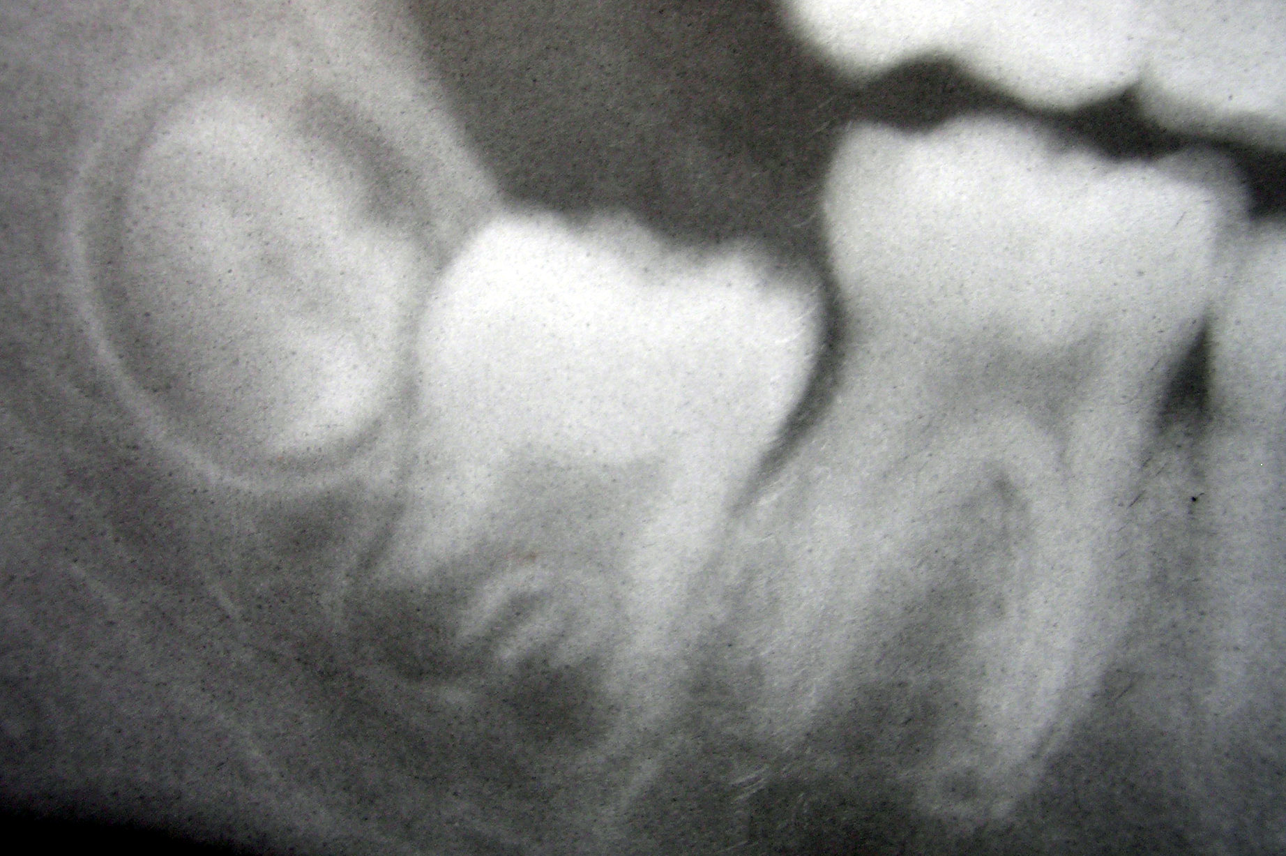
The Hertwig epithelial root sheath (HERS) or epithelial root sheath is a proliferation of epithelial cells located at the cervical loop of the enamel organ in a developing tooth. Hertwig epithelial root sheath initiates the formation of dentin in the root of a tooth by causing the differentiation of odontoblasts from the dental papilla. The root sheath eventually disintegrates with the periodontal ligament, but residual pieces that do not completely disappear are seen as epithelial cell rests of Malassez (ERM). These rests can become cystic, presenting future periodontal infections. Structure Hertwig epithelial root sheath is derived from the inner and outer enamel epithelium of the enamel organ.Illustrated Dental Embryology, Histology, and Anatomy, Bath-Balogh and Fehrenbach, Elsevier, 2011, p. 66 Function The sheath is also responsible for multiple or accessory roots (medial growth) and lateral or accessory canals in the root (break in epithelium).Ten Cate's Oral Histolog ... [...More Info...] [...Related Items...] OR: [Wikipedia] [Google] [Baidu] |
Epithelium
Epithelium or epithelial tissue is one of the four basic types of animal tissue, along with connective tissue, muscle tissue and nervous tissue. It is a thin, continuous, protective layer of compactly packed cells with a little intercellular matrix. Epithelial tissues line the outer surfaces of organs and blood vessels throughout the body, as well as the inner surfaces of cavities in many internal organs. An example is the epidermis, the outermost layer of the skin. There are three principal shapes of epithelial cell: squamous (scaly), columnar, and cuboidal. These can be arranged in a singular layer of cells as simple epithelium, either squamous, columnar, or cuboidal, or in layers of two or more cells deep as stratified (layered), or ''compound'', either squamous, columnar or cuboidal. In some tissues, a layer of columnar cells may appear to be stratified due to the placement of the nuclei. This sort of tissue is called pseudostratified. All glands are made up of epithe ... [...More Info...] [...Related Items...] OR: [Wikipedia] [Google] [Baidu] |
Epithelial Cell Rests Of Malassez
In dentistry, the epithelial cell rests of Malassez (ERM) or epithelial rests of Malassez (''pax epithelialis pediodontii'') are part of the periodontal ligament cells around a tooth. They are discrete clusters of residual cells from Hertwig's epithelial root sheath (HERS) that didn't completely disappear. It is considered that these cell rests proliferate to form epithelial lining of various odontogenic cysts such as radicular cyst under the influence of various stimuli. They are named after Louis-Charles Malassez (1842–1909) who described them. Some rests become calcified in the periodontal ligament (cementicles). ERM plays a role in cementum repair and regeneration. The stem cells in ERM can undergo an epithelial–mesenchymal transition and differentiate into diverse types of cells of mesodermal and ectodermal The ectoderm is one of the three primary germ layers formed in early embryonic development. It is the outermost layer, and is superficial to the mesoderm (the m ... [...More Info...] [...Related Items...] OR: [Wikipedia] [Google] [Baidu] |
Human Tooth Development
Tooth development or odontogenesis is the complex process by which teeth form from embryonic cells, grow, and erupt into the mouth. For human teeth to have a healthy oral environment, all parts of the tooth must develop during appropriate stages of fetal development. Primary (baby) teeth start to form between the sixth and eighth week of prenatal development, and permanent teeth begin to form in the twentieth week.Ten Cate's Oral Histology, Nanci, Elsevier, 2013, pages 70-94 If teeth do not start to develop at or near these times, they will not develop at all, resulting in hypodontia or anodontia. A significant amount of research has focused on determining the processes that initiate tooth development. It is widely accepted that there is a factor within the tissues of the first pharyngeal arch that is necessary for the development of teeth. Overview The tooth germ is an aggregation of cells that eventually forms a tooth.University of Texas Medical Branch. These cells are der ... [...More Info...] [...Related Items...] OR: [Wikipedia] [Google] [Baidu] |
Dentin
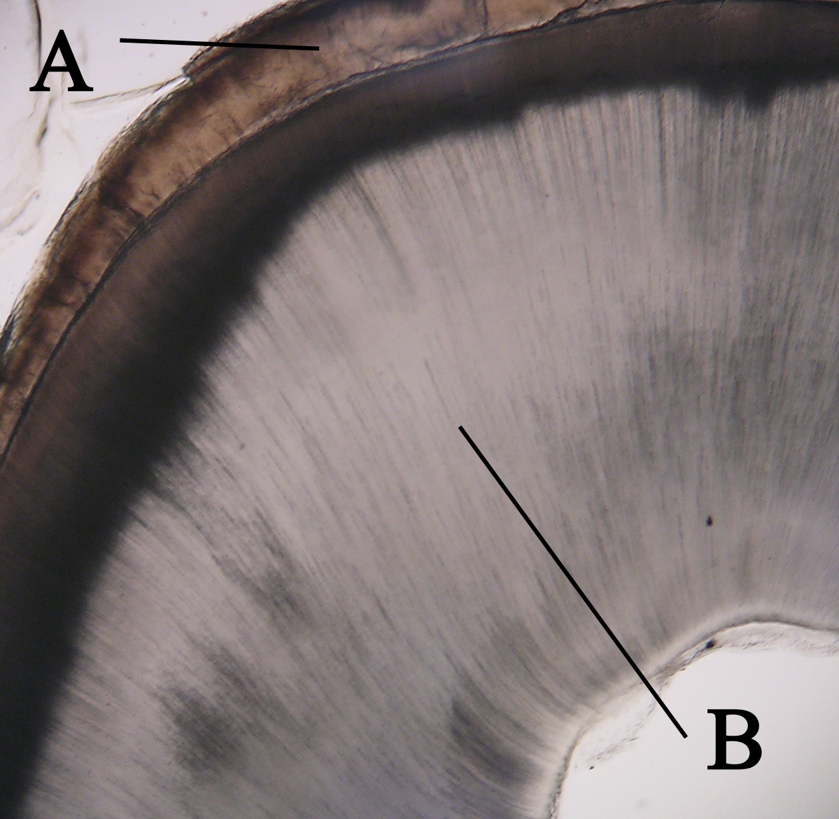
Dentin () (American English) or dentine ( or ) (British English) ( la, substantia eburnea) is a calcified tissue of the body and, along with enamel, cementum, and pulp, is one of the four major components of teeth. It is usually covered by enamel on the crown and cementum on the root and surrounds the entire pulp. By volume, 45% of dentin consists of the mineral hydroxyapatite, 33% is organic material, and 22% is water. Yellow in appearance, it greatly affects the color of a tooth due to the translucency of enamel. Dentin, which is less mineralized and less brittle than enamel, is necessary for the support of enamel. Dentin rates approximately 3 on the Mohs scale of mineral hardness. There are two main characteristics which distinguish dentin from enamel: firstly, dentin forms throughout life; secondly, dentin is sensitive and can become hypersensitive to changes in temperature due to the sensory function of odontoblasts, especially when enamel recedes and dentin channels becom ... [...More Info...] [...Related Items...] OR: [Wikipedia] [Google] [Baidu] |
Cementum
Cementum is a specialized calcified substance covering the root of a tooth. The cementum is the part of the periodontium that attaches the teeth to the alveolar bone by anchoring the periodontal ligament.Illustrated Dental Embryology, Histology, and Anatomy, Bath-Balogh and Fehrenbach, Elsevier, 2011, page 170. Structure The cells of cementum are the entrapped cementoblasts, the cementocytes. Each cementocyte lies in its lacuna, similar to the pattern noted in bone. These lacunae also have canaliculi or canals. Unlike those in bone, however, these canals in cementum do not contain nerves, nor do they radiate outward. Instead, the canals are oriented toward the periodontal ligament and contain cementocytic processes that exist to diffuse nutrients from the ligament because it is vascularized. After the apposition of cementum in layers, the cementoblasts that do not become entrapped in cementum line up along the cemental surface along the length of the outer covering of the perio ... [...More Info...] [...Related Items...] OR: [Wikipedia] [Google] [Baidu] |
Oskar Hertwig
Oscar Hertwig (21 April 1849 in Friedberg – 25 October 1922 in Berlin) was a German embryologist and zoologist known for his research in developmental biology and evolution. Hertwig is credited as the first man to observe sexual reproduction by looking at the cells of sea urchins under the microscope. Biography Hertwig was the elder brother of zoologist-professor Richard Hertwig (1850–1937). The Hertwig brothers were the most eminent scholars of Ernst Haeckel (and Carl Gegenbaur) from the University of Jena. They were independent of Haeckel's philosophical speculations but took his ideas in a positive way to widen their concepts in zoology. Initially, between 1879 and 1883, they performed embryological studies, especially on the theory of the coelom (1881), the fluid-filled body cavity. These problems were based on the phylogenetic theorems of Haeckel, i.e. the biogenic theory (German = biogenetisches Grundgesetz), and the "gastraea theory". Within 10 years, the two broth ... [...More Info...] [...Related Items...] OR: [Wikipedia] [Google] [Baidu] |
Fenestra (histology)
A fenestra (fenestration; plural fenestrae or fenestrations) is any small opening or pore, commonly used as a term in the biological sciences. It is the Latin word for "window", and is used in various fields to describe a pore in an anatomical structure. Biological morphology In morphology, fenestrae are found in cancellous bones, particularly in the skull. In anatomy, the round window and oval window are also known as the ''fenestra rotunda'' and the ''fenestra ovalis''. In microanatomy, fenestrae are found in endothelium of fenestrated capillaries, enabling the rapid exchange of molecules between the blood and surrounding tissue. The elastic layer of the tunica intima is a fenestrated membrane. In surgery, a fenestration is a new opening made in a part of the body to enable drainage or access. Plant biology and mycology In plant biology, the perforations in a perforate leaf are also described as fenestrae, and the leaf is called a fenestrate leaf. The leaf window is al ... [...More Info...] [...Related Items...] OR: [Wikipedia] [Google] [Baidu] |
Cementum
Cementum is a specialized calcified substance covering the root of a tooth. The cementum is the part of the periodontium that attaches the teeth to the alveolar bone by anchoring the periodontal ligament.Illustrated Dental Embryology, Histology, and Anatomy, Bath-Balogh and Fehrenbach, Elsevier, 2011, page 170. Structure The cells of cementum are the entrapped cementoblasts, the cementocytes. Each cementocyte lies in its lacuna, similar to the pattern noted in bone. These lacunae also have canaliculi or canals. Unlike those in bone, however, these canals in cementum do not contain nerves, nor do they radiate outward. Instead, the canals are oriented toward the periodontal ligament and contain cementocytic processes that exist to diffuse nutrients from the ligament because it is vascularized. After the apposition of cementum in layers, the cementoblasts that do not become entrapped in cementum line up along the cemental surface along the length of the outer covering of the perio ... [...More Info...] [...Related Items...] OR: [Wikipedia] [Google] [Baidu] |
Cementogenesis
Cementogenesis is the formation of cementum, one of the three mineralized substances of a tooth. Cementum covers the roots of teeth and serves to anchor gingival and periodontal fibers of the periodontal ligament by the fibers to the alveolar bone (some types of cementum may also form on the surface of the enamel of the crown at the cementoenamel junction (CEJ)). Process For cementogenesis to begin, Hertwig epithelial root sheath (HERS) must fragment. HERS is a collar of epithelial cells derived from the apical prolongation of the enamel organ. Once the root sheath disintegrates, the newly formed surface of root dentin comes into contact with the undifferentiated cells of the dental sac (dental follicle). This then stimulates the activation of cementoblasts to begin cementogenesis. The external shape of each root is fully determined by the position of the surrounding Hertwig epithelial root sheath. It is believed that either 1) HERS becomes interrupted; 2) infiltrating dental sac ... [...More Info...] [...Related Items...] OR: [Wikipedia] [Google] [Baidu] |
Dental Papilla
In embryology and prenatal development, the dental papilla is a condensation of ectomesenchymal cells called odontoblasts, seen in histologic sections of a developing tooth. It lies below a cellular aggregation known as the enamel organ. The dental papilla appears after 8–10 weeks intra uteral life. The dental papilla gives rise to the dentin and pulp of a tooth. The enamel organ, dental papilla, and dental follicle together forms one unit, called the tooth germ. This is of importance because all the tissues of a tooth and its supporting structures form from these distinct cellular aggregations. Similar to dental follicle, the dental papilla has a very rich blood supply and provides nutrition to the enamel organ. Embryology Formation of dental papilla occurs in the Cap stage of Odontogenesis. The cap stage The cap stage is the second stage of tooth development and occurs during the ninth or tenth week of prenatal development. Unequal proliferation of the tooth bu ... [...More Info...] [...Related Items...] OR: [Wikipedia] [Google] [Baidu] |
Cell (biology)
The cell is the basic structural and functional unit of life forms. Every cell consists of a cytoplasm enclosed within a membrane, and contains many biomolecules such as proteins, DNA and RNA, as well as many small molecules of nutrients and metabolites.Cell Movements and the Shaping of the Vertebrate Body in Chapter 21 of Molecular Biology of the Cell '' fourth edition, edited by Bruce Alberts (2002) published by Garland Science. The Alberts text discusses how the "cellular building blocks" move to shape developing embryos. It is also common to describe small molecules such as ... [...More Info...] [...Related Items...] OR: [Wikipedia] [Google] [Baidu] |
Odontoblast
In vertebrates, an odontoblast is a cell of neural crest origin that is part of the outer surface of the dental pulp, and whose biological function is dentinogenesis, which is the formation of dentin, the substance beneath the tooth enamel on the crown and the cementum on the root. Structure Odontoblasts are large columnar cells, whose cell bodies are arranged along the interface between dentin and pulp, from the crown to cervix to the root apex in a mature tooth. The cell is rich in endoplasmic reticulum and Golgi complex, especially during primary dentin formation, which allows it to have a high secretory capacity; it first forms the collagenous matrix to form predentin, then mineral levels to form the mature dentin. Odontoblasts form approximately 4 μm of predentin daily during tooth development.Ten Cate's Oral Histology, Nanci, Elsevier, 2013, page 170 During secretion after differentiation from the outer cells of the dental papilla, it is noted that it is polarized so its nu ... [...More Info...] [...Related Items...] OR: [Wikipedia] [Google] [Baidu] |


