|
Epiphyseal Dysgenesis
The epiphysis () is the rounded end of a long bone, at its joint with adjacent bone(s). Between the epiphysis and diaphysis (the long midsection of the long bone) lies the metaphysis, including the epiphyseal plate (growth plate). At the joint, the epiphysis is covered with articular cartilage; below that covering is a zone similar to the epiphyseal plate, known as subchondral bone. The epiphysis is filled with red bone marrow, which produces erythrocytes (red blood cells). Structure There are four types of epiphysis: # Pressure epiphysis: The region of the long bone that forms the joint is a pressure epiphysis (e.g. the head of the femur, part of the hip joint complex). Pressure epiphyses assist in transmitting the weight of the human body and are the regions of the bone that are under pressure during movement or locomotion. Another example of a pressure epiphysis is the head of the humerus The humerus (; ) is a long bone in the arm that runs from the shoulder ... [...More Info...] [...Related Items...] OR: [Wikipedia] [Google] [Baidu] |
Endocrine
The endocrine system is a messenger system comprising feedback loops of the hormones released by internal glands of an organism directly into the circulatory system, regulating distant target organs. In vertebrates, the hypothalamus is the neural control center for all endocrine systems. In humans, the major endocrine glands are the thyroid gland and the adrenal glands. The study of the endocrine system and its disorders is known as endocrinology. Glands that signal each other in sequence are often referred to as an axis, such as the hypothalamic-pituitary-adrenal axis. In addition to the specialized endocrine organs mentioned above, many other organs that are part of other body systems have secondary endocrine functions, including bone, kidneys, liver, heart and gonads. For example, the kidney secretes the endocrine hormone erythropoietin. Hormones can be amino acid complexes, steroids, eicosanoids, leukotrienes, or prostaglandins. The endocrine system can be contrasted ... [...More Info...] [...Related Items...] OR: [Wikipedia] [Google] [Baidu] |
Coracoid Process
The coracoid process (from Greek κόραξ, raven) is a small hook-like structure on the lateral edge of the superior anterior portion of the scapula (hence: coracoid, or "like a raven's beak"). Pointing laterally forward, it, together with the acromion, serves to stabilize the shoulder joint. It is palpable in the deltopectoral groove between the deltoid and pectoralis major muscles. Structure The coracoid process is a thick curved process attached by a broad base to the upper part of the neck of the scapula; it runs at first upward and medialward; then, becoming smaller, it changes its direction, and projects forward and lateralward. Anatomically it is divided into intervals of: base of coracoid process, angle of coracoid process, shaft and the apex of the coracoid process. The coracoglenoid notch is an indentation localized between the coracoid process and the glenoid. As the coracoid process projects laterally, it house underneath it the subcoracoid space. The ''ascend ... [...More Info...] [...Related Items...] OR: [Wikipedia] [Google] [Baidu] |
Metaphysis
The metaphysis is the neck portion of a long bone between the epiphysis and the diaphysis. It contains the growth plate, the part of the bone that grows during childhood, and as it grows it ossifies near the diaphysis and the epiphyses. The metaphysis contains a diverse population of cells including mesenchymal stem cells, which give rise to bone and fat cells, as well as hematopoietic stem cells which give rise to a variety of blood cells as well as bone-destroying cells called osteoclasts. Thus the metaphysis contains a highly metabolic set of tissues including trabecular (spongy) bone, blood vessels , as well as Marrow Adipose Tissue (MAT). The metaphysis may be divided anatomically into three components based on tissue content: a cartilaginous component (epiphyseal plate), a bony component (metaphysis) and a fibrous component surrounding the periphery of the plate. The growth plate synchronizes chondrogenesis with osteogenesis or interstitial cartilage growth with both appos ... [...More Info...] [...Related Items...] OR: [Wikipedia] [Google] [Baidu] |
Epiphyseal Plate
The epiphyseal plate (or epiphysial plate, physis, or growth plate) is a hyaline cartilage plate in the metaphysis at each end of a long bone. It is the part of a long bone where new bone growth takes place; that is, the whole bone is alive, with maintenance remodeling throughout its existing bone tissue, but the growth plate is the place where the long bone grows longer (adds length). The plate is only found in children and adolescents; in adults, who have stopped growing, the plate is replaced by an epiphyseal line. This replacement is known as epiphyseal closure or growth plate fusion. Complete fusion can occur as early as 12 for girls (with the most common being 14-15 years for girls) and as early as 14 for boys (with the most common being 15–17 years for boys). Structure Development Endochondral ossification is responsible for the initial bone development from cartilage in utero and infants and the longitudinal growth of long bones in the epiphyseal plate. The plate' ... [...More Info...] [...Related Items...] OR: [Wikipedia] [Google] [Baidu] |
Diaphysis
The diaphysis is the main or midsection (shaft) of a long bone. It is made up of cortical bone and usually contains bone marrow and adipose tissue (fat). It is a middle tubular part composed of compact bone which surrounds a central marrow cavity which contains red or yellow marrow. In diaphysis, primary ossification occurs. Ewing sarcoma tends to occur at the diaphysis.Physical Medicine and Rehabilitation Board Review, Cuccurullo Additional images Illu long bone.jpg File:EpiMetaDiaphyse.jpg, Long bone See also *Epiphysis *Metaphysis The metaphysis is the neck portion of a long bone between the epiphysis and the diaphysis. It contains the growth plate, the part of the bone that grows during childhood, and as it grows it ossifies near the diaphysis and the epiphyses. The metap ... References Skeletal system Long bones {{musculoskeletal-stub ... [...More Info...] [...Related Items...] OR: [Wikipedia] [Google] [Baidu] |
Giant-cell Tumor Of Bone
Giant-cell tumor of the bone (GCTOB), is a relatively uncommon tumor of the bone. It is characterized by the presence of multinucleated giant cells (osteoclast-like cells). Malignancy in giant-cell tumor is uncommon and occurs in about 2% of all cases. However, if malignant degeneration does occur, it is likely to metastasize to the lungs. Giant-cell tumors are normally benign, with unpredictable behavior. It is a heterogeneous tumor composed of three different cell populations. The giant-cell tumour stromal cells (GCTSC) constitute the neoplastic cells, which are from an osteoblastic origin and are classified based on expression of osteoblast cell markers such as alkaline phosphatase and osteocalcin. In contrast, the mononuclear histiocytic cells (MNHC) and multinucleated giant cell (MNGC) fractions are secondarily recruited and comprise the non-neoplastic cell population. They are derived from an osteoclast-monocyte lineage determined primarily by expression of ''CD68'', a mar ... [...More Info...] [...Related Items...] OR: [Wikipedia] [Google] [Baidu] |
Chondroblastoma
Chondroblastoma is a rare, benign, locally aggressive bone tumor that typically affects the epiphyses or apophyses of long bones. It is thought to arise from an outgrowth of immature cartilage cells ( chondroblasts) from secondary ossification centers, originating from the epiphyseal plate or some remnant of it. Chondroblastoma is very uncommon, accounting for only 1–2% of all bone tumors. It affects mostly children and young adults with most patients being less than 20 years of age. Chondroblastoma shows a predilection towards the male sex, with a ratio of male to female patients of 2:1. The most commonly affected site is the femur, followed by the humerus and tibia. Less commonly affected sites include the talus and calcaneus of the foot and flat bones. Signs and symptoms The most common symptom is mild to severe pain that is gradually progressive in the affected region and may be initially attributed to a minor injury or sports-related injury. Pain may be present fo ... [...More Info...] [...Related Items...] OR: [Wikipedia] [Google] [Baidu] |
Osteochondritis Dissecans
Osteochondritis dissecans (OCD or OD) is a joint disorder primarily of the subchondral bone in which cracks form in the articular cartilage and the underlying subchondral bone. OCD usually causes pain during and after sports. In later stages of the disorder there will be swelling of the affected joint which catches and locks during movement. Physical examination in the early stages does only show pain as symptom, in later stages there could be an effusion, tenderness, and a crackling sound with joint movement. OCD is caused by blood deprivation of the secondary physes around the bone core of the femoral condyle. This happens to the epiphyseal vessels under the influence of repetitive overloading of the joint during running and jumping sports. During growth such chondronecrotic areas grow into the subchondral bone. There it will show as bone defect area under articular cartilage. The bone will then possibly heal to the surrounding condylar bone in 50% of the cases. Or it wil ... [...More Info...] [...Related Items...] OR: [Wikipedia] [Google] [Baidu] |
Avascular Necrosis
Avascular necrosis (AVN), also called osteonecrosis or bone infarction, is death of bone tissue due to interruption of the blood supply. Early on, there may be no symptoms. Gradually joint pain may develop which may limit the ability to move. Complications may include collapse of the bone or nearby joint surface. Risk factors include bone fractures, joint dislocations, alcoholism, and the use of high-dose steroids. The condition may also occur without any clear reason. The most commonly affected bone is the femur. Other relatively common sites include the upper arm bone, knee, shoulder, and ankle. Diagnosis is typically by medical imaging such as X-ray, CT scan, or MRI. Rarely biopsy may be used. Treatments may include medication, not walking on the affected leg, stretching, and surgery. Most of the time surgery is eventually required and may include core decompression, osteotomy, bone grafts, or joint replacement. About 15,000 cases occur per year in the United States. Peop ... [...More Info...] [...Related Items...] OR: [Wikipedia] [Google] [Baidu] |
Pathology
Pathology is the study of the causes and effects of disease or injury. The word ''pathology'' also refers to the study of disease in general, incorporating a wide range of biology research fields and medical practices. However, when used in the context of modern medical treatment, the term is often used in a narrower fashion to refer to processes and tests that fall within the contemporary medical field of "general pathology", an area which includes a number of distinct but inter-related medical specialties that diagnose disease, mostly through analysis of tissue, cell, and body fluid samples. Idiomatically, "a pathology" may also refer to the predicted or actual progression of particular diseases (as in the statement "the many different forms of cancer have diverse pathologies", in which case a more proper choice of word would be " pathophysiologies"), and the affix ''pathy'' is sometimes used to indicate a state of disease in cases of both physical ailment (as in cardiomy ... [...More Info...] [...Related Items...] OR: [Wikipedia] [Google] [Baidu] |
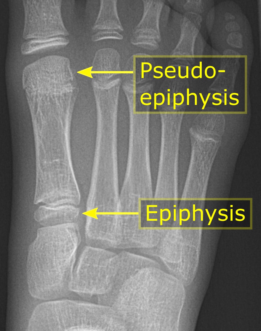
Metatarsal Pseudo-epiphysis
The metatarsal bones, or metatarsus, are a group of five long bones in the foot, located between the tarsal bones of the hind- and mid-foot and the phalanges of the toes. Lacking individual names, the metatarsal bones are numbered from the medial side (the side of the great toe): the first, second, third, fourth, and fifth metatarsal (often depicted with Roman numerals). The metatarsals are analogous to the metacarpal bones of the hand. The lengths of the metatarsal bones in humans are, in descending order, second, third, fourth, fifth, and first. Structure The five metatarsals are dorsal convex long bones consisting of a shaft or body, a base (proximally), and a head (distally).Platzer 2004, p. 220 The body is prismoid in form, tapers gradually from the tarsal to the phalangeal extremity, and is curved longitudinally, so as to be concave below, slightly convex above. The base or posterior extremity is wedge-shaped, articulating proximally with the tarsal bones, and by its ... [...More Info...] [...Related Items...] OR: [Wikipedia] [Google] [Baidu] |
Cuneiform Bones
There are three cuneiform ("wedge-shaped") bones in the human foot: * the first or medial cuneiform * the second or intermediate cuneiform, also known as the middle cuneiform * the third or lateral cuneiform They are located between the navicular bone and the first, second and third metatarsal bones and are medial to the cuboid bone. Structure There are three cuneiform bones: # The medial cuneiform (also known as first cuneiform) is the largest of the cuneiforms. It is situated at the medial side of the foot, anterior to the navicular bone and posterior to the base of the first metatarsal. Lateral to it is the intermediate cuneiform. It articulates with four bones: the navicular, second cuneiform, and first and second metatarsals. The tibialis anterior and fibularis longus muscle inserts at the medial cuneiform bone. # The intermediate cuneiform (second cuneiform or middle cuneiform) is shaped like a wedge, the thin end pointing downwards. The intermediate cuneiform is situate ... [...More Info...] [...Related Items...] OR: [Wikipedia] [Google] [Baidu] |


.jpg)