|
Epilepsy Surgery
Epilepsy surgery involves a neurosurgery, neurosurgical procedure where an area of the brain involved in seizures is either resected, ablative brain surgery, ablated, disconnected or stimulated. The goal is to eliminate seizures or significantly reduce seizure burden. Approximately 60% of all people with epilepsy (0.4% of the population of industrialized countries) have focal epilepsy syndromes. In 15% to 20% of these patients, the condition is not adequately controlled with Anticonvulsant, anticonvulsive drugs. Such patients are potential candidates for surgical epilepsy treatment. First line therapy for epilepsy involves treatment with anticonvulsive drugs, also called antiepileptic drugs. Most patients will respond to one or two different medication trials. The goal of this treatment is the elimination of seizures, since uncontrolled seizures carry significant risks, including injury and sudden death. However, in up to one third of patients, medications alone do not elimina ... [...More Info...] [...Related Items...] OR: [Wikipedia] [Google] [Baidu] |
Neurology
Neurology (from el, wikt:╬Į╬Ąß┐”Žü╬┐╬Į, ╬Į╬Ąß┐”Žü╬┐╬Į (ne├╗ron), "string, nerve" and the suffix wikt:-logia, -logia, "study of") is the branch of specialty (medicine), medicine dealing with the diagnosis and treatment of all categories of conditions and disease involving the brain, the spinal cord and the peripheral nerves. Neurological practice relies heavily on the field of neuroscience, the scientific study of the nervous system. A neurologist is a physician specializing in neurology and trained to investigate, diagnose and treat neurological disorders. Neurologists treat a myriad of neurologic conditions, including stroke, seizures, movement disorders such as Parkinson's disease, autoimmune neurologic disorders such as multiple sclerosis, headache disorders like migraine and dementias such as Alzheimer's disease. Neurologists may also be involved in clinical research, clinical trials, and basic research, basic or translational research. While neurology is a nonsurgical sp ... [...More Info...] [...Related Items...] OR: [Wikipedia] [Google] [Baidu] |
Wada Test
The Wada test, also known as the intracarotid sodium amobarbital procedure (ISAP), establishes cerebral language and memory representation of each hemisphere. Method Medical professionals conduct the test with the patient awake. Essentially, they introduce a barbiturate (usually sodium amobarbital) into one of the internal carotid arteries via a cannula or intra-arterial catheter from the femoral artery. They inject the drug into one hemisphere at a time into the right or left internal carotid artery. If the right carotid is injected, the right side of the brain is inhibited and cannot communicate with the left side. The effect shuts down any language and/or memory function in that hemisphere in order to evaluate the other hemisphere ("half of the brain"). An EEG recording at the same time confirms that the injected side of the brain is inactive as a neurologist performs a neurological examination. The neurologist engages the patient in a series of language and memory related te ... [...More Info...] [...Related Items...] OR: [Wikipedia] [Google] [Baidu] |
Parietal Lobe
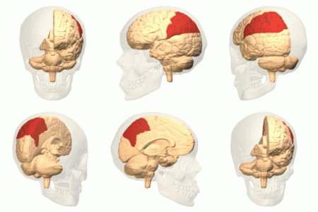
The parietal lobe is one of the four major lobes of the cerebral cortex in the brain of mammals. The parietal lobe is positioned above the temporal lobe and behind the frontal lobe and central sulcus. The parietal lobe integrates sensory information among various modalities, including spatial sense and navigation (proprioception), the main sensory receptive area for the sense of touch in the somatosensory cortex which is just posterior to the central sulcus in the postcentral gyrus, and the dorsal stream of the visual system. The major sensory inputs from the skin (touch, temperature, and pain receptors), relay through the thalamus to the parietal lobe. Several areas of the parietal lobe are important in language processing. The somatosensory cortex can be illustrated as a distorted figure ŌĆō the cortical homunculus (Latin: "little man") in which the body parts are rendered according to how much of the somatosensory cortex is devoted to them. The superior parietal lobule and in ... [...More Info...] [...Related Items...] OR: [Wikipedia] [Google] [Baidu] |
Occipital Lobes
The occipital lobe is one of the four major lobes of the cerebral cortex in the brain of mammals. The name derives from its position at the back of the head, from the Latin ''ob'', "behind", and ''caput'', "head". The occipital lobe is the visual processing center of the mammalian brain containing most of the anatomical region of the visual cortex. The primary visual cortex is Brodmann area 17, commonly called V1 (visual one). Human V1 is located on the medial side of the occipital lobe within the calcarine sulcus; the full extent of V1 often continues onto the occipital pole. V1 is often also called striate cortex because it can be identified by a large stripe of myelin, the Stria of Gennari. Visually driven regions outside V1 are called extrastriate cortex. There are many extrastriate regions, and these are specialized for different visual tasks, such as visuospatial processing, color differentiation, and motion perception. Bilateral lesions of the occipital lobe can l ... [...More Info...] [...Related Items...] OR: [Wikipedia] [Google] [Baidu] |
Temporal Lobe
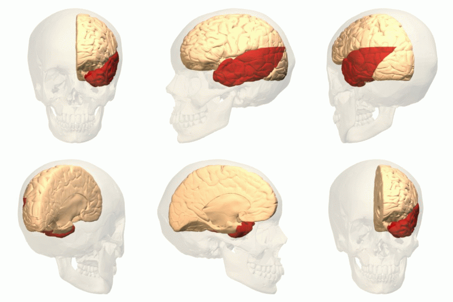
The temporal lobe is one of the four Lobes of the brain, major lobes of the cerebral cortex in the brain of mammals. The temporal lobe is located beneath the lateral fissure on both cerebral hemispheres of the mammalian brain. The temporal lobe is involved in processing sensory input into derived meanings for the appropriate retention of visual memory, language comprehension, and emotion association. ''Temporal'' refers to the head's Temple (anatomy), temples. Structure The Temple (anatomy)#Etymology, temporal Lobe (anatomy), lobe consists of structures that are vital for declarative or long-term memory. Declarative memory, Declarative (denotative) or Explicit memory, explicit memory is conscious memory divided into semantic memory (facts) and episodic memory (events). Medial temporal lobe structures that are critical for long-term memory include the hippocampus, along with the surrounding Hippocampal formation, hippocampal region consisting of the Perirhinal cortex, perirhinal, ... [...More Info...] [...Related Items...] OR: [Wikipedia] [Google] [Baidu] |
Temporal Lobe Epilepsy
Temporal lobe epilepsy (TLE) is a chronic disorder of the nervous system which is characterized by recurrent, unprovoked focal seizures that originate in the temporal lobe of the brain and last about one or two minutes. TLE is the most common form of epilepsy with focal seizures. A focal seizure in the temporal lobe may spread to other areas in the brain when it may become a ''focal to bilateral seizure''. TLE is diagnosed by taking a medical history, blood tests, and brain imaging. It can have a number of causes such as head injury, stroke, brain infections, structural lesions in the brain, brain tumors, or it can be of ''unknown onset''. The first line of treatment is through anticonvulsants. Surgery may be an option, especially when there is an observable abnormality in the brain. Another treatment option is electrical stimulation of the brain through an implanted device called the vagus nerve stimulator (VNS). Types Over forty types of epilepsy are recognized and these ... [...More Info...] [...Related Items...] OR: [Wikipedia] [Google] [Baidu] |
Homonymous Hemianopia
Hemianopsia, or hemianopia, is a visual field loss on the left or right side of the vertical midline. It can affect one eye but usually affects both eyes. Homonymous hemianopsia (or homonymous hemianopia) is hemianopic visual field loss on the same side of both eyes. Homonymous hemianopsia occurs because the right half of the brain has visual pathways for the left hemifield of both eyes, and the left half of the brain has visual pathways for the right hemifield of both eyes. When one of these pathways is damaged, the corresponding visual field is lost. Signs and symptoms Paris as seen with right homonymous hemianopsia Mobility can be difficult for people with homonymous hemianopsia. "Patients frequently complain of bumping into obstacles on the side of the field loss, thereby bruising their arms and legs." People with homonymous hemianopsia often experience discomfort in crowds. "A patient with this condition may be unaware of what he or she cannot see and frequently bumps ... [...More Info...] [...Related Items...] OR: [Wikipedia] [Google] [Baidu] |
Rasmussen's Encephalitis
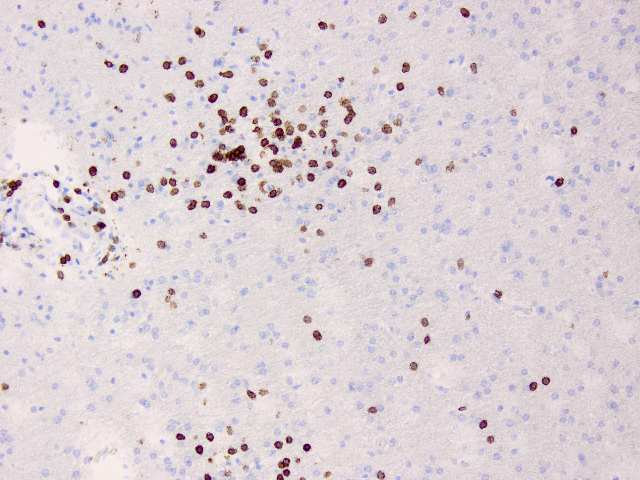
Rasmussen's encephalitis is a rare inflammatory neurological disease, characterized by frequent and severe seizures, loss of motor skills and speech, hemiparesis (weakness on one side of the body), encephalitis (inflammation of the brain), and dementia. The illness affects a single cerebral hemisphere and generally occurs in children under the age of 15. Signs and symptoms The condition mostly affects children, with an average age of 6 years. However, one in ten people with the condition develops it in adulthood. There are two main stages, sometimes preceded by a 'prodromal stage' of a few months. In the ''acute stage'', lasting four to eight months, the inflammation is active and the symptoms become progressively worse. These include weakness of one side of the body (hemiparesis), loss of vision for one side of the visual field (hemianopia), and cognitive difficulties (affecting learning, memory or language, for example). Epileptic seizures are also a major part of the illness, ... [...More Info...] [...Related Items...] OR: [Wikipedia] [Google] [Baidu] |
Functional Disconnection
Lingesha Functional disconnection is the disintegrated function in the brain in the absence of anatomical damage, in distinction to physical disconnection of the cerebral hemispheres by surgical resection, trauma or lesion. Applications have included alexia without agraphia dyslexia, persistent vegetative state and minimally conscious state as well as autistic spectrum disorders. Functional disconnection itself is not a medically recognized condition. It is a theoretical concept used to facilitate research into the causes and symptoms within recognized conditions. History In 1977, Witleson reported that developmental dyslexia may be associated with (i) bi-hemisphere representation of spatial functions, in contrast to the unitary right hemisphere control of these functions observed in normal individuals. The bilateral neural involvement in spatial processing may interfere with the left hemisphere's processing of its own specialized functions and result in deficient linguistic ... [...More Info...] [...Related Items...] OR: [Wikipedia] [Google] [Baidu] |
Hemispherectomy
Hemispherectomy is a neurosurgical procedure in which a cerebral hemisphere (half of the upper brain, or cerebrum) is removed or disconnected that is used to treat a variety of refractory or drug-resistant seizure disorders (epilepsy). Refractory or drug-resistant epilepsy is defined as seizures that fail to be controlled using 2 or more appropriate anti-seizure medications. About one in three patients with epilepsy have drug-resistant epilepsy and of those, about half of them have focal epilepsy that can potentially be treated with epilepsy surgery. In drug-resistant epilepsy where all or most seizures arise from one hemisphere, hemispherectomy is a highly effective procedure producing seizure freedom in about 80-90% of patients. In addition to controlling seizures and as a result of that, improved development and cognition is also very frequently achieved after hemispherectomy. Most patients who qualify for hemispherectomy already have neurological deficits such as hemibody weakne ... [...More Info...] [...Related Items...] OR: [Wikipedia] [Google] [Baidu] |
Callosotomy
Corpus callosotomy is a palliative surgical procedure for the treatment of medically refractory epilepsy. In this procedure the corpus callosum is cut through in an effort to limit the spread of epileptic activity between the two halves of the brain. After the operation the brain has much more difficulty sending messages between the hemispheres. Although the corpus callosum is the largest white matter tract connecting the hemispheres, some limited interhemispheric communication is still possible via the anterior commissure and posterior commissure. "Efficacy and relatively low permanent morbidity in corpus callosotomy for medically intractable epilepsy have been demonstrated by more than six decades of experience. In addition to seizure reduction, behavior and quality of life may improve." History The first examples of corpus callosotomy were performed in the 1940s by Dr. William P. van Wagenen, who co-founded and served as president of the American Association of Neurological ... [...More Info...] [...Related Items...] OR: [Wikipedia] [Google] [Baidu] |
Electrocorticography
Electrocorticography (ECoG), or intracranial electroencephalography (iEEG), is a type of electrophysiological monitoring that uses electrodes placed directly on the exposed surface of the brain to record electrical activity from the cerebral cortex. In contrast, conventional electroencephalography (EEG) electrodes monitor this activity from outside the skull. ECoG may be performed either in the operating room during surgery (intraoperative ECoG) or outside of surgery (extraoperative ECoG). Because a craniotomy (a surgical incision into the skull) is required to implant the electrode grid, ECoG is an invasive procedure. History ECoG was pioneered in the early 1950s by Wilder Penfield and Herbert Jasper, neurosurgeons at the Montreal Neurological Institute. The two developed ECoG as part of their groundbreakinMontreal procedure a surgical protocol used to treat patients with severe epilepsy. The cortical potentials recorded by ECoG were used to identify epileptogenic zones ŌĆ ... [...More Info...] [...Related Items...] OR: [Wikipedia] [Google] [Baidu] |


