|
Embryomics
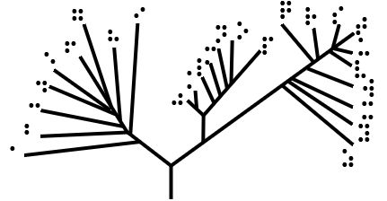
Embryomics is the identification, characterization and study of the diverse cell types which arise during embryogenesis, especially as this relates to the location and developmental history of cells in the embryo. Cell type may be determined according to several criteria: location in the developing embryo, gene expression as indicated by protein and nucleic acid markers and surface antigens, and also position on the embryogenic tree. Embryome There are many cell markers useful in distinguishing, classifying, separating and purifying the numerous cell types present at any given time in a developing organism. These cell markers consist of select RNAs and proteins present inside, and surface antigens present on the surface of, the cells making up the embryo. For any given cell type, these RNA and protein markers reflect the genes characteristically active in that cell type. The catalog of all these cell types and their characteristic markers is known as the organism's embryome. The ... [...More Info...] [...Related Items...] OR: [Wikipedia] [Google] [Baidu] |
Human Embryogenesis
Human embryonic development, or human embryogenesis, is the development and formation of the human embryo. It is characterised by the processes of cell division and cellular differentiation of the embryo that occurs during the early stages of development. In biological terms, the development of the human body entails growth from a one-celled zygote to an adult human being. Fertilization occurs when the sperm cell successfully enters and fuses with an egg cell (ovum). The genetic material of the sperm and egg then combine to form the single cell zygote and the germinal stage of development commences. Embryonic development in the human, covers the first eight weeks of development; at the beginning of the ninth week the embryo is termed a fetus. Human embryology is the study of this development during the first eight weeks after fertilization. The normal period of gestation (pregnancy) is about nine months or 40 weeks. The germinal stage refers to the time from fertilization throu ... [...More Info...] [...Related Items...] OR: [Wikipedia] [Google] [Baidu] |
Embryogenesis
An embryo is an initial stage of development of a multicellular organism. In organisms that reproduce sexually, embryonic development is the part of the life cycle that begins just after fertilization of the female egg cell by the male sperm cell. The resulting fusion of these two cells produces a single-celled zygote that undergoes many cell divisions that produce cells known as blastomeres. The blastomeres are arranged as a solid ball that when reaching a certain size, called a morula, takes in fluid to create a cavity called a blastocoel. The structure is then termed a blastula, or a blastocyst in mammals. The mammalian blastocyst hatches before implantating into the endometrial lining of the womb. Once implanted the embryo will continue its development through the next stages of gastrulation, neurulation, and organogenesis. Gastrulation is the formation of the three germ layers that will form all of the different parts of the body. Neurulation forms the ne ... [...More Info...] [...Related Items...] OR: [Wikipedia] [Google] [Baidu] |
Phylogenetic Tree
A phylogenetic tree (also phylogeny or evolutionary tree Felsenstein J. (2004). ''Inferring Phylogenies'' Sinauer Associates: Sunderland, MA.) is a branching diagram or a tree showing the evolutionary relationships among various biological species or other entities based upon similarities and differences in their physical or genetic characteristics. All life on Earth is part of a single phylogenetic tree, indicating common ancestry. In a ''rooted'' phylogenetic tree, each node with descendants represents the inferred most recent common ancestor of those descendants, and the edge lengths in some trees may be interpreted as time estimates. Each node is called a taxonomic unit. Internal nodes are generally called hypothetical taxonomic units, as they cannot be directly observed. Trees are useful in fields of biology such as bioinformatics, systematics, and phylogenetics. ''Unrooted'' trees illustrate only the relatedness of the leaf nodes and do not require the ancestral root ... [...More Info...] [...Related Items...] OR: [Wikipedia] [Google] [Baidu] |
Ectoderms
A germ layer is a primary layer of cells that forms during embryonic development. The three germ layers in vertebrates are particularly pronounced; however, all eumetazoans (animals that are sister taxa to the sponges) produce two or three primary germ layers. Some animals, like cnidarians, produce two germ layers (the ectoderm and endoderm) making them diploblastic. Other animals such as bilaterians produce a third layer (the mesoderm) between these two layers, making them triploblastic. Germ layers eventually give rise to all of an animal’s tissues and organs through the process of organogenesis. History Caspar Friedrich Wolff observed organization of the early embryo in leaf-like layers. In 1817, Heinz Christian Pander discovered three primordial germ layers while studying chick embryos. Between 1850 and 1855, Robert Remak had further refined the germ cell layer (''Keimblatt'') concept, stating that the external, internal and middle layers form respectively the ep ... [...More Info...] [...Related Items...] OR: [Wikipedia] [Google] [Baidu] |
Allantois
The allantois (plural ''allantoides'' or ''allantoises'') is a hollow sac-like structure filled with clear fluid that forms part of a developing amniote's conceptus (which consists of all embryonic and extraembryonic tissues). It helps the embryo exchange gases and handle liquid waste. The allantois, along with the amnion, chorion, and yolk sac (other extraembryonic membranes), identify humans and other mammals, birds, and other reptiles as amniotes. Fish and amphibians are anamniotes, and lack the allantois. In mammals the extraembryonic membranes are known as the fetal membranes. Function This sac-like structure, whose name is the New Latin equivalent of "sausage" (in reference to its shape when first formed) is primarily involved in nutrition and excretion, and is webbed with blood vessels. The function of the allantois is to collect liquid waste from the embryo, as well as to exchange gases used by the embryo. In mammals In mammals excluding egg-laying monotremes, ... [...More Info...] [...Related Items...] OR: [Wikipedia] [Google] [Baidu] |
Yolk Sac
The yolk sac is a membranous sac attached to an embryo, formed by cells of the hypoblast layer of the bilaminar embryonic disc. This is alternatively called the umbilical vesicle by the Terminologia Embryologica (TE), though ''yolk sac'' is far more widely used. In humans, the yolk sac is important in early embryonic blood supply, and much of it is incorporated into the primordial gut during the fourth week of embryonic development. In humans The yolk sac is the first element seen within the gestational sac during pregnancy, usually at 3 days gestation. The yolk sac is situated on the front ( ventral) part of the embryo; it is lined by extra-embryonic endoderm, outside of which is a layer of extra-embryonic mesenchyme, derived from the epiblast. Blood is conveyed to the wall of the yolk sac by the primitive aorta and after circulating through a wide-meshed capillary plexus, is returned by the vitelline veins to the tubular heart of the embryo. This constitutes the vit ... [...More Info...] [...Related Items...] OR: [Wikipedia] [Google] [Baidu] |
Amnion
The amnion is a membrane that closely covers the human and various other embryos when first formed. It fills with amniotic fluid, which causes the amnion to expand and become the amniotic sac that provides a protective environment for the developing embryo. The amnion, along with the chorion, the yolk sac and the allantois protect the embryo. In birds, reptiles and monotremes, the protective sac is enclosed in a shell. In marsupials and placental mammals, it is enclosed in a uterus. The term is from Ancient Greek ἀμνίον 'little lamb', diminutive of ἀμνός 'lamb'. it is cognate with the English verb 'yean', bring forth young (usually lambs). The amnion is a feature of the vertebrate clade ''Amniota'', which includes reptiles, birds, and mammals. Amphibians and fish are not amniotes and thus lack the amnion. The amnion stems from the extra-embryonic somatic mesoderm on the outer side and the extra-embryonic ectoderm or trophoblast on the inner side. In huma ... [...More Info...] [...Related Items...] OR: [Wikipedia] [Google] [Baidu] |
Embryonic Stem Cell
Embryonic stem cells (ESCs) are pluripotent stem cells derived from the inner cell mass of a blastocyst, an early-stage pre- implantation embryo. Human embryos reach the blastocyst stage 4–5 days post fertilization, at which time they consist of 50–150 cells. Isolating the inner cell mass (embryoblast) using immunosurgery results in destruction of the blastocyst, a process which raises ethical issues, including whether or not embryos at the pre-implantation stage have the same moral considerations as embryos in the post-implantation stage of development. Researchers are currently focusing heavily on the therapeutic potential of embryonic stem cells, with clinical use being the goal for many laboratories. Potential uses include the treatment of diabetes and heart disease. The cells are being studied to be used as clinical therapies, models of genetic disorders, and cellular/DNA repair. However, adverse effects in the research and clinical processes such as tumors and un ... [...More Info...] [...Related Items...] OR: [Wikipedia] [Google] [Baidu] |
Placenta
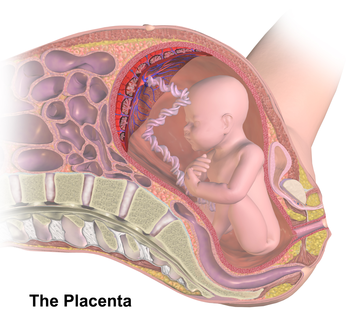
The placenta is a temporary embryonic and later fetal organ (anatomy), organ that begins embryonic development, developing from the blastocyst shortly after implantation (embryology), implantation. It plays critical roles in facilitating nutrient, gas and waste exchange between the physically separate maternal and fetal circulations, and is an important Endocrine system, endocrine organ, producing hormones that regulate both Maternal physiological changes in pregnancy, maternal and fetal physiology during pregnancy. The placenta connects to the fetus via the umbilical cord, and on the opposite aspect to the maternal uterus in a species-dependent manner. In humans, a thin layer of maternal decidual (Endometrium, endometrial) tissue comes away with the placenta when it is expelled from the uterus following birth (sometimes incorrectly referred to as the 'maternal part' of the placenta). Placentas are a defining characteristic of placental mammals, but are also found in marsupials an ... [...More Info...] [...Related Items...] OR: [Wikipedia] [Google] [Baidu] |
Uterus
The uterus (from Latin ''uterus'', plural ''uteri'') or womb () is the organ in the reproductive system of most female mammals, including humans that accommodates the embryonic and fetal development of one or more embryos until birth. The uterus is a hormone-responsive sex organ that contains glands in its lining that secrete uterine milk for embryonic nourishment. In the human, the lower end of the uterus, is a narrow part known as the isthmus that connects to the cervix, leading to the vagina. The upper end, the body of the uterus, is connected to the fallopian tubes, at the uterine horns, and the rounded part above the openings to the fallopian tubes is the fundus. The connection of the uterine cavity with a fallopian tube is called the uterotubal junction. The fertilized egg is carried to the uterus along the fallopian tube. It will have divided on its journey to form a blastocyst that will implant itself into the lining of the uterus – the endometrium, ... [...More Info...] [...Related Items...] OR: [Wikipedia] [Google] [Baidu] |
Implantation (human Embryo)
Implantation (nidation) is the stage in the embryonic development of mammals in which the blastocyst hatches as the embryo, adheres, and invades into the wall of the female's uterus. Implantation is the first stage of gestation, and when successful the female is considered to be pregnant. In a woman, an implanted embryo is detected by the presence of increased levels of human chorionic gonadotropin (hCG) in a pregnancy test. The implanted embryo will receive oxygen and nutrients in order to grow. There is an extensive variation in the type of trophoblast cells, and structures of the placenta across the different species of mammals. Of the five recognised stages of implantation including two pre-implantation stages that precede placentation, the first four are similar across the species. The five stages are migration and hatching, pre-contact, attachment, adhesion, and invasion. The two pre-implantation stages are associated with the pre-implantation embryo. In humans followi ... [...More Info...] [...Related Items...] OR: [Wikipedia] [Google] [Baidu] |
Inner Cell Mass
The inner cell mass (ICM) or embryoblast (known as the pluriblast in marsupials) is a structure in the early development of an embryo. It is the mass of cells inside the blastocyst that will eventually give rise to the definitive structures of the fetus. The inner cell mass forms in the earliest stages of embryonic development, before implantation into the endometrium of the uterus. The ICM is entirely surrounded by the single layer of trophoblast cells of the trophectoderm. Further development The physical and functional separation of the inner cell mass from the trophectoderm (TE) is a special feature of mammalian development and is the first cell lineage specification in these embryos. Following fertilization in the oviduct, the mammalian embryo undergoes a relatively slow round of cleavages to produce an eight-cell morula. Each cell of the morula, called a blastomere, increases surface contact with its neighbors in a process called compaction. This results in a pola ... [...More Info...] [...Related Items...] OR: [Wikipedia] [Google] [Baidu] |


