|
Embolic Stroke Of Undetermined Source
Embolic stroke of undetermined source (ESUS) is a type of ischemic stroke with an unknown origin, defined as a non- lacunar brain infarct without proximal arterial stenosis or cardioembolic sources. As such, it forms a subset of cryptogenic stroke, which is part of the TOAST-classification. The following diagnostic criteria define an ESUS: * Stroke detected by CT or MRI that is not lacunar * No major-risk cardioembolic source of embolism * Absence of extracranial or intracranial atherosclerosis causing 50% luminal stenosis in arteries supplying the area of ischaemia * No other specific cause of stroke identified (e.g., arteritis, dissection, migraine/vasospasm, drug misuse) Signs and symptoms Causes The following factors are suggested as pathogenesis of ESUS: * Subclinical atrial fibrillation: Detectable in ~2.7-30% of ESUS patients, depending on duration and modality of ECG monitoring. * Patent foramen ovale (PFO): Deep vein thrombosis may result in paradoxical embolism in ... [...More Info...] [...Related Items...] OR: [Wikipedia] [Google] [Baidu] |
Ischemic Stroke
A stroke is a medical condition in which poor blood flow to the brain causes cell death. There are two main types of stroke: ischemic, due to lack of blood flow, and hemorrhagic, due to bleeding. Both cause parts of the brain to stop functioning properly. Signs and symptoms of a stroke may include an inability to move or feel on one side of the body, problems understanding or speaking, dizziness, or loss of vision to one side. Signs and symptoms often appear soon after the stroke has occurred. If symptoms last less than one or two hours, the stroke is a transient ischemic attack (TIA), also called a mini-stroke. A hemorrhagic stroke may also be associated with a severe headache. The symptoms of a stroke can be permanent. Long-term complications may include pneumonia and loss of bladder control. The main risk factor for stroke is high blood pressure. Other risk factors include high blood cholesterol, tobacco smoking, obesity, diabetes mellitus, a previous TIA, end-st ... [...More Info...] [...Related Items...] OR: [Wikipedia] [Google] [Baidu] |
Subclinical
In medicine, any disease is classified asymptomatic if a patient tests as carrier for a disease or infection but experiences no symptoms. Whenever a medical condition fails to show noticeable symptoms after a diagnosis it might be considered asymptomatic. Infections of this kind are usually called subclinical infections. Diseases such as mental illnesses or psychosomatic conditions are considered subclinical if they present some individual symptoms but not all those normally required for a clinical diagnosis. The term clinically silent is also found. Producing only a few, mild symptoms, disease is paucisymptomatic. Symptoms appearing later, after an asymptomatic incubation period, mean a pre-symptomatic period has existed. Importance Knowing that a condition is asymptomatic is important because: * It may develop symptoms later and only then require treatment. * It may resolve itself or become benign. * It may be contagious, and the contribution of asymptomatic and pre-symptomati ... [...More Info...] [...Related Items...] OR: [Wikipedia] [Google] [Baidu] |
NT-proBNP
The N-terminal prohormone of brain natriuretic peptide (NT-proBNP or BNPT) is a prohormone with a 76 amino acid N-terminal inactive protein that is cleaved from the molecule to release brain natriuretic peptide. Both BNP and NT-proBNP levels in the blood are used for screening, diagnosis of acute congestive heart failure (CHF) and may be useful to establish prognosis in heart failure, as both markers are typically higher in patients with worse outcome. The plasma concentrations of both BNP and NT-proBNP are also typically increased in patients with asymptomatic or symptomatic left ventricular dysfunction and is associated with coronary artery disease and myocardial ischemia. Blood levels There is no level of BNP that perfectly separates patients with and without heart failure. In screening for congenital heart disease in pediatric patients, an NT-proBNP cut-off value of 91 pg/mL could differentiate an acyanotic heart disease (ACNHD) patient from a healthy patient with a ... [...More Info...] [...Related Items...] OR: [Wikipedia] [Google] [Baidu] |
Supraventricular Tachycardia
Supraventricular tachycardia (SVT) is an umbrella term for fast heart rhythms arising from the upper part of the heart. This is in contrast to the other group of fast heart rhythms – ventricular tachycardia, which start within the lower chambers of the heart. There are four main types of SVT: atrial fibrillation, atrial flutter, paroxysmal supraventricular tachycardia (PSVT), and Wolff–Parkinson–White syndrome. The symptoms of SVT include palpitations, feeling of faintness, sweating, shortness of breath, and/or chest pain. These abnormal rhythms start from either the atria or atrioventricular node. They are generally due to one of two mechanisms: re-entry or increased automaticity. Diagnosis is typically by electrocardiogram (ECG), holter monitor, or event monitor. Blood tests may be done to rule out specific underlying causes such as hyperthyroidism or electrolyte abnormalities. A normal resting heart rate is 60 to 100 beats per minute. A resting heart rate of mo ... [...More Info...] [...Related Items...] OR: [Wikipedia] [Google] [Baidu] |
Cardiopathy
Cardiovascular disease (CVD) is a class of diseases that involve the heart or blood vessels. CVD includes coronary artery diseases (CAD) such as angina and myocardial infarction (commonly known as a heart attack). Other CVDs include stroke, heart failure, hypertensive heart disease, rheumatic heart disease, cardiomyopathy, abnormal heart rhythms, congenital heart disease, valvular heart disease, carditis, aortic aneurysms, peripheral artery disease, thromboembolic disease, and venous thrombosis. The underlying mechanisms vary depending on the disease. It is estimated that dietary risk factors are associated with 53% of CVD deaths. Coronary artery disease, stroke, and peripheral artery disease involve atherosclerosis. This may be caused by high blood pressure, smoking, diabetes mellitus, lack of exercise, obesity, high blood cholesterol, poor diet, excessive alcohol consumption, and poor sleep, among other things. High blood pressure is estimated to account for approximately ... [...More Info...] [...Related Items...] OR: [Wikipedia] [Google] [Baidu] |
Aortic Arch
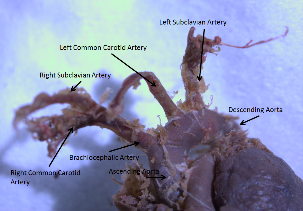
The aortic arch, arch of the aorta, or transverse aortic arch () is the part of the aorta between the ascending and descending aorta. The arch travels backward, so that it ultimately runs to the left of the trachea. Structure The aorta begins at the level of the upper border of the second/third sternocostal articulation of the right side, behind the ventricular outflow tract and pulmonary trunk. The right atrial appendage overlaps it. The first few centimeters of the ascending aorta and pulmonary trunk lies in the same pericardial sheath. and runs at first upward, arches over the pulmonary trunk, right pulmonary artery, and right main bronchus to lie behind the right second coastal cartilage. The right lung and sternum lies anterior to the aorta at this point. The aorta then passes posteriorly and to the left, anterior to the trachea, and arches over left main bronchus and left pulmonary artery, and reaches to the left side of the T4 vertebral body. Apart from T4 vertebral body ... [...More Info...] [...Related Items...] OR: [Wikipedia] [Google] [Baidu] |
Carotid Artery , an artery on each side of the head and neck supplying blood to the brain
{{SIA ...
Carotid artery may refer to: * Common carotid artery, often "carotids" or "carotid", an artery on each side of the neck which divides into the external carotid artery and internal carotid artery * External carotid artery, an artery on each side of the head and neck supplying blood to the face, scalp, skull, neck and meninges * Internal carotid artery The internal carotid artery (Latin: arteria carotis interna) is an artery in the neck which supplies the anterior circulation of the brain. In human anatomy, the internal and external carotids arise from the common carotid arteries, where these b ... [...More Info...] [...Related Items...] OR: [Wikipedia] [Google] [Baidu] |
Ipsilateral
Standard anatomical terms of location are used to unambiguously describe the anatomy of animals, including humans. The terms, typically derived from Latin or Greek roots, describe something in its standard anatomical position. This position provides a definition of what is at the front ("anterior"), behind ("posterior") and so on. As part of defining and describing terms, the body is described through the use of anatomical planes and anatomical axes. The meaning of terms that are used can change depending on whether an organism is bipedal or quadrupedal. Additionally, for some animals such as invertebrates, some terms may not have any meaning at all; for example, an animal that is radially symmetrical will have no anterior surface, but can still have a description that a part is close to the middle ("proximal") or further from the middle ("distal"). International organisations have determined vocabularies that are often used as standard vocabularies for subdisciplines of anatom ... [...More Info...] [...Related Items...] OR: [Wikipedia] [Google] [Baidu] |
Haemorrhage
Bleeding, hemorrhage, haemorrhage or blood loss, is blood escaping from the circulatory system from damaged blood vessels. Bleeding can occur internally, or externally either through a natural opening such as the mouth, nose, ear, urethra, vagina or anus, or through a puncture in the skin. Hypovolemia is a massive decrease in blood volume, and death by excessive loss of blood is referred to as exsanguination. Typically, a healthy person can endure a loss of 10–15% of the total blood volume without serious medical difficulties (by comparison, blood donation typically takes 8–10% of the donor's blood volume). The stopping or controlling of bleeding is called hemostasis and is an important part of both first aid and surgery. Types * Upper head ** Intracranial hemorrhage – bleeding in the skull. ** Cerebral hemorrhage – a type of intracranial hemorrhage, bleeding within the brain tissue itself. ** Intracerebral hemorrhage – bleeding in the brain caused by the rupture of ... [...More Info...] [...Related Items...] OR: [Wikipedia] [Google] [Baidu] |
Atheroma
An atheroma, or atheromatous plaque, is an abnormal and reversible accumulation of material in the inner layer of an arterial wall. The material consists of mostly macrophage cells, or debris, containing lipids, calcium and a variable amount of fibrous connective tissue. The accumulated material forms a swelling in the artery wall, which may intrude into the lumen of the artery, narrowing it and restricting blood flow. Atheroma is the pathological basis for the disease entity atherosclerosis, a subtype of arteriosclerosis. Signs and symptoms For most people, the first symptoms result from atheroma progression within the heart arteries, most commonly resulting in a heart attack and ensuing debility. The heart arteries are difficult to track because they are small (from about 5 mm down to microscopic), they are hidden deep within the chest and they never stop moving. Additionally, all mass-applied clinical strategies focus on both minimal cost and the overall safety of ... [...More Info...] [...Related Items...] OR: [Wikipedia] [Google] [Baidu] |
Paradoxical Embolism
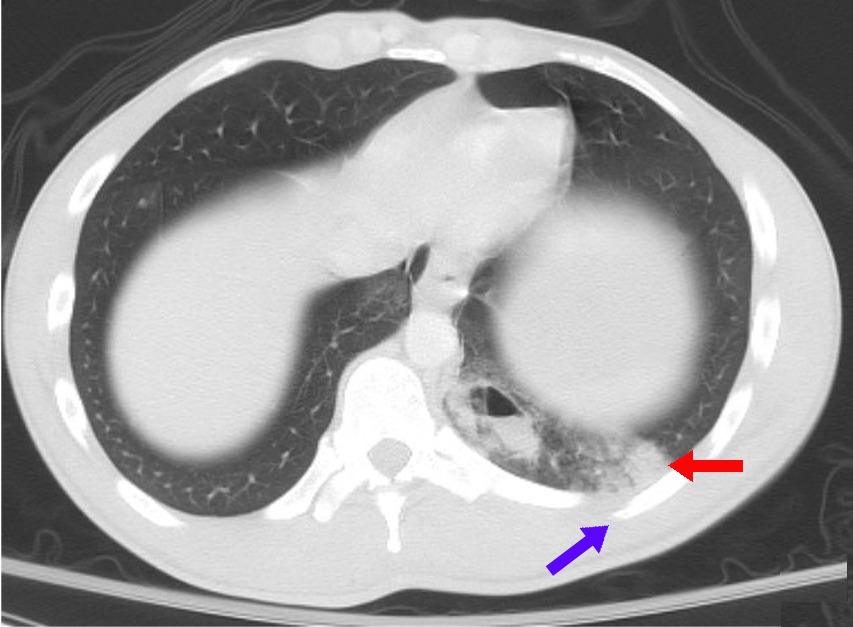
An embolus, is described as a free-floating mass, located inside blood vessels that can travel from one site in the blood stream to another. An embolus can be made up of solid (like a blood clot), liquid (like amniotic fluid), or gas (like air). Once these masses get "stuck" in a different blood vessel, it is then known as an "embolism." An embolism can cause ischemia - or damage to an organ from lack of oxygen. A paradoxical embolism is a specific type of embolism in which the emboli travels from the right side of the heart or "venous circulation," travels to the left side of the heart or "arterial circulation," and lodges itself in a blood vessel known as an artery. Thus, it is termed "paradoxical" because the emboli lands in an artery, rather than a vein. Pathophysiology An embolism may be made from any one of numerous materials that may find itself in a blood vessel, including a piece of a thrombus, known as a thromboembolism, air from an intravenous catheter, fat globules f ... [...More Info...] [...Related Items...] OR: [Wikipedia] [Google] [Baidu] |
Deep Vein Thrombosis
Deep vein thrombosis (DVT) is a type of venous thrombosis involving the formation of a blood clot in a deep vein, most commonly in the legs or pelvis. A minority of DVTs occur in the arms. Symptoms can include pain, swelling, redness, and enlarged veins in the affected area, but some DVTs have no symptoms. The most common life-threatening concern with DVT is the potential for a clot to embolize (detach from the veins), travel as an embolus through the right side of the heart, and become lodged in a pulmonary artery that supplies blood to the lungs. This is called a pulmonary embolism (PE). DVT and PE comprise the cardiovascular disease of venous thromboembolism (VTE). About two-thirds of VTE manifests as DVT only, with one-third manifesting as PE with or without DVT. The most frequent long-term DVT complication is post-thrombotic syndrome, which can cause pain, swelling, a sensation of heaviness, itching, and in severe cases, ulcers. Recurrent VTE occurs in about 30% of those i ... [...More Info...] [...Related Items...] OR: [Wikipedia] [Google] [Baidu] |


