|
Electronystagmography
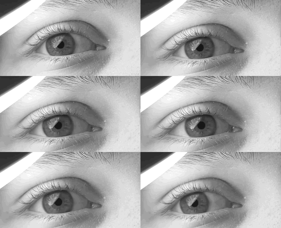
Electronystagmography (ENG) is a diagnostic test to record involuntary movements of the eye caused by a condition known as nystagmus. It can also be used to diagnose the cause of vertigo, dizziness or balance dysfunction by testing the vestibular system. Electronystagmography is used to assess voluntary and involuntary eye movements. It evaluates the cochlear nerve and the oculomotor nerve (CN III). The ENG can be used to determine the origin of various eye and ear disorders. Technique and results Electrodes applied around the eyes record eye movements using the corneo-retinal potential. Some portions of the ENG measure a patient's ability to track a moving stimuli while others observe the presence of nystagmus. The vestibular system monitors the position and movements of the head to stabilize retinal images. This information is integrated with the visual system and spinal afferents in the brain stem to produce the vestibulo-ocular reflex (VOR). ENG provides an objective assess ... [...More Info...] [...Related Items...] OR: [Wikipedia] [Google] [Baidu] |
Electrography (other)
Electrography often refers to electrophotography, that is, Kirlian photography. Electrography may also refer to: * Measurement and recording of electrophysiology, electrophysiologic activity for diagnostic purposes ** Electrocardiography (ECG or EKG), electrography of heart electrical activity and rhythm ** Electromyography (EMG), electrography of other muscle action potentials throughout the body ** Electroencephalography (EEG), electrography of brain waves (from outside the skull) *** Electrocorticography or intracranial EEG (iEEG or ECoG), EEG with direct contact to the cerebral cortex ** Electrooculography (EOG), electrography of intraocular potential differences ** Electro-olfactography, Electroolfactography (EOG), electrography of olfaction (smell) ** Electroretinography (ERG), electrography of retinal cell action potentials ** Electronystagmography (ENG), electrography of eye muscle movements ** Electrocochleography (ECOG), electrography of cochlear auditory activity ** E ... [...More Info...] [...Related Items...] OR: [Wikipedia] [Google] [Baidu] |
Electrodes
An electrode is an electrical conductor used to make contact with a nonmetallic part of a circuit (e.g. a semiconductor, an electrolyte, a vacuum or air). Electrodes are essential parts of batteries that can consist of a variety of materials depending on the type of battery. The electrophore, invented by Johan Wilcke, was an early version of an electrode used to study static electricity. Anode and cathode in electrochemical cells Electrodes are an essential part of any battery. The first electrochemical battery made was devised by Alessandro Volta and was aptly named the Voltaic cell. This battery consisted of a stack of copper and zinc electrodes separated by brine-soaked paper disks. Due to fluctuation in the voltage provided by the voltaic cell it wasn't very practical. The first practical battery was invented in 1839 and named the Daniell cell after John Frederic Daniell. Still making use of the zinc–copper electrode combination. Since then many more batteries have be ... [...More Info...] [...Related Items...] OR: [Wikipedia] [Google] [Baidu] |
Peripheral Nervous System
The peripheral nervous system (PNS) is one of two components that make up the nervous system of bilateral animals, with the other part being the central nervous system (CNS). The PNS consists of nerves and ganglia, which lie outside the brain and the spinal cord. The main function of the PNS is to connect the CNS to the limbs and organs, essentially serving as a relay between the brain and spinal cord and the rest of the body. Unlike the CNS, the PNS is not protected by the vertebral column and skull, or by the blood–brain barrier, which leaves it exposed to toxins. The peripheral nervous system can be divided into the somatic nervous system and the autonomic nervous system. In the somatic nervous system, the cranial nerves are part of the PNS with the exception of the optic nerve (cranial nerve II), along with the retina. The second cranial nerve is not a true peripheral nerve but a tract of the diencephalon. Cranial nerve ganglia, as with all ganglia, are part of the P ... [...More Info...] [...Related Items...] OR: [Wikipedia] [Google] [Baidu] |
Central Nervous System
The central nervous system (CNS) is the part of the nervous system consisting primarily of the brain and spinal cord. The CNS is so named because the brain integrates the received information and coordinates and influences the activity of all parts of the bodies of bilaterally symmetric and triploblastic animals—that is, all multicellular animals except sponges and diploblasts. It is a structure composed of nervous tissue positioned along the rostral (nose end) to caudal (tail end) axis of the body and may have an enlarged section at the rostral end which is a brain. Only arthropods, cephalopods and vertebrates have a true brain (precursor structures exist in onychophorans, gastropods and lancelets). The rest of this article exclusively discusses the vertebrate central nervous system, which is radically distinct from all other animals. Overview In vertebrates, the brain and spinal cord are both enclosed in the meninges. The meninges provide a barrier to chemicals dissolv ... [...More Info...] [...Related Items...] OR: [Wikipedia] [Google] [Baidu] |
Saccade
A saccade ( , French for ''jerk'') is a quick, simultaneous movement of both eyes between two or more phases of fixation in the same direction.Cassin, B. and Solomon, S. ''Dictionary of Eye Terminology''. Gainesville, Florida: Triad Publishing Company, 1990. In contrast, in smooth pursuit movements, the eyes move smoothly instead of in jumps. The phenomenon can be associated with a shift in frequency of an emitted signal or a movement of a body part or device. Controlled cortically by the frontal eye fields (FEF), or subcortically by the superior colliculus, saccades serve as a mechanism for fixation, rapid eye movement, and the fast phase of optokinetic nystagmus. The word appears to have been coined in the 1880s by French ophthalmologist Émile Javal, who used a mirror on one side of a page to observe eye movement in silent reading, and found that it involves a succession of discontinuous individual movements. Function Humans and many animals do not look at a scene in f ... [...More Info...] [...Related Items...] OR: [Wikipedia] [Google] [Baidu] |
Caloric Reflex Test
In medicine, the caloric reflex test (sometimes termed vestibular caloric stimulation) is a test of the vestibulo-ocular reflex that involves irrigating cold or warm water or air into the external auditory canal. This method was developed by Robert Bárány, who won a Nobel prize in 1914 for this discovery. Utility The test is commonly used by physicians, audiologists and other trained professionals to validate a diagnosis of asymmetric function in the peripheral vestibular system. Calorics are usually a subtest of the electronystagmography (ENG) battery of tests. It is one of several tests which can be used to test for brain stem death. One novel use of this test has been to provide temporary pain relief from phantom limb pains in amputees and paraplegics. It can also induce a temporary remission of anosognosia, the visual and personal aspects of hemispatial neglect, hemianesthesia, and other consequences of right hemispheric damage. Technique and results Ice cold or warm wa ... [...More Info...] [...Related Items...] OR: [Wikipedia] [Google] [Baidu] |
Videonystagmography
Videonystagmography (VNG) is a technology for testing inner ear and central motor functions, a process known as vestibular assessment. It involves the use of infrared goggles to trace eye movements during visual stimulation and positional changes. VNG can determine whether dizziness is caused by inner ear disease, particularly benign paroxysmal positional vertigo (BPPV), as opposed to some other cause such as low blood pressure or anxiety Anxiety is an emotion which is characterized by an unpleasant state of inner turmoil and includes feelings of dread over anticipated events. Anxiety is different than fear in that the former is defined as the anticipation of a future threat wh .... VNG testing is made up of several components. Patients are asked to wear goggles with sensitive video ca ... [...More Info...] [...Related Items...] OR: [Wikipedia] [Google] [Baidu] |
Oculomotor Nerve
The oculomotor nerve, also known as the third cranial nerve, cranial nerve III, or simply CN III, is a cranial nerve that enters the orbit through the superior orbital fissure and innervates extraocular muscles that enable most movements of the eye and that raise the eyelid. The nerve also contains fibers that innervate the intrinsic eye muscles that enable pupillary constriction and accommodation (ability to focus on near objects as in reading). The oculomotor nerve is derived from the basal plate of the embryonic midbrain. Cranial nerves IV and VI also participate in control of eye movement. Structure The oculomotor nerve originates from the third nerve nucleus at the level of the superior colliculus in the midbrain. The third nerve nucleus is located ventral to the cerebral aqueduct, on the pre-aqueductal grey matter. The fibers from the two third nerve nuclei located laterally on either side of the cerebral aqueduct then pass through the red nucleus. From the red nuc ... [...More Info...] [...Related Items...] OR: [Wikipedia] [Google] [Baidu] |
Pathologic Nystagmus
Nystagmus is a condition of involuntary (or voluntary, in some cases) eye movement. Infants can be born with it but more commonly acquire it in infancy or later in life. In many cases it may result in reduced or limited vision. Due to the involuntary movement of the eye, it has been called "dancing eyes". In normal eyesight, while the head rotates about an axis, distant visual images are sustained by rotating eyes in the opposite direction of the respective axis. The semicircular canals in the vestibule of the ear sense angular acceleration, and send signals to the nuclei for eye movement in the brain. From here, a signal is relayed to the extraocular muscles to allow one's gaze to fix on an object as the head moves. Nystagmus occurs when the semicircular canals are stimulated (e.g., by means of the caloric test, or by disease) while the head is stationary. The direction of ocular movement is related to the semicircular canal that is being stimulated. There are two key forms of ... [...More Info...] [...Related Items...] OR: [Wikipedia] [Google] [Baidu] |
Cochlear Nerve
The cochlear nerve (also auditory nerve or acoustic nerve) is one of two parts of the vestibulocochlear nerve, a cranial nerve present in amniotes, the other part being the vestibular nerve. The cochlear nerve carries auditory sensory information from the cochlea of the inner ear directly to the brain. The other portion of the vestibulocochlear nerve is the vestibular nerve, which carries spatial orientation information to the brain from the semicircular canals, also known as semicircular ducts. Anatomy and connections In terms of anatomy, an auditory nerve fiber is either bipolar or unipolar, with its distal projection being called the peripheral process, and its proximal projection being called the axon; these two projections are also known as the "peripheral axon" and the "central axon", respectively. The peripheral process is sometimes referred to as a dendrite, although that term is somewhat inaccurate. Unlike the typical dendrite, the peripheral process generates and cond ... [...More Info...] [...Related Items...] OR: [Wikipedia] [Google] [Baidu] |
Eye Movement
Eye movement includes the voluntary or involuntary movement of the eyes. Eye movements are used by a number of organisms (e.g. primates, rodents, flies, birds, fish, cats, crabs, octopus) to fixate, inspect and track visual objects of interests. A special type of eye movement, rapid eye movement, occurs during REM sleep. The eyes are the visual organs of the human body, and move using a system of six muscles. The retina, a specialised type of tissue containing photoreceptors, senses light. These specialised cells convert light into electrochemical signals. These signals travel along the optic nerve fibers to the brain, where they are interpreted as vision in the visual cortex. Primates and many other vertebrates use three types of voluntary eye movement to track objects of interest: smooth pursuit, vergence shifts and saccades. These types of movements appear to be initiated by a small cortical region in the brain's frontal lobe. This is corroborated by removal of the frontal ... [...More Info...] [...Related Items...] OR: [Wikipedia] [Google] [Baidu] |


