|
Electroneuronography
Electroneuronography or electroneurography (ENoG) is a neurological non-invasive test used to study the facial nerve in cases of muscle weakness in one side of the face (Bell's palsy). The technique of electroneuronography was first used by Esslen and Fisch in 1979 to describe a technique that examines the integrity and conductivity of peripheral nerves. In modern use, ENoG is used to describe study of the facial nerve, while the term nerve conduction study is employed for other nerves. It consists of a brief electrical stimulation of the nerve in one point underneath the skin, and at the same time recording the electrical activity (compound action potentials) at another point of the nerve's trajectory in the body. The response is displayed in a cathode ray tube (CRT) or through the video monitor of a computer. The stimulation as well as the recording are carried out by disc electrodes taped to the skin, and the technician may use electrically conducting gel or paste to bolster ... [...More Info...] [...Related Items...] OR: [Wikipedia] [Google] [Baidu] |
Non-invasive (medical)
A medical procedure is defined as ''non-invasive'' when no break in the skin is created and there is no contact with the mucosa, or skin break, or internal body cavity beyond a natural or artificial body orifice. For example, deep palpation and percussion (medicine), percussion are non-invasive but a rectal examination is invasive. Likewise, examination of the ear-drum or inside the nose or a wound dressing change all fall outside the definition of ''non-invasive procedure''. There are many non-invasive procedures, ranging from simple observation, to specialised forms of surgery, such as radiosurgery. Extracorporeal shock wave lithotripsy is a non-invasive treatment of calculus (medicine), stones in the kidney, gallbladder or liver, using an acoustic pulse. For centuries, physicians have employed many simple non-invasive methods based on physical parameters in order to assess body function in health and disease (physical examination and inspection (medicine), inspection), such as pu ... [...More Info...] [...Related Items...] OR: [Wikipedia] [Google] [Baidu] |
Microneurography
Microneurography is a neurophysiological method employed to visualize and record the traffic of nerve impulses that are conducted in peripheral nerves of waking human subjects. It can also be used in animal recordings. The method has been successfully employed to reveal functional properties of a number of neural systems, e.g. sensory systems related to touch, pain, and muscle sense as well as sympathetic activity controlling the constriction state of blood vessels. To study nerve impulses of an identified nerve, a fine tungsten needle microelectrode is inserted into the nerve and connected to a high input impedance differential amplifier. The exact position of the electrode tip within the nerve is then adjusted in minute steps until the electrode discriminates nerve impulses of interest. A unique feature and a significant strength of the microneurography method is that subjects are fully awake and able to cooperate in tests requiring mental attention, while impulses in a represen ... [...More Info...] [...Related Items...] OR: [Wikipedia] [Google] [Baidu] |
Neurology
Neurology (from el, wikt:νεῦρον, νεῦρον (neûron), "string, nerve" and the suffix wikt:-logia, -logia, "study of") is the branch of specialty (medicine), medicine dealing with the diagnosis and treatment of all categories of conditions and disease involving the brain, the spinal cord and the peripheral nerves. Neurological practice relies heavily on the field of neuroscience, the scientific study of the nervous system. A neurologist is a physician specializing in neurology and trained to investigate, diagnose and treat neurological disorders. Neurologists treat a myriad of neurologic conditions, including stroke, seizures, movement disorders such as Parkinson's disease, autoimmune neurologic disorders such as multiple sclerosis, headache disorders like migraine and dementias such as Alzheimer's disease. Neurologists may also be involved in clinical research, clinical trials, and basic research, basic or translational research. While neurology is a nonsurgical sp ... [...More Info...] [...Related Items...] OR: [Wikipedia] [Google] [Baidu] |
Electromyoneurography
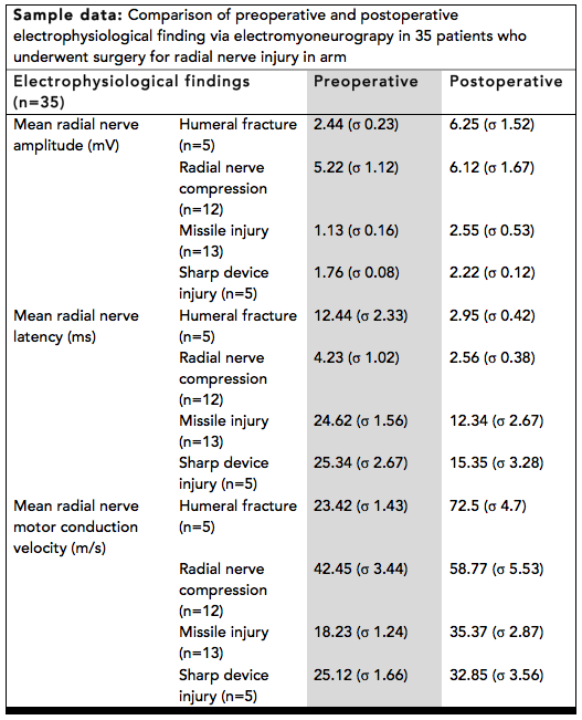
Electromyoneurography (EMNG) is the combined use of electromyography and electroneurography This technique allows for the measurement of a peripheral nerve’s conduction velocity upon stimulation (electroneurography) alongside electrical recording of muscular activity (electromyography). Their combined use proves to be clinically relevant by allowing for both the source and location of a particular neuromuscular disease to be known, and for more accurate diagnoses. Characteristics Electromyoneurography is a technique that uses surface electrical probes to obtain electrophysiological readings from nerve and muscle cells. The nerve activity is generally recorded using surface electrodes, stimulating the nerve at one site and recording from another with a minimum distance between the two. The time difference of the potential is a measure of the time taken for the potential to travel the distance across the two sites and is a measure of the conduction velocity along the nerve. The ... [...More Info...] [...Related Items...] OR: [Wikipedia] [Google] [Baidu] |
Electromyography
Electromyography (EMG) is a technique for evaluating and recording the electrical activity produced by skeletal muscles. EMG is performed using an instrument called an electromyograph to produce a record called an electromyogram. An electromyograph detects the electric potential generated by muscle cells when these cells are electrically or neurologically activated. The signals can be analyzed to detect abnormalities, activation level, or recruitment order, or to analyze the biomechanics of human or animal movement. Needle EMG is an electrodiagnostic medicine technique commonly used by neurologists. Surface EMG is a non-medical procedure used to assess muscle activation by several professionals, including physiotherapists, kinesiologists and biomedical engineers. In Computer Science, EMG is also used as middleware in gesture recognition towards allowing the input of physical action to a computer as a form of human-computer interaction. Clinical uses EMG testing has a variety of ... [...More Info...] [...Related Items...] OR: [Wikipedia] [Google] [Baidu] |
Axonotmesis
Axonotmesis is an injury to the peripheral nerve of one of the extremities of the body. The axons and their myelin sheath are damaged in this kind of injury, but the endoneurium, perineurium and epineurium remain intact. Motor and sensory functions distal to the point of injury are completely lost over time leading to Wallerian degeneration due to ischemia, or loss of blood supply. Axonotmesis is usually the result of a more severe crush or contusion than neurapraxia. Axonotmesis mainly follows a stretch injury. These stretch injuries can either dislocate joints or fracture a limb, due to which peripheral nerves are severed. If the sharp pain from the exposed axon of the nerve is not observed, one can identify a nerve injury from abnormal sensations in their limb. A doctor may ask for a nerve conduction velocity (NCV) test to completely diagnose the issue. If diagnosed as nerve injury, electromyography performed after 3 to 4 weeks shows signs of denervations and fibrillations, or ir ... [...More Info...] [...Related Items...] OR: [Wikipedia] [Google] [Baidu] |
Neurotmesis
Neurotmesis (in Greek tmesis signifies "to cut") is part of Seddon's classification scheme used to classify nerve damage. It is the most serious nerve injury in the scheme. In this type of injury, both the nerve and the nerve sheath are disrupted. While partial recovery may occur, complete recovery is impossible. Symptoms Symptoms of neurotmesis include but are not limited to pain, dysesthesias (uncomfortable sensations), and complete loss of sensory and motor function of the affected nerve. Anatomy Neurotmesis occurs in the peripheral nervous system and most often in the upper-limb (arms), accounting for 73.5% of all peripheral nerve injury cases. Of these cases, the ulnar nerve was most often injured. Peripheral nerves are structured so that the axons are surrounded by most often a myelinated sheath and then an endoneurium. A perineurium surrounds that and the outermost layer is considered the epineurium. When injury occurs, "local vascular trauma leads to hemorrhage and ... [...More Info...] [...Related Items...] OR: [Wikipedia] [Google] [Baidu] |
Neuropraxia
Neurapraxia is a disorder of the peripheral nervous system in which there is a temporary loss of motor and sensory function due to blockage of nerve conduction, usually lasting an average of six to eight weeks before full recovery. Neurapraxia is derived from the word apraxia, meaning “loss or impairment of the ability to execute complex coordinated movements without muscular or sensory impairment”. This condition is typically caused by a blunt neural injury due to external blows or shock-like injuries to muscle fibers and motor neuron, skeletal nerve fibers, which leads to repeated or prolonged pressure buildup on the nerve. As a result of this pressure, ischemia occurs, a neural lesion results, and the human body naturally responds with edema extending in all directions from the source of the pressure. This lesion causes a complete or partial action potential conduction block over a segment of a nerve fiber and thus a reduction or loss of function in parts of the neural conn ... [...More Info...] [...Related Items...] OR: [Wikipedia] [Google] [Baidu] |
Wallerian Degeneration
Wallerian degeneration is an active process of degeneration that results when a nerve fiber is cut or crushed and the part of the axon distal to the injury (i.e. farther from the neuron's cell body) degenerates. A related process of dying back or retrograde degeneration known as 'Wallerian-like degeneration' occurs in many neurodegenerative diseases, especially those where axonal transport is impaired such as ALS and Alzheimer's disease. Primary culture studies suggest that a failure to deliver sufficient quantities of the essential axonal protein NMNAT2 is a key initiating event. Wallerian degeneration occurs after axonal injury in both the peripheral nervous system (PNS) and central nervous system (CNS). It occurs in the section of the axon distal to the site of injury and usually begins within 24–36 hours of a lesion. Prior to degeneration, the distal section of the axon tends to remain electrically excitable. After injury, the axonal skeleton disintegrates, and th ... [...More Info...] [...Related Items...] OR: [Wikipedia] [Google] [Baidu] |
House–Brackmann Score
The House–Brackmann score is a score to grade the degree of nerve damage in a facial nerve palsy. The measurement is determined by measuring the upwards (superior) movement of the mid-portion of the top of the eyebrow, and the outwards (lateral) movement of the angle of the mouth. Each reference point scores 1 point for each 0.25 cm movement, up to a maximum of 1 cm. The scores are then added together, to give a number out of 8. The score predicts recovery in those with Bell's palsy Bell's palsy is a type of facial paralysis that results in a temporary inability to control the facial muscles on the affected side of the face. In most cases, the weakness is temporary and significantly improves over weeks. Symptoms can vary fr .... The score carries the name of the Dr John W. House and Dr Derald E. Brackmann, otolaryngologists in Los Angeles, California, who first described the system in 1985. It is one of a number of facial nerve scoring systems, such as Burres-Fisch ... [...More Info...] [...Related Items...] OR: [Wikipedia] [Google] [Baidu] |
Transcranial Magnetic Stimulation
Transcranial magnetic stimulation (TMS) is a noninvasive form of brain stimulation in which a changing magnetic field is used to induce an electric current at a specific area of the brain through electromagnetic induction. An electric pulse generator, or stimulator, is connected to a magnetic coil connected to the scalp. The stimulator generates a changing electric current within the coil which creates a varying magnetic field, inducing a current within a region in the brain itself.NICE. January 201Transcranial magnetic stimulation for treating and preventing migraine/ref>Michael Craig Miller for Harvard Health Publications. July 26, 201Magnetic stimulation: a new approach to treating depression?/ref> TMS has shown diagnostic and therapeutic potential in the central nervous system with a wide variety of disease states in neurology and mental health, with research still evolving. Adverse effects of TMS appear rare and include fainting and seizure. Other potential issues include ... [...More Info...] [...Related Items...] OR: [Wikipedia] [Google] [Baidu] |
Computed Tomography
A computed tomography scan (CT scan; formerly called computed axial tomography scan or CAT scan) is a medical imaging technique used to obtain detailed internal images of the body. The personnel that perform CT scans are called radiographers or radiology technologists. CT scanners use a rotating X-ray tube and a row of detectors placed in a gantry to measure X-ray attenuations by different tissues inside the body. The multiple X-ray measurements taken from different angles are then processed on a computer using tomographic reconstruction algorithms to produce tomographic (cross-sectional) images (virtual "slices") of a body. CT scans can be used in patients with metallic implants or pacemakers, for whom magnetic resonance imaging (MRI) is contraindicated. Since its development in the 1970s, CT scanning has proven to be a versatile imaging technique. While CT is most prominently used in medical diagnosis, it can also be used to form images of non-living objects. The 1979 Nob ... [...More Info...] [...Related Items...] OR: [Wikipedia] [Google] [Baidu] |


