|
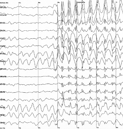
Electroencephalograph
Electroencephalography (EEG) is a method to record an electrogram of the spontaneous electrical activity of the brain. The biosignals detected by EEG have been shown to represent the postsynaptic potentials of pyramidal neurons in the neocortex and allocortex. It is typically non-invasive, with the EEG electrodes placed along the scalp (commonly called "scalp EEG") using the International 10-20 system, or variations of it. Electrocorticography, involving surgical placement of electrodes, is sometimes called " intracranial EEG". Clinical interpretation of EEG recordings is most often performed by visual inspection of the tracing or quantitative EEG analysis. Voltage fluctuations measured by the EEG bioamplifier and electrodes allow the evaluation of normal brain activity. As the electrical activity monitored by EEG originates in neurons in the underlying brain tissue, the recordings made by the electrodes on the surface of the scalp vary in accordance with their orientation and ... [...More Info...] [...Related Items...] OR: [Wikipedia] [Google] [Baidu] |
10–20 System (EEG)
The 10–20 system or International 10–20 system is an internationally recognized method to describe and apply the location of scalp electrodes in the context of an EEG exam, polysomnograph sleep study, or voluntary lab research. This method was developed to maintain standardized testing methods ensuring that a subject's study outcomes (clinical or research) could be compiled, reproduced, and effectively analyzed and compared using the scientific method. The system is based on the relationship between the location of an electrode and the underlying area of the brain, specifically the cerebral cortex. During sleep and wake cycles, the brain produces different, objectively recognized and distinguishable electrical patterns, which can be detected by electrodes on the skin. (These patterns might vary, and can be affected by multiple extrinsic factors, i.e. age, prescription drugs, somatic diagnoses, hx of neurologic insults/injury/trauma, and substance abuse) The "10" and "20" ref ... [...More Info...] [...Related Items...] OR: [Wikipedia] [Google] [Baidu] |
Evoked Potential
An evoked potential or evoked response is an electrical potential in a specific pattern recorded from a specific part of the nervous system, especially the brain, of a human or other animals following presentation of a Stimulus (physiology), stimulus such as a light flash or a pure tone. Different types of potentials result from stimuli of different Stimulus modality, modalities and types. Evoked potential is distinct from spontaneous potentials as detected by electroencephalography (EEG), electromyography (EMG), or other electrophysiology, electrophysiologic recording method. Such potentials are useful for electrodiagnostic medicine, electrodiagnosis and monitoring (medicine), monitoring that include detections of disease and drug-related sensory dysfunction and intraoperative monitoring of sensory pathway integrity. Evoked potential amplitudes tend to be low, ranging from less than a microvolt to several microvolts, compared to tens of microvolts for EEG, millivolts for EMG, and ... [...More Info...] [...Related Items...] OR: [Wikipedia] [Google] [Baidu] |
Spike-and-wave
Spike-and-wave is a pattern of the electroencephalogram (EEG) typically observed during epileptic seizures. A spike-and-wave discharge is a regular, symmetrical, generalized EEG pattern seen particularly during absence epilepsy, also known as ‘petit mal’ epilepsy. The basic mechanisms underlying these patterns are complex and involve part of the cerebral cortex, the thalamocortical network, and intrinsic neuronal mechanisms. The first spike-and-wave pattern was recorded in the early twentieth century by Hans Berger. Many aspects of the pattern are still being researched and discovered, and still many aspects are uncertain. The spike-and-wave pattern is most commonly researched in absence epilepsy, but is common in several epilepsies such as Lennox-Gastaut syndrome (LGS) and Ohtahara syndrome. Antiepileptic drugs (AEDs) are commonly prescribed to treat epileptic seizures, and new ones are being discovered with fewer adverse effects. Today, most of the research is focused on t ... [...More Info...] [...Related Items...] OR: [Wikipedia] [Google] [Baidu] |
Electrogram
An electrogram (EGM) is a recording of electrical activity of organs such as the brain and heart, measured by monitoring changes in electric potential. Brain Electroencephalography (EEG) An electroencephalogram (EEG) is an electrical recording of the activity of the brain taken from the scalp. An EEG can be used to diagnose seizures, sleep disorders, and for monitoring of level of anesthesia during surgery. Electrocorticography (ECoG or iEEG) An electrocorticogram is an electrical recording of the brain measured intracranially, that is, from within the brain. Eye Electrooculography (EOG) An electrooculogram (EOG) is an electrical recording of the potential between the cornea and the retina, and does not change with visual stimuli. An EOG can measure movements of the eyes and can help in diagnosis of nystagmus. Electroretinography (ERG) An electroretinogram (ERG) is an electrical recording of the electrical activity of the retina. Heart Electrocardiogram (ECG) An elect ... [...More Info...] [...Related Items...] OR: [Wikipedia] [Google] [Baidu] |
Bioamplifier
A Bioamplifier is an electrophysiological device, a variation of the instrumentation amplifier, used to gather and increase the signal integrity of physiologic electrical activity for output to various sources. It may be an independent unit, or integrated into the electrodes. History Efforts to amplify biosignals started with the development of electrocardiography. In 1887, Augustus Waller, a British physiologist, successfully measured the electrocardiograph of his dog using two buckets of saline, in which he submerged each of the front and the hind paws.Webster, John G. (2006) Encyclopedia of Medical Devices and Instrumentation Volume I. New Jersey: Wiley-Interscience. . A few months later, Waller successfully recorded the first human electrocardiography using the capillary electrometer. However, at the time of invention, Waller did not envision that electrocardiography would be used extensively in healthcare. The electrocardiograph was impractical to use until Willem Einthoven ... [...More Info...] [...Related Items...] OR: [Wikipedia] [Google] [Baidu] |
Coma
A coma is a deep state of prolonged unconsciousness in which a person cannot be awakened, fails to respond normally to painful stimuli, light, or sound, lacks a normal wake-sleep cycle and does not initiate voluntary actions. Coma patients exhibit a complete absence of wakefulness and are unable to consciously feel, speak or move. Comas can be derived by natural causes, or can be medically induced. Clinically, a coma can be defined as the inability consistently to follow a one-step command. It can also be defined as a score of ≤ 8 on the Glasgow Coma Scale (GCS) lasting ≥ 6 hours. For a patient to maintain consciousness, the components of ''wakefulness'' and ''awareness'' must be maintained. Wakefulness describes the quantitative degree of consciousness, whereas awareness relates to the qualitative aspects of the functions mediated by the cortex, including cognitive abilities such as attention, sensory perception, explicit memory, language, the execution of tasks, temporal ... [...More Info...] [...Related Items...] OR: [Wikipedia] [Google] [Baidu] |
Hippocampus
The hippocampus (via Latin from Greek , 'seahorse') is a major component of the brain of humans and other vertebrates. Humans and other mammals have two hippocampi, one in each side of the brain. The hippocampus is part of the limbic system, and plays important roles in the consolidation of information from short-term memory to long-term memory, and in spatial memory that enables navigation. The hippocampus is located in the allocortex, with neural projections into the neocortex in humans, as well as primates. The hippocampus, as the medial pallium, is a structure found in all vertebrates. In humans, it contains two main interlocking parts: the hippocampus proper (also called ''Ammon's horn''), and the dentate gyrus. In Alzheimer's disease (and other forms of dementia), the hippocampus is one of the first regions of the brain to suffer damage; short-term memory loss and disorientation are included among the early symptoms. Damage to the hippocampus can also result from ... [...More Info...] [...Related Items...] OR: [Wikipedia] [Google] [Baidu] |
Quantitative EEG
Quantitative electroencephalography (qEEG or QEEG) is a field concerned with the numerical analysis of electroencephalography (EEG) data and associated behavioral correlates. Details Techniques used in digital signal analysis are extended to the analysis of electroencephalography (EEG). These include Wavelet transform, wavelet analysis and Fourier Transform, Fourier analysis, with new focus on shared activity between rhythms including phase synchrony (coherence, phase lag) and magnitude synchrony (comodulation/correlation, and asymmetry). The analog signal comprises a microvoltage time series of the EEG, sampled digitally and sampling rates adequate to over-sample the signal (using the Harry Nyquist, Nyquist principle of exceeding twice the highest frequency being detected). Modern EEG amplifiers use adequate sampling to resolve the EEG across the traditional medical band from DC to 70 or 100 Hz, using sample rates of 250/256, 500/512, to over 1000 samples per second, depen ... [...More Info...] [...Related Items...] OR: [Wikipedia] [Google] [Baidu] |
Electrocorticography
Electrocorticography (ECoG), or intracranial electroencephalography (iEEG), is a type of electrophysiological monitoring that uses electrodes placed directly on the exposed surface of the brain to record electrical activity from the cerebral cortex. In contrast, conventional electroencephalography (EEG) electrodes monitor this activity from outside the skull. ECoG may be performed either in the operating room during surgery (intraoperative ECoG) or outside of surgery (extraoperative ECoG). Because a craniotomy (a surgical incision into the skull) is required to implant the electrode grid, ECoG is an invasive procedure. History ECoG was pioneered in the early 1950s by Wilder Penfield and Herbert Jasper, neurosurgeons at the Montreal Neurological Institute. The two developed ECoG as part of their groundbreakinMontreal procedure a surgical protocol used to treat patients with severe epilepsy. The cortical potentials recorded by ECoG were used to identify epileptogenic zones � ... [...More Info...] [...Related Items...] OR: [Wikipedia] [Google] [Baidu] |
Electrography (other)
Electrography often refers to electrophotography, that is, Kirlian photography. Electrography may also refer to: * Measurement and recording of electrophysiology, electrophysiologic activity for diagnostic purposes ** Electrocardiography (ECG or EKG), electrography of heart electrical activity and rhythm ** Electromyography (EMG), electrography of other muscle action potentials throughout the body ** Electroencephalography (EEG), electrography of brain waves (from outside the skull) *** Electrocorticography or intracranial EEG (iEEG or ECoG), EEG with direct contact to the cerebral cortex ** Electrooculography (EOG), electrography of intraocular potential differences ** Electro-olfactography, Electroolfactography (EOG), electrography of olfaction (smell) ** Electroretinography (ERG), electrography of retinal cell action potentials ** Electronystagmography (ENG), electrography of eye muscle movements ** Electrocochleography (ECOG), electrography of cochlear auditory activity ** E ... [...More Info...] [...Related Items...] OR: [Wikipedia] [Google] [Baidu] |
Brain
A brain is an organ that serves as the center of the nervous system in all vertebrate and most invertebrate animals. It is located in the head, usually close to the sensory organs for senses such as vision. It is the most complex organ in a vertebrate's body. In a human, the cerebral cortex contains approximately 14–16 billion neurons, and the estimated number of neurons in the cerebellum is 55–70 billion. Each neuron is connected by synapses to several thousand other neurons. These neurons typically communicate with one another by means of long fibers called axons, which carry trains of signal pulses called action potentials to distant parts of the brain or body targeting specific recipient cells. Physiologically, brains exert centralized control over a body's other organs. They act on the rest of the body both by generating patterns of muscle activity and by driving the secretion of chemicals called hormones. This centralized control allows rapid and coordinated respon ... [...More Info...] [...Related Items...] OR: [Wikipedia] [Google] [Baidu] |
Biosignal
A biosignal is any signal in living beings that can be continually measured and monitored. The term biosignal is often used to refer to bioelectrical signals, but it may refer to both electrical and non-electrical signals. The usual understanding is to refer only to time-varying signals, although spatial parameter variations (e.g. the nucleotide sequence determining the genetic code) are sometimes subsumed as well. Electrical biosignals Electrical biosignals, or bioelectrical time signals, usually refers to the change in electric current produced by the sum of an electrical potential difference across a specialized tissue, organ or cell system like the nervous system. Thus, among the best-known bioelectrical signals are: * Electroencephalogram (EEG) * Electrocardiogram (ECG) * Electromyogram (EMG) * Electrooculogram (EOG) * Electroretinogram (ERG) * Electrogastrogram (EGG) * Galvanic skin response (GSR) or electrodermal activity (EDA) EEG, ECG, EOG and EMG are measured with a ... [...More Info...] [...Related Items...] OR: [Wikipedia] [Google] [Baidu] |


