|
Dorsal Antibrachial Cutaneous Nerve
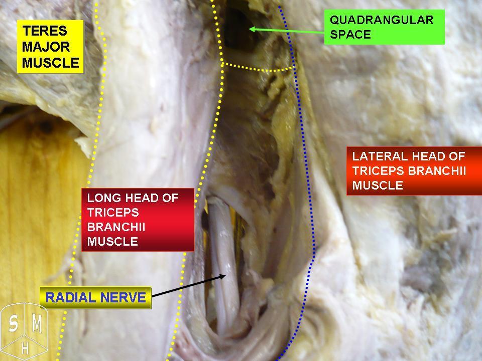
The posterior cutaneous nerve of forearm is a nerve found in humans and other animals. It is also known as the dorsal antebrachial cutaneous nerve, the external cutaneous branch of the musculospiral nerve, and the posterior antebrachial cutaneous nerve. It is a cutaneous nerve (a nerve that supplies skin) of the forearm. Origin It arises from the radial nerve in the posterior compartment of the arm, often along with the posterior cutaneous nerve of the arm. Course It perforates the lateral head of the triceps brachii muscle at the triceps' attachment to the humerus. The upper and smaller branch of the nerve passes to the front of the elbow, lying close to the cephalic vein, and supplies the skin of the lower half of the arm. The lower branch pierces the deep fascia below the insertion of the Deltoideus, and descends along the lateral side of the arm and elbow, and then along the back of the forearm to the wrist, supplying the skin in its course, and joining, near its termination, ... [...More Info...] [...Related Items...] OR: [Wikipedia] [Google] [Baidu] |
Radial Nerve
The radial nerve is a nerve in the human body that supplies the posterior portion of the upper limb. It innervates the medial and lateral heads of the triceps brachii muscle of the arm, as well as all 12 muscles in the posterior osteofascial compartment of the forearm and the associated joints and overlying skin. It originates from the brachial plexus, carrying fibers from the ventral roots of spinal nerves C5, C6, C7, C8 & T1. The radial nerve and its branches provide motor innervation to the dorsal arm muscles (the triceps brachii and the anconeus) and the extrinsic extensors of the wrists and hands; it also provides cutaneous sensory innervation to most of the back of the hand, except for the back of the little finger and adjacent half of the ring finger (which are innervated by the ulnar nerve). The radial nerve divides into a deep branch, which becomes the posterior interosseous nerve, and a superficial branch, which goes on to innervate the dorsum (back) of the hand. Th ... [...More Info...] [...Related Items...] OR: [Wikipedia] [Google] [Baidu] |
Nerve
A nerve is an enclosed, cable-like bundle of nerve fibers (called axons) in the peripheral nervous system. A nerve transmits electrical impulses. It is the basic unit of the peripheral nervous system. A nerve provides a common pathway for the electrochemical nerve impulses called action potentials that are transmitted along each of the axons to peripheral organs or, in the case of sensory nerves, from the periphery back to the central nervous system. Each axon, within the nerve, is an extension of an individual neuron, along with other supportive cells such as some Schwann cells that coat the axons in myelin. Within a nerve, each axon is surrounded by a layer of connective tissue called the endoneurium. The axons are bundled together into groups called fascicles, and each fascicle is wrapped in a layer of connective tissue called the perineurium. Finally, the entire nerve is wrapped in a layer of connective tissue called the epineurium. Nerve cells (often called neurons) are f ... [...More Info...] [...Related Items...] OR: [Wikipedia] [Google] [Baidu] |
Cutaneous Nerve
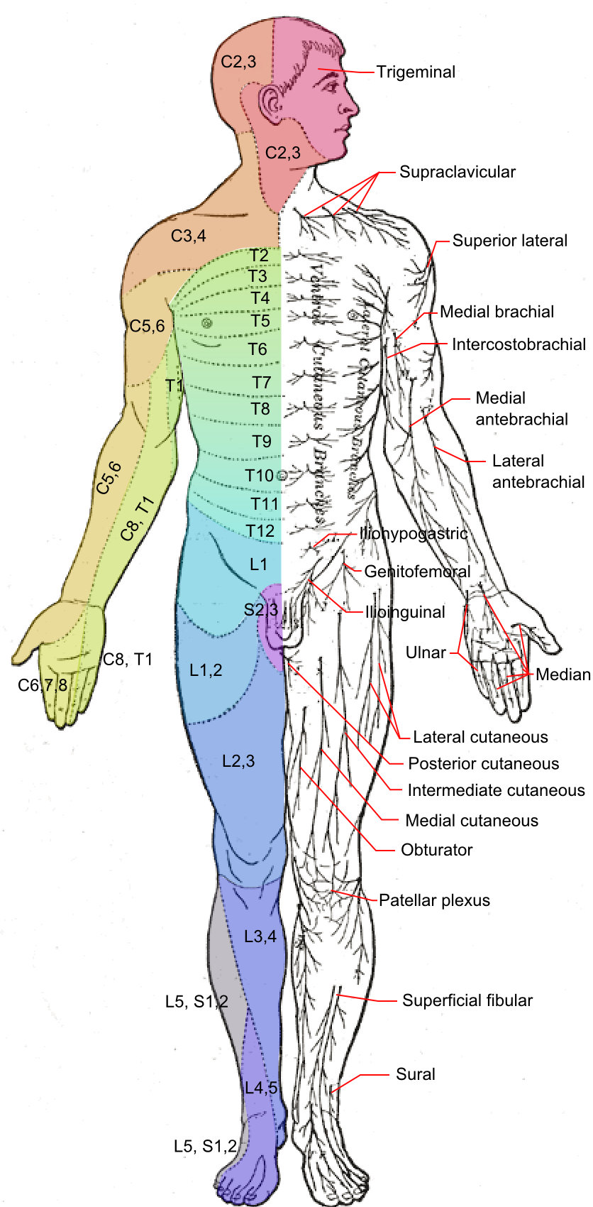
A cutaneous nerve is a nerve that provides nerve supply to the skin. Human anatomy In human anatomy, cutaneous nerves are primarily responsible for providing sensory innervation to the skin. In addition to sympathetic and autonomic afferent (sensory) fibers, most cutaneous nerves also contain sympathetic efferent (visceromotor) fibers, which innervate cutaneous blood vessels, sweat glands, and the arrector pilli muscles of hair follicles. These structures are important to the sympathetic nervous response. There are many cutaneous nerves in the human body, only some of which are named. Some of the larger cutaneous nerves are as follows: Upper body * In the arm (proper) ** Superior lateral cutaneous nerve of arm (Superior LCNOA) ** Inferior lateral cutaneous nerve of arm (Inferior LCNOA) ** Posterior cutaneous nerve of arm (PCNOA) ** Medial cutaneous nerve of arm (MCNOA) * In the forearm ** Lateral cutaneous nerve of forearm (LCNOF) ** Posterior cutaneous nerve of forearm (PCNOF ... [...More Info...] [...Related Items...] OR: [Wikipedia] [Google] [Baidu] |
Forearm
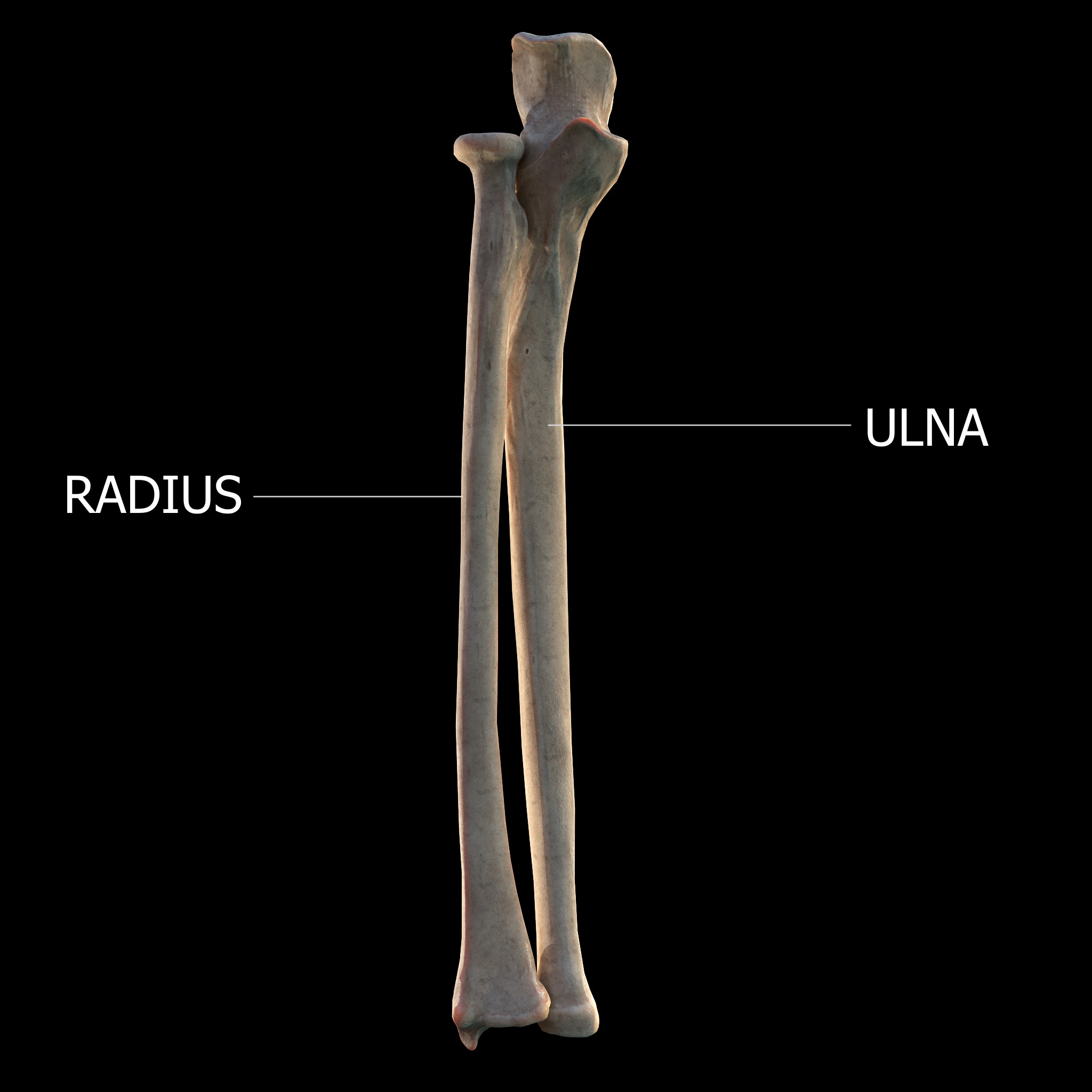
The forearm is the region of the upper limb between the elbow and the wrist. The term forearm is used in anatomy to distinguish it from the arm, a word which is most often used to describe the entire appendage of the upper limb, but which in anatomy, technically, means only the region of the upper arm, whereas the lower "arm" is called the forearm. It is homologous to the region of the leg that lies between the knee and the ankle joints, the crus. The forearm contains two long bones, the radius and the ulna, forming the two radioulnar joints. The interosseous membrane connects these bones. Ultimately, the forearm is covered by skin, the anterior surface usually being less hairy than the posterior surface. The forearm contains many muscles, including the flexors and extensors of the wrist, flexors and extensors of the digits, a flexor of the elbow (brachioradialis), and pronators and supinators that turn the hand to face down or upwards, respectively. In cross-section, the for ... [...More Info...] [...Related Items...] OR: [Wikipedia] [Google] [Baidu] |
Radial Nerve
The radial nerve is a nerve in the human body that supplies the posterior portion of the upper limb. It innervates the medial and lateral heads of the triceps brachii muscle of the arm, as well as all 12 muscles in the posterior osteofascial compartment of the forearm and the associated joints and overlying skin. It originates from the brachial plexus, carrying fibers from the ventral roots of spinal nerves C5, C6, C7, C8 & T1. The radial nerve and its branches provide motor innervation to the dorsal arm muscles (the triceps brachii and the anconeus) and the extrinsic extensors of the wrists and hands; it also provides cutaneous sensory innervation to most of the back of the hand, except for the back of the little finger and adjacent half of the ring finger (which are innervated by the ulnar nerve). The radial nerve divides into a deep branch, which becomes the posterior interosseous nerve, and a superficial branch, which goes on to innervate the dorsum (back) of the hand. Th ... [...More Info...] [...Related Items...] OR: [Wikipedia] [Google] [Baidu] |
Triceps Brachii
The triceps, or triceps brachii (Latin for "three-headed muscle of the arm"), is a large muscle on the back of the upper limb of many vertebrates Vertebrates () comprise all animal taxa within the subphylum Vertebrata () ( chordates with backbones), including all mammals, birds, reptiles, amphibians, and fish. Vertebrates represent the overwhelming majority of the phylum Chordata, .... It consists of 3 parts: the medial, lateral, and long head. It is the muscle principally responsible for Extension (kinesiology), extension of the elbow joint (straightening of the arm). Structure The long head arises from the infraglenoid tubercle of the scapula. It extends distally anterior to the Teres minor muscle, teres minor and posterior to the Teres major muscle, teres major. The medial head arises proximally in the humerus, just inferior to the Radial sulcus, groove of the radial nerve; from the dorsal (back) surface of the humerus; from the Medial intermuscular septum of arm ... [...More Info...] [...Related Items...] OR: [Wikipedia] [Google] [Baidu] |
Humerus
The humerus (; ) is a long bone in the arm that runs from the shoulder to the elbow. It connects the scapula and the two bones of the lower arm, the radius and ulna, and consists of three sections. The humeral upper extremity consists of a rounded head, a narrow neck, and two short processes (tubercles, sometimes called tuberosities). The body is cylindrical in its upper portion, and more prismatic below. The lower extremity consists of 2 epicondyles, 2 processes (trochlea & capitulum), and 3 fossae (radial fossa, coronoid fossa, and olecranon fossa). As well as its true anatomical neck, the constriction below the greater and lesser tubercles of the humerus is referred to as its surgical neck due to its tendency to fracture, thus often becoming the focus of surgeons. Etymology The word "humerus" is derived from la, humerus, umerus meaning upper arm, shoulder, and is linguistically related to Gothic ''ams'' shoulder and Greek ''ōmos''. Structure Upper extremity The upper or pr ... [...More Info...] [...Related Items...] OR: [Wikipedia] [Google] [Baidu] |
Elbow-joint
The elbow is the region between the arm and the forearm that surrounds the elbow joint. The elbow includes prominent landmarks such as the olecranon, the cubital fossa (also called the chelidon, or the elbow pit), and the lateral and the medial epicondyles of the humerus. The elbow joint is a hinge joint between the arm and the forearm; more specifically between the humerus in the upper arm and the radius and ulna in the forearm which allows the forearm and hand to be moved towards and away from the body. The term ''elbow'' is specifically used for humans and other primates, and in other vertebrates forelimb plus joint is used. The name for the elbow in Latin is ''cubitus'', and so the word cubital is used in some elbow-related terms, as in ''cubital nodes'' for example. Structure Joint The elbow joint has three different portions surrounded by a common joint capsule. These are joints between the three bones of the elbow, the humerus of the upper arm, and the radius and th ... [...More Info...] [...Related Items...] OR: [Wikipedia] [Google] [Baidu] |
Cephalic Vein
In human anatomy, the cephalic vein is a superficial vein in the arm. It originates from the radial end of the dorsal venous network of hand, and ascends along the radial (lateral) side of the arm before emptying into the axillary vein. At the elbow, it communicates with the basilic vein via the median cubital vein. Anatomy The cephalic vein is situated within the superficial fascia along the anterolateral surface of the biceps. Origin The cephalic vein forms over the anatomical snuffbox at the radial end of the dorsal venous network of hand. Course and relations From its origin, it ascends ascends up the lateral aspect of the radius. Near the shoulder, the cephalic vein passes between the deltoid and pectoralis major muscles (deltopectoral groove) and through the clavipectoral triangle, where it empties into the axillary vein. Anastomoses It communicates with the basilic vein via the median cubital vein at the elbow. Clinical significance The cephalic vein ... [...More Info...] [...Related Items...] OR: [Wikipedia] [Google] [Baidu] |
Deep Fascia
Deep fascia (or investing fascia) is a fascia, a layer of dense connective tissue that can surround individual muscles and groups of muscles to separate into fascial compartments. This fibrous connective tissue interpenetrates and surrounds the muscles, bones, nerves, and blood vessels of the body. It provides connection and communication in the form of aponeuroses, ligaments, tendons, retinacula, joint capsules, and septa. The deep fasciae envelop all bone (periosteum and endosteum); cartilage (perichondrium), and blood vessels (tunica externa) and become specialized in muscles (epimysium, perimysium, and endomysium) and nerves (epineurium, perineurium, and endoneurium). The high density of collagen fibers gives the deep fascia its strength and integrity. The amount of elastin fiber determines how much extensibility and resilience it will have. Examples Examples include: * Fascia lata * Deep fascia of leg * Brachial fascia * Buck's fascia Fascial dynamics Deep fascia is l ... [...More Info...] [...Related Items...] OR: [Wikipedia] [Google] [Baidu] |
Deltoideus
The deltoid muscle is the muscle forming the rounded contour of the human shoulder. It is also known as the 'common shoulder muscle', particularly in other animals such as the domestic cat. Anatomically, the deltoid muscle appears to be made up of three distinct sets of muscle fibers, namely the # anterior or clavicular part (pars clavicularis) # posterior or scapular part (pars scapularis) # intermediate or acromial part (pars acromialis) However, electromyography suggests that it consists of at least seven groups that can be independently coordinated by the nervous system. It was previously called the deltoideus (plural ''deltoidei'') and the name is still used by some anatomists. It is called so because it is in the shape of the Greek capital letter delta (Δ). Deltoid is also further shortened in slang as "delt". A study of 30 shoulders revealed an average mass of in humans, ranging from to . Structure Previous studies showed that the insertions of the tendons of the delto ... [...More Info...] [...Related Items...] OR: [Wikipedia] [Google] [Baidu] |
Medial Cutaneous Nerve Of Forearm
The medial cutaneous nerve of the forearm (medial antebrachial cutaneous nerve) branches from the medial cord of the brachial plexus. It contains axons from the ventral rami of the eighth cervical (C8) and first thoracic (T1) nerves. It gives off a branch near the axilla, which pierces the fascia and supplies the skin covering the biceps brachii, nearly as far as the elbow. The nerve then runs down the ulnar side of the arm medial to the brachial artery, pierces the deep fascia with the basilic vein, about the middle of the arm, and divides into a volar and an ulnar branch. Volar branch The ''volar branch'' (ramus volaris; anterior branch), the larger, passes usually in front of, but occasionally behind, the vena mediana cubiti (median basilic vein). It then descends on the front of the ulnar side of the forearm, distributing filaments to the skin as far as the wrist, and communicating with the palmar cutaneous branch of the ulnar nerve. Ulnar branch The ''ulnar branch'' (r ... [...More Info...] [...Related Items...] OR: [Wikipedia] [Google] [Baidu] |


