|
Diad
Within the muscle tissue of animals and humans, contraction and relaxation of the muscle cells (myocytes) is a highly regulated and rhythmic process. In cardiomyocytes, or cardiac muscle cells, muscular contraction takes place due to movement at a structure referred to as the diad, sometimes spelled "dyad." The dyad is the connection of transverse- tubules ( t-tubules) and the junctional sarcoplasmic reticulum (jSR). Like skeletal muscle contractions, Calcium (Ca2+) ions are required for polarization and depolarization through a voltage-gated calcium channel. The rapid influx of calcium into the cell signals for the cells to contract. When the calcium intake travels through an entire muscle, it will trigger a united muscular contraction. This process is known as excitation-contraction coupling. This contraction pushes blood inside the heart and from the heart to other regions of the body. Myocyte Anatomy Myocytes are incredibly specialized cells with only a select number of diffe ... [...More Info...] [...Related Items...] OR: [Wikipedia] [Google] [Baidu] |
Myocytes
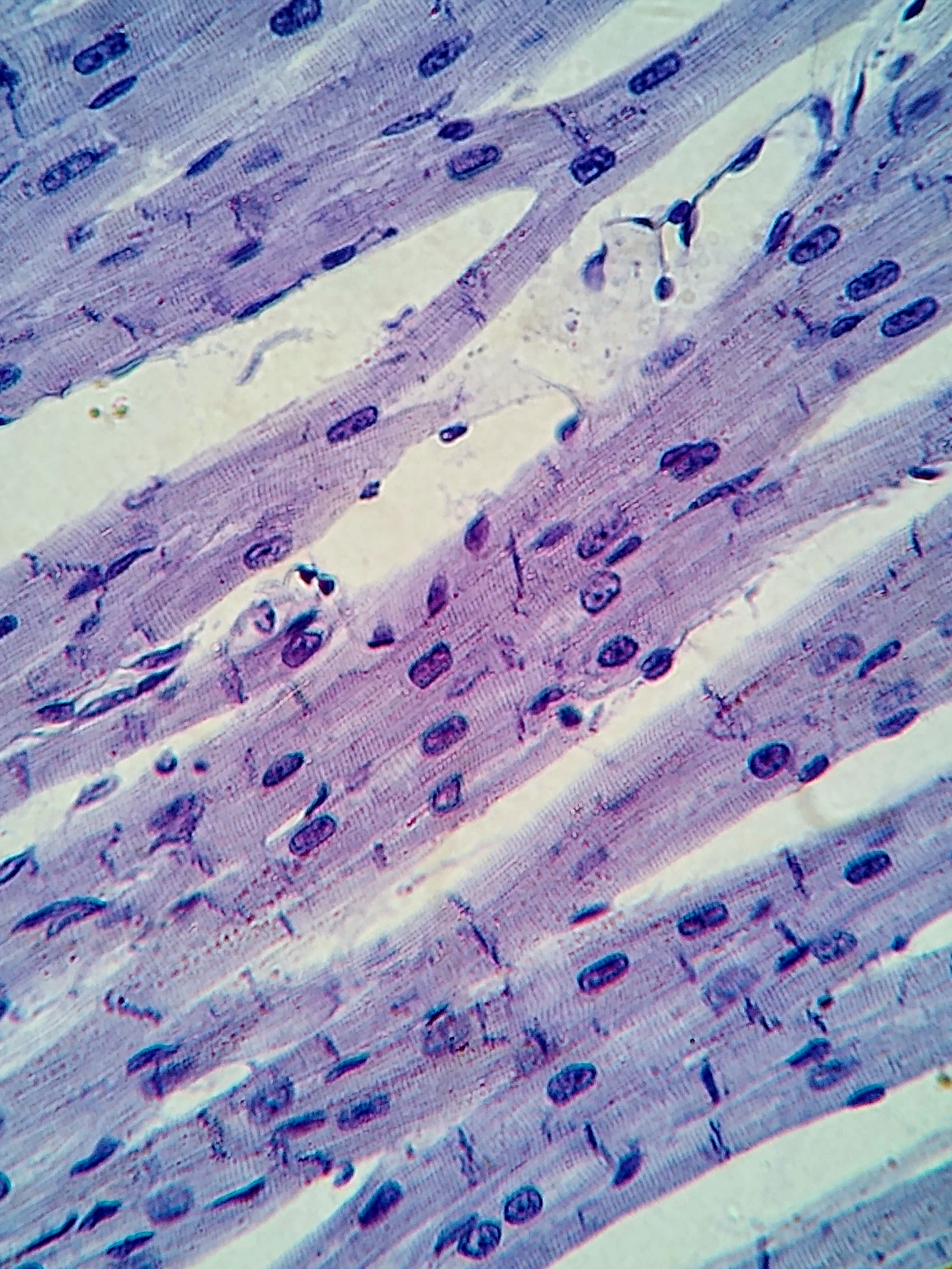
A muscle cell is also known as a myocyte when referring to either a cardiac muscle cell (cardiomyocyte), or a smooth muscle cell as these are both small cells. A skeletal muscle cell is long and threadlike with many nuclei and is called a muscle fiber. Muscle cells (including myocytes and muscle fibers) develop from embryonic precursor cells called myoblasts. Myoblasts fuse to form multinucleated skeletal muscle cells known as syncytia in a process known as myogenesis. Skeletal muscle cells and cardiac muscle cells both contain myofibrils and sarcomeres and form a striated muscle tissue. Cardiac muscle cells form the cardiac muscle in the walls of the heart chambers, and have a single central nucleus. Cardiac muscle cells are joined to neighboring cells by intercalated discs, and when joined in a visible unit they are described as a ''cardiac muscle fiber''. Smooth muscle cells control involuntary movements such as the peristalsis contractions in the esophagus and stomach. Sm ... [...More Info...] [...Related Items...] OR: [Wikipedia] [Google] [Baidu] |
Pacemaker Cell
350px, Image showing the cardiac pacemaker or SA node, the primary pacemaker within the electrical_conduction_system_of_the_heart">SA_node,_the_primary_pacemaker_within_the_electrical_conduction_system_of_the_heart. The_muscle_contraction.html" "title="electrical conduction system of the heart.">electrical conduction system of the heart">SA node, the primary pacemaker within the electrical conduction system of the heart. The muscle contraction">contraction of cardiac muscle (heart muscle) in all animals is initiated by electrical impulses known as action potentials that in the heart are known as cardiac action potentials. The rate at which these impulses fire controls the rate of cardiac contraction, that is, the heart rate. The cells that create these rhythmic impulses, setting the pace for blood pumping, are called pacemaker cells, and they directly control the heart rate. They make up the cardiac pacemaker, that is, the natural pacemaker of the heart. In most humans, the h ... [...More Info...] [...Related Items...] OR: [Wikipedia] [Google] [Baidu] |
Cardiomyopathy
Cardiomyopathy is a group of diseases that affect the heart muscle. Early on there may be few or no symptoms. As the disease worsens, shortness of breath, feeling tired, and swelling of the legs may occur, due to the onset of heart failure. An irregular heart beat and fainting may occur. Those affected are at an increased risk of sudden cardiac death. Types of cardiomyopathy include hypertrophic cardiomyopathy, dilated cardiomyopathy, restrictive cardiomyopathy, arrhythmogenic right ventricular dysplasia, and Takotsubo cardiomyopathy (broken heart syndrome). In hypertrophic cardiomyopathy the heart muscle enlarges and thickens. In dilated cardiomyopathy the ventricles enlarge and weaken. In restrictive cardiomyopathy the ventricle stiffens. In many cases, the cause cannot be determined. Hypertrophic cardiomyopathy is usually inherited, whereas dilated cardiomyopathy is inherited in about one third of cases. Dilated cardiomyopathy may also result from alcohol, h ... [...More Info...] [...Related Items...] OR: [Wikipedia] [Google] [Baidu] |
Hypertension
Hypertension (HTN or HT), also known as high blood pressure (HBP), is a long-term medical condition in which the blood pressure in the arteries is persistently elevated. High blood pressure usually does not cause symptoms. Long-term high blood pressure, however, is a major risk factor for stroke, coronary artery disease, heart failure, atrial fibrillation, peripheral arterial disease, vision loss, chronic kidney disease, and dementia. Hypertension is a major cause of premature death worldwide. High blood pressure is classified as primary (essential) hypertension or secondary hypertension. About 90–95% of cases are primary, defined as high blood pressure due to nonspecific lifestyle and genetic factors. Lifestyle factors that increase the risk include excess salt in the diet, excess body weight, smoking, and alcohol use. The remaining 5–10% of cases are categorized as secondary high blood pressure, defined as high blood pressure due to an identifiable cause, such ... [...More Info...] [...Related Items...] OR: [Wikipedia] [Google] [Baidu] |
Heart Failure
Heart failure (HF), also known as congestive heart failure (CHF), is a syndrome, a group of signs and symptoms caused by an impairment of the heart's blood pumping function. Symptoms typically include shortness of breath, excessive fatigue, and leg swelling. The shortness of breath may occur with exertion or while lying down, and may wake people up during the night. Chest pain, including angina, is not usually caused by heart failure, but may occur if the heart failure was caused by a heart attack. The severity of the heart failure is measured by the severity of symptoms during exercise. Other conditions that may have symptoms similar to heart failure include obesity, kidney failure, liver disease, anemia, and thyroid disease. Common causes of heart failure include coronary artery disease, heart attack, high blood pressure, atrial fibrillation, valvular heart disease, excessive alcohol consumption, infection, and cardiomyopathy. These cause heart failure by alterin ... [...More Info...] [...Related Items...] OR: [Wikipedia] [Google] [Baidu] |
T-type Calcium Channel
T-type calcium channels are low voltage activated calcium channels that become inactivated during cell membrane hyperpolarization but then open to depolarization. The entry of calcium into various cells has many different physiological responses associated with it. Within cardiac muscle cell and smooth muscle cells voltage-gated calcium channel activation initiates contraction directly by allowing the cytosolic concentration to increase. Not only are T-type calcium channels known to be present within cardiac and smooth muscle, but they also are present in many neuronal cells within the central nervous system. Different experimental studies within the 1970s allowed for the distinction of T-type calcium channels (transient opening calcium channels) from the already well-known L-type calcium channels (Long-Lasting calcium channels). The new T-type channels were much different from the L-type calcium channels due to their ability to be activated by more negative membrane potentials, ... [...More Info...] [...Related Items...] OR: [Wikipedia] [Google] [Baidu] |
L-type Calcium Channel
The L-type calcium channel (also known as the dihydropyridine channel, or DHP channel) is part of the high-voltage activated family of voltage-dependent calcium channel. "L" stands for long-lasting referring to the length of activation. This channel has four isoforms: Cav1.1, Cav1.2, Cav1.3, and Cav1.4. L-type calcium channels are responsible for the excitation-contraction coupling of skeletal, smooth, cardiac muscle, and for aldosterone secretion in endocrine cells of the adrenal cortex. They are also found in neurons, and with the help of L-type calcium channels in endocrine cells, they regulate neurohormones and neurotransmitters. They have also been seen to play a role in gene expression, mRNA stability, neuronal survival, ischemic-induced axonal injury, synaptic efficacy, and both activation and deactivation of other ion channels. In cardiac myocytes, the L-type calcium channel passes inward Ca2+ current (ICaL) and triggers calcium release from the sarcoplasmic reti ... [...More Info...] [...Related Items...] OR: [Wikipedia] [Google] [Baidu] |
Action Potential
An action potential occurs when the membrane potential of a specific cell location rapidly rises and falls. This depolarization then causes adjacent locations to similarly depolarize. Action potentials occur in several types of animal cells, called excitable cells, which include neurons, muscle cells, and in some plant cells. Certain endocrine cells such as pancreatic beta cells, and certain cells of the anterior pituitary gland are also excitable cells. In neurons, action potentials play a central role in cell-cell communication by providing for—or with regard to saltatory conduction, assisting—the propagation of signals along the neuron's axon toward synaptic boutons situated at the ends of an axon; these signals can then connect with other neurons at synapses, or to motor cells or glands. In other types of cells, their main function is to activate intracellular processes. In muscle cells, for example, an action potential is the first step in the chain of events l ... [...More Info...] [...Related Items...] OR: [Wikipedia] [Google] [Baidu] |
Extracellular Space
Extracellular space refers to the part of a multicellular organism outside the cells, usually taken to be outside the plasma membranes, and occupied by fluid. This is distinguished from intracellular space, which is inside the cells. The composition of the extracellular space includes metabolites, ions, proteins, and many other substances that might affect cellular function. For example, neurotransmitters "jump" from cell to cell to facilitate the transmission of an electric current in the nervous system. Hormones also act by travelling the extracellular space towards cell receptors. In cell biology, molecular biology and related fields, the word extracellular (or sometimes extracellular space) means "outside the cell". This space is usually taken to be outside the plasma membranes, and occupied by fluid (see extracellular matrix). The term is used in contrast to intracellular (inside the cell). According to the Gene Ontology, the extracellular space is a cellular component ... [...More Info...] [...Related Items...] OR: [Wikipedia] [Google] [Baidu] |
Blausen 0801 SkeletalMuscle
Blausen Medical Communications, Inc. is the creator and owner of a library of two- and three-dimensional medical and scientific images and animations, a developer of information technology allowing access to that content, and a business focused on licensing and distributing the content. It was founded by Bruce Blausen in Houston, Texas, in 1991, and is privately held. Background Blausen Medical Communications, Inc. (BMC) is a privately held company founded by Bruce Blausen in Houston, Texas in 1991. BMC created and owns a library of medical and scientific images and animations, and has developed information technology tools allowing access to the library; as well, it licenses and otherwise works to distribute the content. As of this date, BMC's animation library comprised approximately 1,500 animations and over 27,000 two- and three-dimensional images designed for point-of-care patient education, which could be accessed by consumers or professional caregivers (primarily via h ... [...More Info...] [...Related Items...] OR: [Wikipedia] [Google] [Baidu] |
Sinoatrial Node
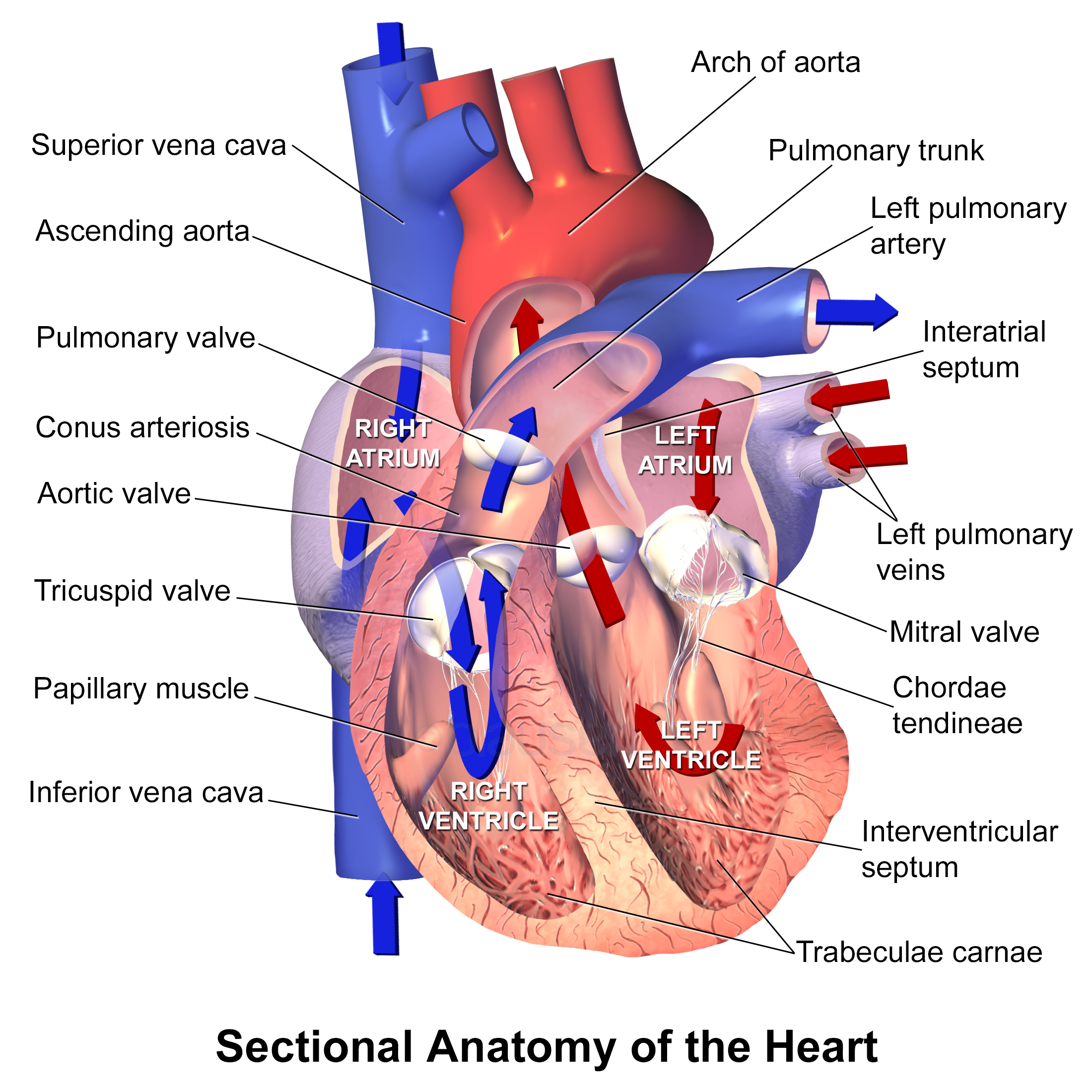
The sinoatrial node (also known as the sinuatrial node, SA node or sinus node) is an oval shaped region of special cardiac muscle in the upper back wall of the right atrium made up of cells known as pacemaker cells. The sinus node is approximately fifteen mm long, three mm wide, and one mm thick, located directly below and to the side of the superior vena cava. These cells can produce an electrical impulse an action potential known as a cardiac action potential that travels through the electrical conduction system of the heart, causing it to contract. In a healthy heart, the SA node continuously produces action potentials, setting the rhythm of the heart (sinus rhythm), and so is known as the heart's natural pacemaker. The rate of action potentials produced (and therefore the heart rate) is influenced by the nerves that supply it. Structure The sinoatrial node is a oval-shaped structure that is approximately fifteen mm long, three mm wide, and one mm thick, located directl ... [...More Info...] [...Related Items...] OR: [Wikipedia] [Google] [Baidu] |


