|
Degmacyte
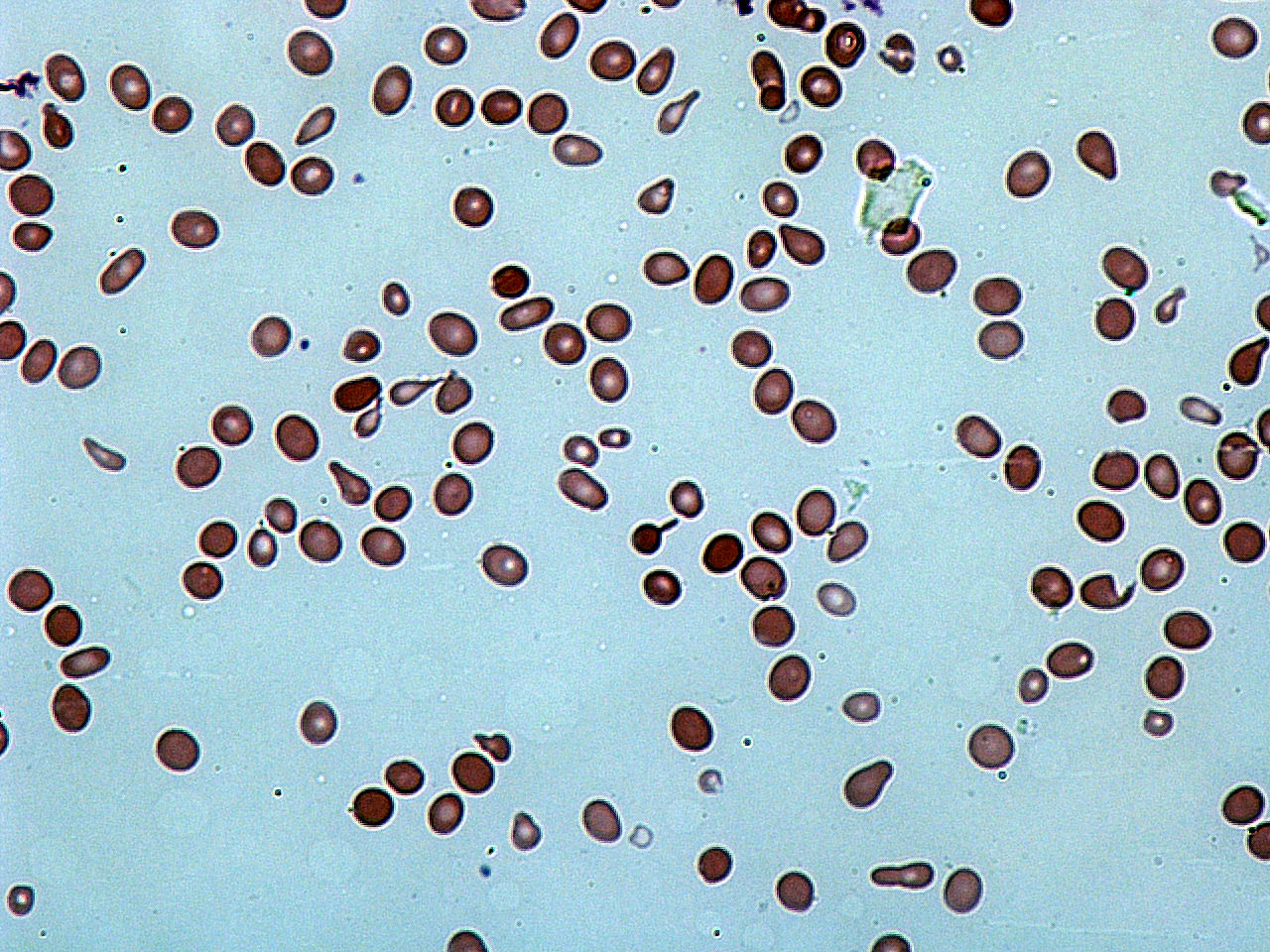
A degmacyte or bite cell is an abnormally shaped mature red blood cell with one or more semicircular portions removed from the cell margin, known as "bites". These "bites" result from the mechanical removal of denatured hemoglobin during splenic filtration as red cells attempt to migrate through endothelial slits from splenic cords into the splenic sinuses. Bite cells are known to be a result from processes of oxidative hemolysis, such as Glucose-6-phosphate dehydrogenase deficiency, in which uncontrolled oxidative stress causes hemoglobin to denature and form Heinz bodies. Bite cells can contain more than one "bite." The "bites" in degmacytes are smaller than the missing red blood cell fragments seen in schistocytes. Degmacytes usually appear smaller, denser, and more contracted than a normal red blood cell due to the bites. The appearance of the "bites" in red blood cells may vary in number, smoothness, and size. These cells can also exhibit other peripheral effects. Blist ... [...More Info...] [...Related Items...] OR: [Wikipedia] [Google] [Baidu] |
Poikilocytosis
Poikilocytosis is variation in the shapes of red blood cells. Poikilocytes may be oval, teardrop-shaped, sickle-shaped or irregularly contracted. Normal red blood cells are round, flattened disks that are thinner in the middle than at the edges. A ''poikilocyte'' is an abnormally-shaped red blood cell. Generally, poikilocytosis can refer to an increase in abnormal red blood cells of any shape, where they make up 10% or more of the total population of red blood cells. Types Membrane abnormalities # Acanthocytes or Spur/Spike cells # Codocytes or Target cells # Echinocytes and Burr cells # Elliptocytes and Ovalocytes # Spherocytes # Stomatocytes or Mouth cells # Drepanocytes or Sickle Cells # Degmacytes or "bite cells" Trauma # Dacrocytes or Teardrop Cells # Keratocytes # Microspherocytes and Pyropoikilocytes # Schistocytes # Semilunar bodies Diagnosis Poikilocytosis may be diagnosed with a test called a blood smear. During a blood smear, a medical technologist spreads ... [...More Info...] [...Related Items...] OR: [Wikipedia] [Google] [Baidu] |
BITE CELLS
A degmacyte or bite cell is an abnormally shaped mature red blood cell with one or more semicircular portions removed from the cell margin, known as "bites". These "bites" result from the mechanical removal of denatured hemoglobin during splenic filtration as red cells attempt to migrate through endothelial slits from splenic cords into the splenic sinuses. Bite cells are known to be a result from processes of oxidative hemolysis, such as Glucose-6-phosphate dehydrogenase deficiency, in which uncontrolled oxidative stress causes hemoglobin to denature and form Heinz bodies. Bite cells can contain more than one "bite." The "bites" in degmacytes are smaller than the missing red blood cell fragments seen in schistocytes. Degmacytes usually appear smaller, denser, and more contracted than a normal red blood cell due to the bites. The appearance of the "bites" in red blood cells may vary in number, smoothness, and size. These cells can also exhibit other peripheral effects. Blis ... [...More Info...] [...Related Items...] OR: [Wikipedia] [Google] [Baidu] |
Blister Cell
A degmacyte or bite cell is an abnormally shaped mature red blood cell with one or more semicircular portions removed from the cell margin, known as "bites". These "bites" result from the mechanical removal of denatured hemoglobin during splenic filtration as red cells attempt to migrate through endothelial slits from splenic cords into the splenic sinuses. Bite cells are known to be a result from processes of oxidative hemolysis, such as Glucose-6-phosphate dehydrogenase deficiency, in which uncontrolled oxidative stress causes hemoglobin to denature and form Heinz bodies. Bite cells can contain more than one "bite." The "bites" in degmacytes are smaller than the missing red blood cell fragments seen in schistocytes. Degmacytes usually appear smaller, denser, and more contracted than a normal red blood cell due to the bites. The appearance of the "bites" in red blood cells may vary in number, smoothness, and size. These cells can also exhibit other peripheral effects. Blist ... [...More Info...] [...Related Items...] OR: [Wikipedia] [Google] [Baidu] |
Pentose Phosphate Shunt
The pentose phosphate pathway (also called the phosphogluconate pathway and the hexose monophosphate shunt and the HMP Shunt) is a metabolic pathway parallel to glycolysis. It generates NADPH and pentoses (5-carbon sugars) as well as ribose 5-phosphate, a precursor for the synthesis of nucleotides. While the pentose phosphate pathway does involve oxidation of glucose, its primary role is anabolic rather than catabolic. The pathway is especially important in red blood cells (erythrocytes). There are two distinct phases in the pathway. The first is the oxidative phase, in which NADPH is generated, and the second is the non-oxidative synthesis of 5-carbon sugars. For most organisms, the pentose phosphate pathway takes place in the cytosol; in plants, most steps take place in plastids. Like glycolysis, the pentose phosphate pathway appears to have a very ancient evolutionary origin. The reactions of this pathway are mostly enzyme-catalyzed in modern cells, however, they also o ... [...More Info...] [...Related Items...] OR: [Wikipedia] [Google] [Baidu] |
Blood Transfusions
Blood transfusion is the process of transferring blood products into a person's circulation intravenously. Transfusions are used for various medical conditions to replace lost components of the blood. Early transfusions used whole blood, but modern medical practice commonly uses only components of the blood, such as red blood cells, white blood cells, plasma, clotting factors and platelets. Red blood cells (RBC) contain hemoglobin, and supply the cells of the body with oxygen. White blood cells are not commonly used during transfusion, but they are part of the immune system, and also fight infections. Plasma is the "yellowish" liquid part of blood, which acts as a buffer, and contains proteins and important substances needed for the body's overall health. Platelets are involved in blood clotting, preventing the body from bleeding. Before these components were known, doctors believed that blood was homogeneous. Because of this scientific misunderstanding, many patients died becau ... [...More Info...] [...Related Items...] OR: [Wikipedia] [Google] [Baidu] |
Hemolytic Anemia
Hemolytic anemia or haemolytic anaemia is a form of anemia due to hemolysis, the abnormal breakdown of red blood cells (RBCs), either in the blood vessels (intravascular hemolysis) or elsewhere in the human body (extravascular). This most commonly occurs within the spleen, but also can occur in the reticuloendothelial system or mechanically (prosthetic valve damage). Hemolytic anemia accounts for 5% of all existing anemias. It has numerous possible consequences, ranging from general symptoms to life-threatening systemic effects. The general classification of hemolytic anemia is either intrinsic or extrinsic. Treatment depends on the type and cause of the hemolytic anemia. Symptoms of hemolytic anemia are similar to other forms of anemia (fatigue and shortness of breath), but in addition, the breakdown of red cells leads to jaundice and increases the risk of particular long-term complications, such as gallstones and pulmonary hypertension. Signs and symptoms Symptoms of hemolytic ... [...More Info...] [...Related Items...] OR: [Wikipedia] [Google] [Baidu] |
Hypoxia (medical)
Hypoxia is a condition in which the body or a region of the body is deprived of adequate oxygen supply at the tissue level. Hypoxia may be classified as either '' generalized'', affecting the whole body, or ''local'', affecting a region of the body. Although hypoxia is often a pathological condition, variations in arterial oxygen concentrations can be part of the normal physiology, for example, during strenuous physical exercise.. Hypoxia differs from hypoxemia and anoxemia, in that hypoxia refers to a state in which oxygen present in a tissue or the whole body is insufficient, whereas hypoxemia and anoxemia refer specifically to states that have low or no oxygen in the blood. Hypoxia in which there is complete absence of oxygen supply is referred to as anoxia. Hypoxia can be due to external causes, when the breathing gas is hypoxic, or internal causes, such as reduced effectiveness of gas transfer in the lungs, reduced capacity of the blood to carry oxygen, compromised gene ... [...More Info...] [...Related Items...] OR: [Wikipedia] [Google] [Baidu] |
G6PD Deficiency
Glucose-6-phosphate dehydrogenase deficiency (G6PDD), which is the most common enzyme deficiency worldwide, is an inborn error of metabolism that predisposes to red blood cell breakdown. Most of the time, those who are affected have no symptoms. Following a specific trigger, symptoms such as yellowish skin, dark urine, shortness of breath, and feeling tired may develop. Complications can include anemia and newborn jaundice. Some people never have symptoms. It is an X-linked recessive disorder that results in defective glucose-6-phosphate dehydrogenase enzyme. Glucose-6-phosphate dehydrogenase is an enzyme which protects red blood cells, which carry oxygen from the lungs to tissues throughout the body. A defect of the enzyme results in the premature breakdown of red blood cells. This destruction of red blood cells is called hemolysis. Red blood cell breakdown may be triggered by infections, certain medication, stress, or foods such as fava beans. Depending on the specific mutati ... [...More Info...] [...Related Items...] OR: [Wikipedia] [Google] [Baidu] |
Hemolysis
Hemolysis or haemolysis (), also known by several other names, is the rupturing ( lysis) of red blood cells (erythrocytes) and the release of their contents (cytoplasm) into surrounding fluid (e.g. blood plasma). Hemolysis may occur in vivo or in vitro. One cause of hemolysis is the action of hemolysins, toxins that are produced by certain pathogenic bacteria or fungi. Another cause is intense physical exercise. Hemolysins damage the red blood cell's cytoplasmic membrane, causing lysis and eventually cell death. Etymology From hemo- + -lysis, from , "blood") + , "loosening"). Inside the body Hemolysis inside the body can be caused by a large number of medical conditions, including some parasites (''e.g.'', '' Plasmodium''), some autoimmune disorders (''e.g.'', autoimmune haemolytic anaemia, drug-induced hemolytic anemia, atypical hemolytic uremic syndrome (aHUS)), some genetic disorders (''e.g.'', Sickle-cell disease or G6PD deficiency), or blood with too low a solute ... [...More Info...] [...Related Items...] OR: [Wikipedia] [Google] [Baidu] |
Schistocyte
A schistocyte or schizocyte (from Greek for "divided" and for "hollow" or "cell") is a fragmented part of a red blood cell. Schistocytes are typically irregularly shaped, jagged, and have two pointed ends. Several microangiopathic diseases, including disseminated intravascular coagulation and thrombotic microangiopathies, generate fibrin strands that sever red blood cells as they try to move past a thrombus, creating schistocytes. Schistocytes are often seen in patients with hemolytic anemia. They are frequently a consequence of mechanical artificial heart valves, hemolytic uremic syndrome, and thrombotic thrombocytopenic purpura, among other causes. Excessive schistocytes present in blood can be a sign of microangiopathic hemolytic anemia (MAHA). Appearance Schistocytes are fragmented red blood cells that can take on different shapes. They can be found as triangular, helmet shaped, or comma shaped with pointed edges. Schistocytes are most often found to be microcytic w ... [...More Info...] [...Related Items...] OR: [Wikipedia] [Google] [Baidu] |
Blood Smear
A blood smear, peripheral blood smear or blood film is a thin layer of blood smeared on a glass microscope slide and then stained in such a way as to allow the various blood cells to be examined microscopically. Blood smears are examined in the investigation of hematological (blood) disorders and are routinely employed to look for blood parasites, such as those of malaria and filariasis. Preparation A blood smear is made by placing a drop of blood on one end of a slide, and using a ''spreader slide'' to disperse the blood over the slide's length. The aim is to get a region, called a monolayer, where the cells are spaced far enough apart to be counted and differentiated. The monolayer is found in the "feathered edge" created by the spreader slide as it draws the blood forward. The slide is left to air dry, after which the blood is fixed to the slide by immersing it briefly in methanol. The fixative is essential for good staining and presentation of cellular detail. After fix ... [...More Info...] [...Related Items...] OR: [Wikipedia] [Google] [Baidu] |
Vacuole
A vacuole () is a membrane-bound organelle which is present in plant and fungal cells and some protist, animal, and bacterial cells. Vacuoles are essentially enclosed compartments which are filled with water containing inorganic and organic molecules including enzymes in solution, though in certain cases they may contain solids which have been engulfed. Vacuoles are formed by the fusion of multiple membrane vesicles and are effectively just larger forms of these. The organelle has no basic shape or size; its structure varies according to the requirements of the cell. Discovery Contractile vacuoles ("stars") were first observed by Spallanzani (1776) in protozoa, although mistaken for respiratory organs. Dujardin (1841) named these "stars" as ''vacuoles''. In 1842, Schleiden applied the term for plant cells, to distinguish the structure with cell sap from the rest of the protoplasm. In 1885, de Vries named the vacuole membrane as tonoplast. Function The function and sig ... [...More Info...] [...Related Items...] OR: [Wikipedia] [Google] [Baidu] |


