|
DSG1
Desmoglein-1 is a protein that in humans is encoded by the ''DSG1'' gene. Desmoglein-1 is expressed everywhere in the skin epidermis, but mainly it is expressed in the superficial upper layers of the skin epidermis. Function Desmosomes are cell-cell junctions between epithelial, myocardial and certain other cell types. Desmoglein-1 is a calcium-binding transmembrane glycoprotein component of desmosomes in vertebrate epithelial cells. Currently, four desmoglein subfamily members have been identified and all are members of the cadherin cell adhesion molecule superfamily. These desmoglein gene family members are located in a cluster on chromosome 18. The protein encoded by this gene has been identified as the autoantigen of the autoimmune skin blistering disease pemphigus foliaceus. It has been found that desmoglein-1 is the target antigen in majority of the cases linked to IgG/IgA pemphigus, which is an autoimmune IgG/ IgA antibody mediated response. Desmoglein-1 is also a ta ... [...More Info...] [...Related Items...] OR: [Wikipedia] [Google] [Baidu] |
Desmoglein
The desmogleins are a family of desmosomal cadherins consisting of proteins DSG1, DSG2, DSG3, and DSG4. They play a role in the formation of desmosomes that join cells to one another. Pathology Desmogleins are targeted in the autoimmune disease pemphigus. Desmoglein proteins are a type of cadherin, which is a transmembrane protein A transmembrane protein (TP) is a type of integral membrane protein that spans the entirety of the cell membrane. Many transmembrane proteins function as gateways to permit the transport of specific substances across the membrane. They frequent ... that binds with other cadherins to form junctions known as desmosomes between cells. These desmoglein proteins thus hold cells together, but, when the body starts producing antibodies against desmoglein, these junctions break down, and this results in subsequent blister or vesicle formation.Bolognia JL, Jorizzo JL, Schaffer JV, editors. Dermatology. 3rd ed. Philadelphia: Elsevier Saunders; 2012 Re ... [...More Info...] [...Related Items...] OR: [Wikipedia] [Google] [Baidu] |
PKP2
Plakophilin-2 is a protein that in humans is encoded by the ''PKP2'' gene. Plakophilin 2 is expressed in skin and cardiac muscle, where it functions to link cadherins to intermediate filaments in the cytoskeleton. In cardiac muscle, plakophilin-2 is found in desmosome structures located within intercalated discs. Mutations in ''PKP2'' have been shown to be causal in arrhythmogenic right ventricular cardiomyopathy. Structure Two splice variants of the ''PKP2'' gene have been identified. The first has a molecular weight of 97.4 kDa (881 amino acids) and the second of molecular weight of 92.7 kDa (837 amino acids). A processed pseudogene with high similarity to this locus has been mapped to chromosome 12p13. Plakophilin-2 is a member of the armadillo repeat and plakophilin protein family. Plakophilin proteins contain nine central, conserved armadillo repeat domains flanked by N-terminal and C-terminal domains. Alternately spliced transcripts encoding protein isoforms have been ident ... [...More Info...] [...Related Items...] OR: [Wikipedia] [Google] [Baidu] |
Striate Palmoplantar Keratoderma
Palmoplantar keratodermas are a heterogeneous group of disorders characterized by abnormal thickening of the stratum corneum of the palms and soles. Autosomal recessive, dominant, X-linked, and acquired forms have all been described. Types Clinically, three distinct patterns of palmoplantar keratoderma may be identified: diffuse, focal, and punctate. Diffuse Diffuse palmoplantar keratoderma is a type of palmoplantar keratoderma that is characterized by an even, thick, symmetric hyperkeratosis over the whole of the palm and sole, usually evident at birth or in the first few months of life. Restated, diffuse palmoplantar keratoderma is an autosomal dominant disorder in which hyperkeratosis is confined to the palms and soles. The two major types can have a similar clinical appearance: *''Diffuse epidermolytic palmoplantar keratoderma'' (also known as "Palmoplantar keratoderma cum degeneratione granulosa Vörner," "Vörner's epidermolytic palmoplantar keratoderma", and "Vörn ... [...More Info...] [...Related Items...] OR: [Wikipedia] [Google] [Baidu] |
Desmosomes
A desmosome (; "binding body"), also known as a macula adherens (plural: maculae adherentes) (Latin for ''adhering spot''), is a cell structure specialized for cell-to-cell adhesion. A type of junctional complex, they are localized spot-like adhesions randomly arranged on the lateral sides of plasma membranes. Desmosomes are one of the stronger cell-to-cell adhesion types and are found in tissue that experience intense mechanical stress, such as cardiac muscle tissue, bladder tissue, gastrointestinal mucosa, and epithelia. Structure Desmosomes are composed of desmosome-intermediate filament complexes (DIFC), which is a network of cadherin proteins, linker proteins and intermediate filaments. The DIFCs can be broken into three regions: the extracellular core region, or desmoglea, the outer dense plaque, or ODP, and the inner dense plaque, or IDP. The extracellular core region, approximately 34 nm in length, contains desmoglein and desmocollin, which are in the cadherin famil ... [...More Info...] [...Related Items...] OR: [Wikipedia] [Google] [Baidu] |
Cadherin
Cadherins (named for "calcium-dependent adhesion") are a type of cell adhesion molecule (CAM) that is important in the formation of adherens junctions to allow cells to adhere to each other . Cadherins are a class of type-1 transmembrane proteins, and they are dependent on calcium (Ca2+) ions to function, hence their name. Cell-cell adhesion is mediated by extracellular cadherin domains, whereas the intracellular cytoplasmic tail associates with numerous adaptors and signaling proteins, collectively referred to as the cadherin adhesome. The cadherin family is essential in maintaining the cell-cell contact and regulating cytoskeletal complexes. The cadherin superfamily includes cadherins, protocadherins, desmogleins, desmocollins, and more. In structure, they share ''cadherin repeats'', which are the extracellular Ca2+-binding domains. There are multiple classes of cadherin molecules, each designated with a prefix (in general, noting the types of tissue with which it is associated). ... [...More Info...] [...Related Items...] OR: [Wikipedia] [Google] [Baidu] |
Exotoxin
An exotoxin is a toxin secreted by bacteria. An exotoxin can cause damage to the host by destroying cells or disrupting normal cellular metabolism. They are highly potent and can cause major damage to the host. Exotoxins may be secreted, or, similar to endotoxins, may be released during lysis of the cell. Gram negative pathogens may secrete outer membrane vesicles containing lipopolysaccharide endotoxin and some virulence proteins in the bounding membrane along with some other toxins as intra-vesicular contents, thus adding a previously unforeseen dimension to the well-known eukaryote process of membrane vesicle trafficking, which is quite active at the host–pathogen interface. They may exert their effect locally or produce systemic effects. Well-known exotoxins include: botulinum toxin produced by ''Clostridium botulinum''; '' Corynebacterium diphtheriae'' toxin, produced during life-threatening symptoms of diphtheria; tetanospasmin produced by ''Clostridium tetani''. The toxi ... [...More Info...] [...Related Items...] OR: [Wikipedia] [Google] [Baidu] |
PTPRT
Receptor-type tyrosine-protein phosphatase T is an enzyme that in humans is encoded by the ''PTPRT'' gene. PTPRT is also known as PTPrho, PTPρ and human accelerated region 9. The human accelerated regions are 49 regions of the human genome that are conserved among vertebrates, but in humans show significant distinction from other vertebrates. This region may, therefore, have played a key role in differentiating humans from apes. Function The protein encoded by this gene is a member of the protein tyrosine phosphatase (PTP) family. PTPs are known to be signaling molecules that regulate a variety of cellular processes including cell growth, differentiation, mitotic cycle, and oncogenic transformation. PTPrho has been proposed to function during development of the nervous system and as a tumor suppressor in cancer. Structure This PTP possesses an extracellular region, a single transmembrane region, and two tandem intracellular catalytic domains, and thus represents a recept ... [...More Info...] [...Related Items...] OR: [Wikipedia] [Google] [Baidu] |
Autosomal Dominant
In genetics, dominance is the phenomenon of one variant (allele) of a gene on a chromosome masking or overriding the effect of a different variant of the same gene on the other copy of the chromosome. The first variant is termed dominant and the second recessive. This state of having two different variants of the same gene on each chromosome is originally caused by a mutation in one of the genes, either new (''de novo'') or inherited. The terms autosomal dominant or autosomal recessive are used to describe gene variants on non-sex chromosomes ( autosomes) and their associated traits, while those on sex chromosomes (allosomes) are termed X-linked dominant, X-linked recessive or Y-linked; these have an inheritance and presentation pattern that depends on the sex of both the parent and the child (see Sex linkage). Since there is only one copy of the Y chromosome, Y-linked traits cannot be dominant or recessive. Additionally, there are other forms of dominance such as incomplete d ... [...More Info...] [...Related Items...] OR: [Wikipedia] [Google] [Baidu] |
Haploinsufficient
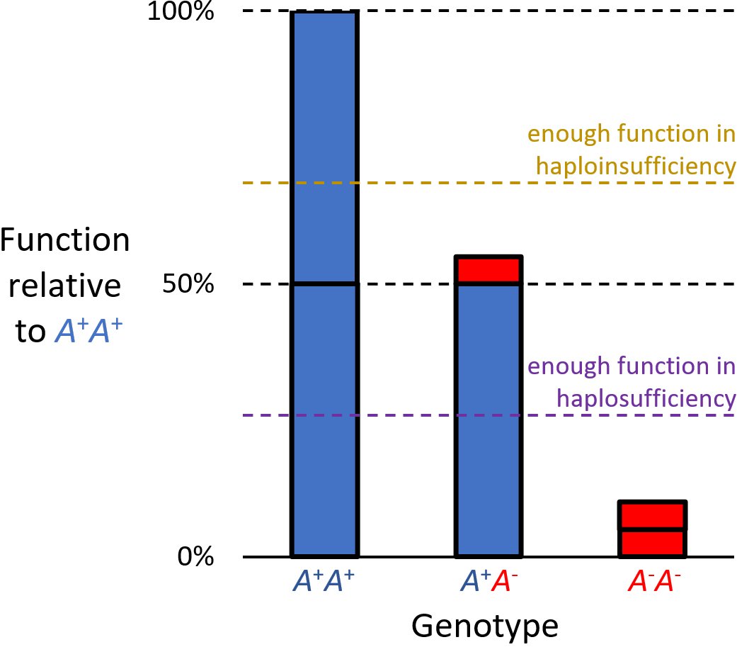
Haploinsufficiency in genetics describes a model of dominant gene action in diploid organisms, in which a single copy of the wild-type allele at a locus in heterozygous combination with a variant allele is insufficient to produce the wild-type phenotype. Haploinsufficiency may arise from a ''de novo'' or inherited loss-of-function mutation in the variant allele, such that it yields little or no gene product (often a protein). Although the other, standard allele still produces the standard amount of product, the total product is insufficient to produce the standard phenotype. This heterozygous genotype may result in a non- or sub-standard, deleterious, and (or) disease phenotype. Haploinsufficiency is the standard explanation for dominant deleterious alleles. In the alternative case of haplosufficiency, the loss-of-function allele behaves as above, but the single standard allele in the heterozygous genotype produces sufficient gene product to produce the same, standard phenotype ... [...More Info...] [...Related Items...] OR: [Wikipedia] [Google] [Baidu] |
Cytokine
Cytokines are a broad and loose category of small proteins (~5–25 kDa) important in cell signaling. Cytokines are peptides and cannot cross the lipid bilayer of cells to enter the cytoplasm. Cytokines have been shown to be involved in autocrine, paracrine and endocrine signaling as immunomodulating agents. Cytokines include chemokines, interferons, interleukins, lymphokines, and tumour necrosis factors, but generally not hormones or growth factors (despite some overlap in the terminology). Cytokines are produced by a broad range of cells, including immune cells like macrophages, B lymphocytes, T lymphocytes and mast cells, as well as endothelial cells, fibroblasts, and various stromal cells; a given cytokine may be produced by more than one type of cell. They act through cell surface receptors and are especially important in the immune system; cytokines modulate the balance between humoral and cell-based immune responses, and they regulate the maturation, growth, and res ... [...More Info...] [...Related Items...] OR: [Wikipedia] [Google] [Baidu] |


