|
Dorsal Cochlear Nucleus
The dorsal cochlear nucleus (DCN, also known as the "tuberculum acusticum"), is a cortex-like structure on the dorso-lateral surface of the brainstem. Along with the ventral cochlear nucleus (VCN), it forms the cochlear nucleus (CN), where all auditory nerve fibers from the cochlea form their first synapses. Anatomy The DCN differs from the ventral portion of the CN as it not only projects to the central nucleus (a subdivision) of the inferior colliculus (CIC), but also receives efferent innervation from the auditory cortex, superior olivary complex and the inferior colliculus. The cytoarchitecture and neurochemistry of the DCN is similar to that of the cerebellum, an important concept in theories of DCN function. Thus, the DCN is thought to be involved with more complex auditory processing, rather than merely transferring information. The pyramidal cells or giant cells are a major cell grouping of the DCN. These cells are the target of two different input systems. The first ... [...More Info...] [...Related Items...] OR: [Wikipedia] [Google] [Baidu] |
Ventral Cochlear Nucleus
In the ventral cochlear nucleus (VCN), auditory nerve fibers enter the brain via the nerve root in the VCN. The ventral cochlear nucleus is divided into the anterior ventral (anteroventral) cochlear nucleus (AVCN) and the posterior ventral (posteroventral) cochlear nucleus (PVCN). In the VCN, auditory nerve fibers bifurcate, the ascending branch innervates the AVCN and the descending branch innervates the PVCN and then continue to the dorsal cochlear nucleus. The orderly innervation by auditory nerve fibers gives the AVCN a tonotopic organization along the dorsoventral axis. Fibers that carry information from the apex of the cochlea that are tuned to low frequencies contact neurons in the ventral part of the AVCN; those that carry information from the base of the cochlea that are tuned to high frequencies contact neurons in the dorsal part of the AVCN. Several populations of neurons populate the AVCN. Bushy cells receive input from auditory nerve fibers through particularly larg ... [...More Info...] [...Related Items...] OR: [Wikipedia] [Google] [Baidu] |
Cochlear Nucleus
The cochlear nuclear (CN) complex comprises two cranial nerve nuclei in the human brainstem, the ventral cochlear nucleus (VCN) and the dorsal cochlear nucleus (DCN). The ventral cochlear nucleus is unlayered whereas the dorsal cochlear nucleus is layered. Auditory nerve fibers, fibers that travel through the auditory nerve (also known as the cochlear nerve or eighth cranial nerve) carry information from the inner ear, the cochlea, on the same side of the head, to the nerve root in the ventral cochlear nucleus. At the nerve root the fibers branch to innervate the ventral cochlear nucleus and the deep layer of the dorsal cochlear nucleus. All acoustic information thus enters the brain through the cochlear nuclei, where the processing of acoustic information begins. The outputs from the cochlear nuclei are received in higher regions of the auditory brainstem. Structure The cochlear nuclei (CN) are located at the dorso-lateral side of the brainstem, spanning the junction of ... [...More Info...] [...Related Items...] OR: [Wikipedia] [Google] [Baidu] |
Inferior Colliculus
The inferior colliculus (IC) (Latin for ''lower hill'') is the principal midbrain nucleus of the auditory pathway and receives input from several peripheral brainstem nuclei in the auditory pathway, as well as inputs from the auditory cortex. The inferior colliculus has three subdivisions: the central nucleus, a dorsal cortex by which it is surrounded, and an external cortex which is located laterally. Its bimodal neurons are implicated in auditory- somatosensory interaction, receiving projections from somatosensory nuclei. This multisensory integration may underlie a filtering of self-effected sounds from vocalization, chewing, or respiration activities. The inferior colliculi together with the superior colliculi form the eminences of the corpora quadrigemina, and also part of the tectal region of the midbrain. The inferior colliculus lies caudal to its counterpart – the superior colliculus – above the trochlear nerve, and at the base of the projection of the medial ge ... [...More Info...] [...Related Items...] OR: [Wikipedia] [Google] [Baidu] |
Auditory Cortex
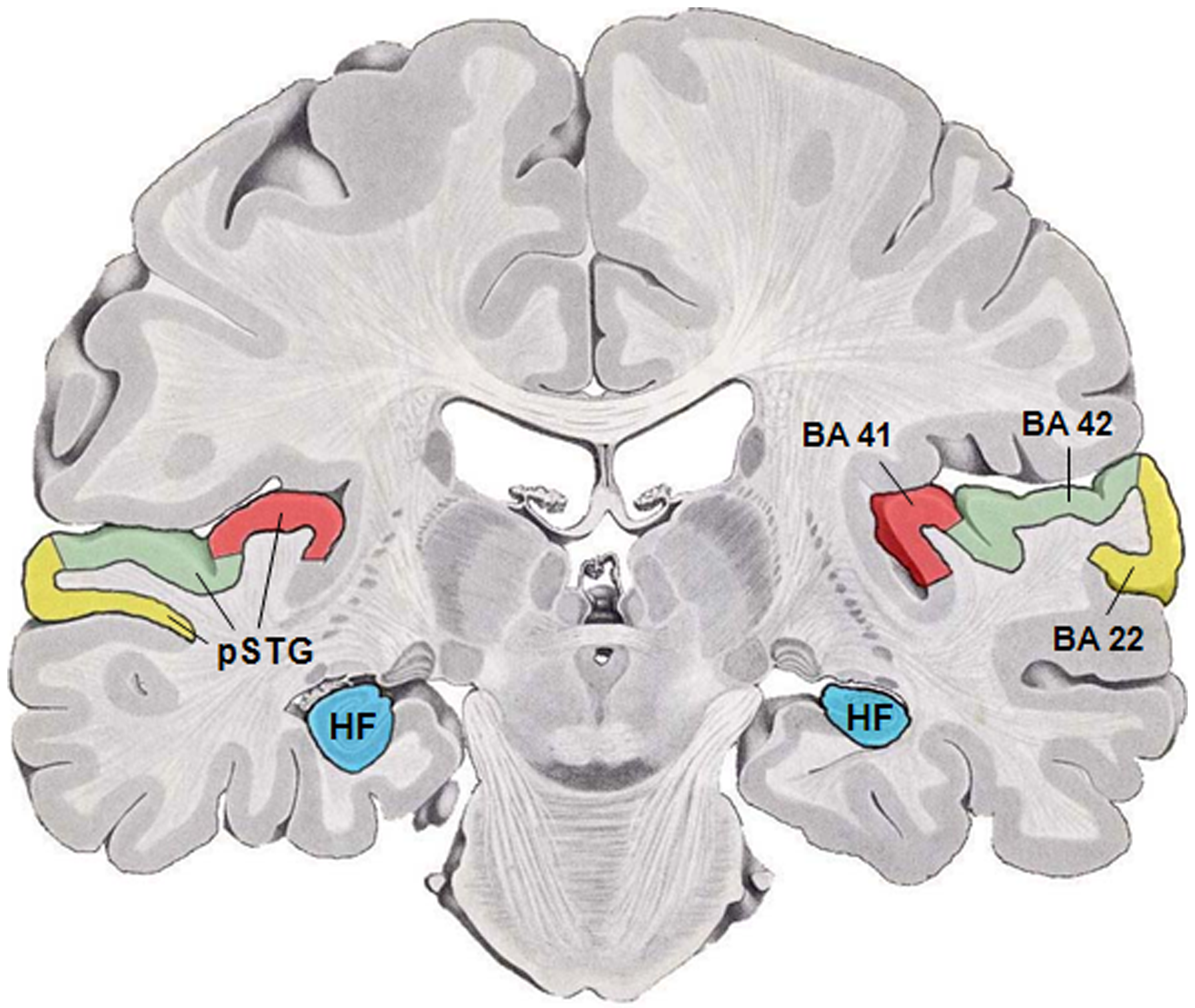
The auditory cortex is the part of the temporal lobe that processes auditory information in humans and many other vertebrates. It is a part of the auditory system, performing basic and higher functions in hearing, such as possible relations to language switching.Cf. Pickles, James O. (2012). ''An Introduction to the Physiology of Hearing'' (4th ed.). Bingley, UK: Emerald Group Publishing Limited, p. 238. It is located bilaterally, roughly at the upper sides of the temporal lobes – in humans, curving down and onto the medial surface, on the superior temporal plane, within the lateral sulcus and comprising parts of the transverse temporal gyri, and the superior temporal gyrus, including the planum polare and planum temporale (roughly Brodmann areas 41 and 42, and partially 22). The auditory cortex takes part in the spectrotemporal, meaning involving time and frequency, analysis of the inputs passed on from the ear. The cortex then filters and passes on the information to ... [...More Info...] [...Related Items...] OR: [Wikipedia] [Google] [Baidu] |
Superior Olivary Complex
The superior olivary complex (SOC) or superior olive is a collection of brainstem nuclei that functions in multiple aspects of hearing and is an important component of the ascending and descending auditory pathways of the auditory system. The SOC is intimately related to the trapezoid body: most of the cell groups of the SOC are dorsal (posterior in primates) to this axon bundle while a number of cell groups are embedded in the trapezoid body. Overall, the SOC displays a significant interspecies variation, being largest in bats and rodents and smaller in primates. Physiology The superior olivary nucleus plays a number of roles in hearing. The medial superior olive (MSO) is a specialized nucleus that is believed to measure the time difference of arrival of sounds between the ears (the interaural time difference or ITD). The ITD is a major cue for determining the azimuth of sounds, i.e., localising them on the azimuthal plane – their degree to the left or the right. The later ... [...More Info...] [...Related Items...] OR: [Wikipedia] [Google] [Baidu] |
Cytoarchitecture
Cytoarchitecture ( Greek '' κύτος''= "cell" + '' ἀρχιτεκτονική''= "architecture"), also known as cytoarchitectonics, is the study of the cellular composition of the central nervous system's tissues under the microscope. Cytoarchitectonics is one of the ways to parse the brain, by obtaining sections of the brain using a microtome and staining them with chemical agents which reveal where different neurons are located. The study of the parcellation of ''nerve fibers'' (primarily axons) into layers forms the subject of myeloarchitectonics ( History of the cerebral cytoarchitecture Defining cerebral cytoarchitecture began with the advent of histology—the science of slicing and staining brain slices fo ...[...More Info...] [...Related Items...] OR: [Wikipedia] [Google] [Baidu] |
Neurochemistry
Neurochemistry is the study of chemicals, including neurotransmitters and other molecules such as psychopharmaceuticals and neuropeptides, that control and influence the physiology of the nervous system. This particular field within neuroscience examines how neurochemicals influence the operation of neurons, synapses, and neural networks. Neurochemists analyze the biochemistry and molecular biology of organic compounds in the nervous system, and their roles in such neural processes including cortical plasticity, neurogenesis, and neural differentiation. History While neurochemistry as a recognized science is relatively new, the idea behind neurochemistry has been around since the 18th century. Originally, the brain had been thought to be a separate entity apart from the peripheral nervous system. Beginning in 1856, there was a string of research that refuted that idea. The chemical makeup of the brain was nearly identical to the makeup of the peripheral nervous system. The first ... [...More Info...] [...Related Items...] OR: [Wikipedia] [Google] [Baidu] |
Cerebellum
The cerebellum (Latin for "little brain") is a major feature of the hindbrain of all vertebrates. Although usually smaller than the cerebrum, in some animals such as the mormyrid fishes it may be as large as or even larger. In humans, the cerebellum plays an important role in motor control. It may also be involved in some cognitive functions such as attention and language as well as emotional control such as regulating fear and pleasure responses, but its movement-related functions are the most solidly established. The human cerebellum does not initiate movement, but contributes to coordination, precision, and accurate timing: it receives input from sensory systems of the spinal cord and from other parts of the brain, and integrates these inputs to fine-tune motor activity. Cerebellar damage produces disorders in fine movement, equilibrium, posture, and motor learning in humans. Anatomically, the human cerebellum has the appearance of a separate structure attached to th ... [...More Info...] [...Related Items...] OR: [Wikipedia] [Google] [Baidu] |
Pyramidal Cells
Pyramidal cells, or pyramidal neurons, are a type of multipolar neuron found in areas of the brain including the cerebral cortex, the hippocampus, and the amygdala. Pyramidal neurons are the primary excitation units of the mammalian prefrontal cortex and the corticospinal tract. Pyramidal neurons are also one of two cell types where the characteristic sign, Negri bodies, are found in post-mortem rabies infection. Pyramidal neurons were first discovered and studied by Santiago Ramón y Cajal. Since then, studies on pyramidal neurons have focused on topics ranging from neuroplasticity to cognition. Structure File:GFPneuron.png, Pyramidal neuron visualized by green fluorescent protein (gfp) File:Hippocampal-pyramidal-cell.png, A hippocampal pyramidal cell One of the main structural features of the pyramidal neuron is the conic shaped soma, or cell body, after which the neuron is named. Other key structural features of the pyramidal cell are a single axon, a large apical dend ... [...More Info...] [...Related Items...] OR: [Wikipedia] [Google] [Baidu] |
Cartwheel Cell
Cartwheel cells are neurons of the dorsal cochlear nucleus (DCN) where they greatly outnumber the other inhibitory interneurons of the DCN. Their somas lie on the superficial side of the pyramidal layer of the DCN, and their dendrites receive input from the parallel fibres of the granule cell layer. Their axons do not extend beyond the dorsal cochlear nucleus but synapse In the nervous system, a synapse is a structure that permits a neuron (or nerve cell) to pass an electrical or chemical signal to another neuron or to the target effector cell. Synapses are essential to the transmission of nervous impulses from ... with other cartwheel cells and pyramidal cells within the DCN releasing GABA and glycine onto their targets. Cartwheel cells have similar spiking patterns to Purkinje cells, firing complex spike bursts as well as simple spikes. They are also seen to share other features common to the cerebellar Purkinje cells. Other data supports the structural and functio ... [...More Info...] [...Related Items...] OR: [Wikipedia] [Google] [Baidu] |
Lateral Superior Olive
The superior olivary complex (SOC) or superior olive is a collection of brainstem nuclei that functions in multiple aspects of hearing and is an important component of the ascending and descending auditory pathways of the auditory system. The SOC is intimately related to the trapezoid body: most of the cell groups of the SOC are dorsal (posterior in primates) to this axon bundle while a number of cell groups are embedded in the trapezoid body. Overall, the SOC displays a significant interspecies variation, being largest in bats and rodents and smaller in primates. Physiology The superior olivary nucleus plays a number of roles in hearing. The medial superior olive (MSO) is a specialized nucleus that is believed to measure the time difference of arrival of sounds between the ears (the interaural time difference or ITD). The ITD is a major cue for determining the azimuth of sounds, i.e., localising them on the azimuthal plane – their degree to the left or the right. The latera ... [...More Info...] [...Related Items...] OR: [Wikipedia] [Google] [Baidu] |
Action Potential
An action potential occurs when the membrane potential of a specific cell location rapidly rises and falls. This depolarization then causes adjacent locations to similarly depolarize. Action potentials occur in several types of animal cells, called excitable cells, which include neurons, muscle cells, and in some plant cells. Certain endocrine cells such as pancreatic beta cells, and certain cells of the anterior pituitary gland are also excitable cells. In neurons, action potentials play a central role in cell-cell communication by providing for—or with regard to saltatory conduction, assisting—the propagation of signals along the neuron's axon toward synaptic boutons situated at the ends of an axon; these signals can then connect with other neurons at synapses, or to motor cells or glands. In other types of cells, their main function is to activate intracellular processes. In muscle cells, for example, an action potential is the first step in the chain of event ... [...More Info...] [...Related Items...] OR: [Wikipedia] [Google] [Baidu] |


