|
Diatomys
''Diatomys'' is an extinct rodent genus known from Miocene deposits in China, Japan, Pakistan, and Thailand. The fossil range is from the late Early Miocene to the Middle Miocene (22.5–11 Ma). Specimens Specifically the strata and regions from which ''Diatomys'' has been collected are: Shanwang series in Shandong province, China, Jiangsu province in China, Kyūshū in Japan, the Siwaliks in northern Pakistan, and Li Basin in Lamphun Province, Thailand. Li (1974) described ''Diatomys shantungensis'' on the basis of two moderately complete specimens from Shandong. This material had good preservation of dental characters, but much of the skull was difficult to interpret due to flattening. Dawson ''et al.'' (2006) reported the finding of another ''D. shantungensis'' fossil from Shandong that showed much improved preservation of cranial and skeletal characters. Impressions of hair and whiskers were observable in the specimen. Mein and Ginsburg (1985) described ''Diatomys liens ... [...More Info...] [...Related Items...] OR: [Wikipedia] [Google] [Baidu] |
Miocene
The Miocene ( ) is the first geological epoch of the Neogene Period and extends from about (Ma). The Miocene was named by Scottish geologist Charles Lyell; the name comes from the Greek words (', "less") and (', "new") and means "less recent" because it has 18% fewer modern marine invertebrates than the Pliocene has. The Miocene is preceded by the Oligocene and is followed by the Pliocene. As Earth went from the Oligocene through the Miocene and into the Pliocene, the climate slowly cooled towards a series of ice ages. The Miocene boundaries are not marked by a single distinct global event but consist rather of regionally defined boundaries between the warmer Oligocene and the cooler Pliocene Epoch. During the Early Miocene, the Arabian Peninsula collided with Eurasia, severing the connection between the Mediterranean and Indian Ocean, and allowing a faunal interchange to occur between Eurasia and Africa, including the dispersal of proboscideans into Eurasia. During the ... [...More Info...] [...Related Items...] OR: [Wikipedia] [Google] [Baidu] |
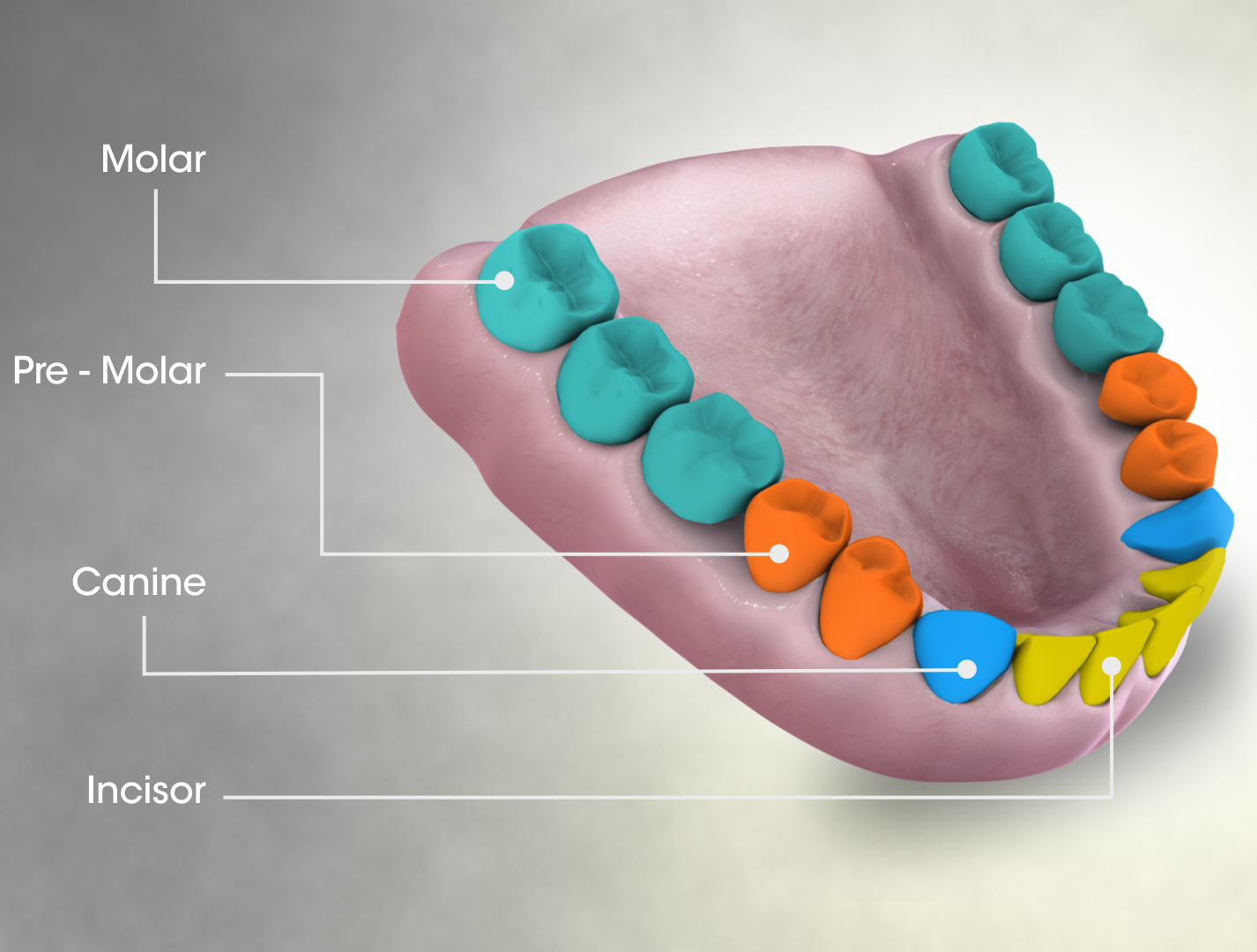
Premolar
The premolars, also called premolar teeth, or bicuspids, are transitional teeth located between the canine and molar teeth. In humans, there are two premolars per quadrant in the permanent set of teeth, making eight premolars total in the mouth. They have at least two cusps. Premolars can be considered transitional teeth during chewing, or mastication. They have properties of both the canines, that lie anterior and molars that lie posterior, and so food can be transferred from the canines to the premolars and finally to the molars for grinding, instead of directly from the canines to the molars. Human anatomy The premolars in humans are the maxillary first premolar, maxillary second premolar, mandibular first premolar, and the mandibular second premolar. Premolar teeth by definition are permanent teeth distal to the canines, preceded by deciduous molars. Morphology There is always one large buccal cusp, especially so in the mandibular first premolar. The lower second ... [...More Info...] [...Related Items...] OR: [Wikipedia] [Google] [Baidu] |
Fossorial
A fossorial () animal is one adapted to digging which lives primarily, but not solely, underground. Some examples are badgers, naked mole-rats, clams, meerkats, and mole salamanders, as well as many beetles, wasps, and bees. Prehistoric evidence The physical adaptation of fossoriality is widely accepted as being widespread among many prehistoric phyla and taxa, such as bacteria and early eukaryotes. Furthermore, fossoriality has evolved independently multiple times, even within a single family. Fossorial animals appeared simultaneously with the colonization of land by arthropods in the late Ordovician period (over 440 million years ago). Other notable early burrowers include ''Eocaecilia'' and possibly ''Dinilysia''. The oldest example of burrowing in synapsids, the lineage which includes modern mammals and their ancestors, is a cynodont, ''Thrinaxodon liorhinus'', found in the Karoo of South Africa, estimated to be 251 million years old. Evidence shows that this ... [...More Info...] [...Related Items...] OR: [Wikipedia] [Google] [Baidu] |
Coronoid Process Of The Mandible
In human anatomy, the mandible's coronoid process (from Greek ''korōnē'', denoting something hooked) is a thin, triangular eminence, which is flattened from side to side and varies in shape and size. Its anterior border is convex and is continuous below with the anterior border of the ramus. Its ''posterior border'' is concave and forms the anterior boundary of the mandibular notch. The ''lateral surface'' is smooth, and affords insertion to the temporalis and masseter muscles. Its ''medial surface'' gives insertion to the temporalis, and presents a ridge which begins near the apex of the process and runs downward and forward to the inner side of the last molar tooth. Between this ridge and the anterior border is a grooved triangular area, the upper part of which gives attachment to the temporalis, the lower part to some fibers of the buccinator. Clinical significance Fractures of the mandible are common. However, coronoid process fractures are very rare. Isolated fractures of th ... [...More Info...] [...Related Items...] OR: [Wikipedia] [Google] [Baidu] |
Mandible
In anatomy, the mandible, lower jaw or jawbone is the largest, strongest and lowest bone in the human facial skeleton. It forms the lower jaw and holds the lower tooth, teeth in place. The mandible sits beneath the maxilla. It is the only movable bone of the skull (discounting the ossicles of the middle ear). It is connected to the temporal bones by the temporomandibular joints. The bone is formed prenatal development, in the fetus from a fusion of the left and right mandibular prominences, and the point where these sides join, the mandibular symphysis, is still visible as a faint ridge in the midline. Like other symphyses in the body, this is a midline articulation where the bones are joined by fibrocartilage, but this articulation fuses together in early childhood.Illustrated Anatomy of the Head and Neck, Fehrenbach and Herring, Elsevier, 2012, p. 59 The word "mandible" derives from the Latin word ''mandibula'', "jawbone" (literally "one used for chewing"), from ''wikt:mandere ... [...More Info...] [...Related Items...] OR: [Wikipedia] [Google] [Baidu] |
Infraorbital Canal
The infraorbital canal is a canal found at the base of the orbit that opens on to the maxilla. It is continuous with the infraorbital groove and opens onto the maxilla at the infraorbital foramen. The infraorbital nerve and infraorbital artery travel through the canal. Structure One of the canals of the orbital surface of the maxilla, the infraorbital canal, opens just below the margin of the orbit, the area of the skull containing the eye and related structures. It should not be confused with the infraorbital foramen, with which it is continuous. Function It transmits the infraorbital nerve as well as infraorbital artery, both of which enter this canal at the infraorbital groove and after coursing through the maxillary sinus exit via the infraorbital foramen. Before exiting, the anterior superior alveolar nerve, middle superior alveolar nerve The middle superior alveolar nerve is a nerve that drops from the infraorbital portion of the maxillary nerve to supply the sinus mu ... [...More Info...] [...Related Items...] OR: [Wikipedia] [Google] [Baidu] |
Horseshoe
A horseshoe is a fabricated product designed to protect a horse hoof from wear. Shoes are attached on the palmar surface (ground side) of the hooves, usually nailed through the insensitive hoof wall that is anatomically akin to the human toenail, although much larger and thicker. However, there are also cases where shoes are glued. Horseshoes are available in a wide variety of materials and styles, developed for different types of horse and for the work they do. The most common materials are steel and aluminium, but specialized shoes may include use of rubber, plastic, magnesium, titanium, or copper.Price, Steven D. (ed.) ''The Whole Horse Catalog: Revised and Updated'' New York:Fireside 1998 , pp. 84–87. Steel tends to be preferred in sports in which a strong, long-wearing shoe is needed, such as polo, eventing, show jumping, and western riding events. Aluminium shoes are lighter, making them common in horse racing where a lighter shoe is desired, and often facilitate ce ... [...More Info...] [...Related Items...] OR: [Wikipedia] [Google] [Baidu] |
Bilophodont
The molars or molar teeth are large, flat teeth at the back of the mouth. They are more developed in mammals. They are used primarily to grind food during chewing. The name ''molar'' derives from Latin, ''molaris dens'', meaning "millstone tooth", from ''mola'', millstone and ''dens'', tooth. Molars show a great deal of diversity in size and shape across mammal groups. The third molar of humans is sometimes vestigial. Human anatomy In humans, the molar teeth have either four or five cusps. Adult humans have 12 molars, in four groups of three at the back of the mouth. The third, rearmost molar in each group is called a wisdom tooth. It is the last tooth to appear, breaking through the front of the gum at about the age of 20, although this varies from individual to individual. Race can also affect the age at which this occurs, with statistical variations between groups. In some cases, it may not even erupt at all. The human mouth contains upper (maxillary) and lower (mandibul ... [...More Info...] [...Related Items...] OR: [Wikipedia] [Google] [Baidu] |
Cheek Tooth
Cheek teeth or post-canines comprise the molar and premolar teeth in mammals. Cheek teeth are multicuspidate (having many folds or tubercles). Mammals have multicuspidate molars (three in placentals, four in marsupials, in each jaw quadrant) and premolars situated between canines and molars whose shape and number varies considerably among particular groups. For example, many modern Carnivora possess carnassials, or secodont teeth. This scissor-like pairing of the last upper premolar and first lower molar is adapted for shearing meat. In contrast, the cheek teeth of deer and cattle are selenodont. Viewed from the side, these teeth have a series of triangular cusps or ridges, enabling the ruminants' sideways jaw motions to break down tough vegetable matter. Cheek teeth are sometimes separated from the incisors by a gap called a diastema. Cheek teeth in reptiles are much simpler as compared to mammals. Roles and significance Apart from helping grind the food to properly reduce th ... [...More Info...] [...Related Items...] OR: [Wikipedia] [Google] [Baidu] |
Hunter-Schreger Bands
Hunter-Schreger bands, commonly abbreviated as HSB, are features of the enamel of the teeth in mammals, mostly placentals.Line and Bergqvist, 2005, p. 924 In HSB, enamel prisms are arranged in layers of varying thickness at about right angles to each other. HSB strengthen the enamel and prevent cracks from propagating through the tooth.Martin, 1994, p. 121 HSB are first observed in early Paleocene mammals, but at this time the HSB occupy only a small portion of the incisor and the angle between the bands is low. By the late Paleocene, HSB is seen to extend throughout the enamel and the bands are located at nearly right angles to each other. Under oblique reflected light HSB can be seen as dark and light strips of variable width. Among Glires, the group containing rodent Rodents (from Latin , 'to gnaw') are mammals of the order Rodentia (), which are characterized by a single pair of continuously growing incisors in each of the upper and lower jaws. About 40% ... [...More Info...] [...Related Items...] OR: [Wikipedia] [Google] [Baidu] |


