|
Determination Of Sex
Determination of sex is a process by which scientists and medical professionals determine the biological sex of a person or other animal using genetics and biological sexual traits. It is not to be confused with sex assignment which is a more recent colloquial term that allows for the use of non-sexual or non-genetic traits to define a person's sex. Primary sex determination Primary sex determination is the determination of the gonads. In mammals, including humans, primary sex determination is strictly chromosomal and is not usually influenced by the environment. Hence, the gonads are usually indicative of the biological sex. This direct correlation allows scientists and medical professionals the option to determine biological sex using gonads. When the purpose is to distinguish male vs. female in animals, this is sexing. Genetic sequencing is a second way for a scientist to determine biological sex in both humans and animals (distinct from sexing). It became widely available and ... [...More Info...] [...Related Items...] OR: [Wikipedia] [Google] [Baidu] |
Sex Assignment
Sex assignment (sometimes known as gender assignment) is the discernment of an infant's sex at or before birth. A relative, midwife, nurse or physician inspects the external genitalia when the baby is delivered and, in more than 99.95% of births, sex is assigned without ambiguity. Assignment may also be done prior to birth through prenatal sex discernment. The sex assignment at or before birth usually aligns with a child's anatomical sex and phenotype. The number of births where the baby is intersex—where they do not fit into typical definitions of male and female at birth—has been reported to be as low as 0.018%, but is often estimated at around 0.2%. The number of births with ambiguous genitals is in the range of 0.02% to 0.05%. These conditions may complicate sex assignment. Other intersex conditions involve atypical chromosomes, gonads or hormones. Reinforcing sex assignments through surgical or hormonal interventions is often considered to violate the individual's huma ... [...More Info...] [...Related Items...] OR: [Wikipedia] [Google] [Baidu] |
Sexing
Through sexing, biologists and agricultural workers determine the sex of livestock and other animals they work with. The specialized trade of chicken sexing has a particular importance in the poultry industry. The sex of mammals can often be determined using sexually dimorphic characteristics. Assisted physical sexing is relevant in vertebrates with cloacae (e.g. birds, reptiles or amphibians) when there is no external sexual dimorphism. In veterinary practice, fibroscopy is used under general anaesthesia in birds such as parrots. Molecular sexing is a set of techniques that use DNA for determining sex in wild or domestic species (population studies, farming, genetics) or humans (archaeology, forensic medicine). Markers commonly used include amelogenin, SRY and ZFX/ZFY. Various techniques have been developed using simple polymerase chain reaction product size dimorphism, presence/absence, restriction Restriction, restrict or restrictor may refer to: Science and techn ... [...More Info...] [...Related Items...] OR: [Wikipedia] [Google] [Baidu] |
Whole Genome Sequencing
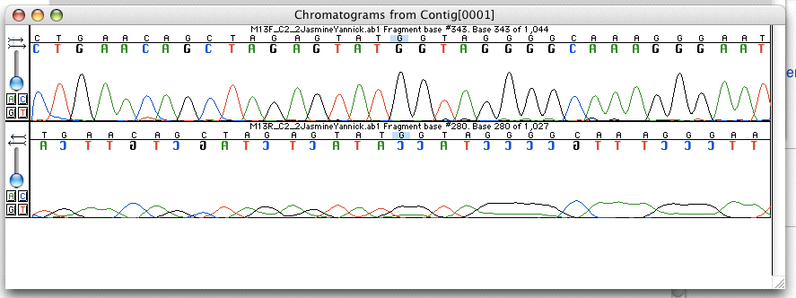
Whole genome sequencing (WGS), also known as full genome sequencing, complete genome sequencing, or entire genome sequencing, is the process of determining the entirety, or nearly the entirety, of the DNA sequence of an organism's genome at a single time. This entails sequencing all of an organism's chromosomal DNA as well as DNA contained in the mitochondrial DNA, mitochondria and, for plants, in the chloroplast. Whole genome sequencing has largely been used as a research tool, but was being introduced to clinics in 2014. In the future of personalized medicine, whole genome sequence data may be an important tool to guide therapeutic intervention. The tool of DNA sequencing, gene sequencing at Single-nucleotide polymorphism, SNP level is also used to pinpoint functional variants from association studies and improve the knowledge available to researchers interested in evolutionary biology, and hence may lay the foundation for predicting disease susceptibility and drug response. ... [...More Info...] [...Related Items...] OR: [Wikipedia] [Google] [Baidu] |
Prenatal Test
Prenatal testing consists of prenatal screening and prenatal diagnosis, which are aspects of prenatal care that focus on detecting problems with the pregnancy as early as possible. These may be anatomic and physiologic problems with the health of the zygote, embryo, or fetus, either before gestation even starts (as in preimplantation genetic diagnosis) or as early in gestation as practicable. Screening can detect problems such as neural tube defects, chromosome abnormalities, and gene mutations that would lead to genetic disorders and birth defects, such as spina bifida, cleft palate, Down syndrome, Tay–Sachs disease, sickle cell anemia, thalassemia, cystic fibrosis, muscular dystrophy, and fragile X syndrome. Some tests are designed to discover problems which primarily affect the health of the mother, such as PAPP-A to detect pre-eclampsia or glucose tolerance tests to diagnose gestational diabetes. Screening can also detect anatomical defects such as hydrocephalus, anencepha ... [...More Info...] [...Related Items...] OR: [Wikipedia] [Google] [Baidu] |
Cell-free Fetal DNA
Cell-free fetal DNA (cffDNA) is fetal DNA that circulates freely in the maternal blood. Maternal blood is sampled by venipuncture. Analysis of cffDNA is a method of non-invasive prenatal diagnosis frequently ordered for pregnant women of advanced maternal age. Two hours after delivery, cffDNA is no longer detectable in maternal blood. Background cffDNA originates from placental trophoblasts. Fetal DNA is fragmented when placental microparticles are shed into the maternal blood circulation. cffDNA fragments are approximately 200 base pairs (bp) in length. They are significantly smaller than maternal DNA fragments. The difference in size allows cffDNA to be distinguished from maternal DNA fragments. Approximately 11 to 13.4 percent of the cell-free DNA in maternal blood is of fetal origin. The amount varies widely from one pregnant woman to another. cffDNA is present after five to seven weeks gestation. The amount of cffDNA increases as the pregnancy progresses. The quantity of ... [...More Info...] [...Related Items...] OR: [Wikipedia] [Google] [Baidu] |
Venipuncture
In medicine, venipuncture or venepuncture is the process of obtaining intravenous access for the purpose of venous blood sampling (also called ''phlebotomy'') or intravenous therapy. In healthcare, this procedure is performed by medical laboratory scientists, medical practitioners, some EMTs, paramedics, phlebotomists, dialysis technicians, and other nursing staff. In veterinary medicine, the procedure is performed by veterinarians and veterinary technicians. It is essential to follow a standard procedure for the collection of blood specimens to get accurate laboratory results. Any error in collecting the blood or filling the test tubes may lead to erroneous laboratory results. Venipuncture is one of the most routinely performed invasive procedures and is carried out for any of five reasons: # to obtain blood for diagnostic purposes; # to monitor levels of blood components; # to administer therapeutic treatments including medications, nutrition, or chemotherapy; # to remov ... [...More Info...] [...Related Items...] OR: [Wikipedia] [Google] [Baidu] |
Chorionic Villus Sampling
Chorionic villus sampling (CVS), sometimes called "chorionic ''villous'' sampling" (as "villous" is the adjectival form of the word "villus"), is a form of prenatal diagnosis done to determine chromosomal or genetic disorders in the fetus. It entails sampling of the chorionic villus (placental tissue) and testing it for chromosomal abnormalities, usually with FISH or PCR. CVS usually takes place at 10–12 weeks' gestation, earlier than amniocentesis or percutaneous umbilical cord blood sampling. It is the preferred technique before 15 weeks. CVS was performed for the first time in Milan by Italian biologist Giuseppe Simoni, scientific director of Biocell Center, in 1983. Use as early as eight weeks in special circumstances has been described. It can be performed in a transcervical or transabdominal manner. Although this procedure is mostly associated with testing for Down syndrome, overall, CVS can detect more than 200 disorders. Indications Poss ... [...More Info...] [...Related Items...] OR: [Wikipedia] [Google] [Baidu] |
Amniocentesis
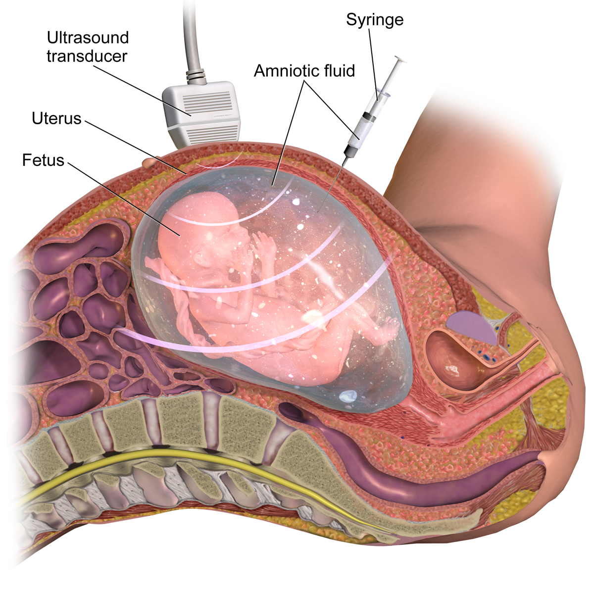
Amniocentesis is a medical procedure used primarily in the prenatal diagnosis of genetic conditions. It has other uses such as in the assessment of infection and fetal lung maturity. Prenatal diagnostic testing, which includes amniocentesis, is necessary to conclusively diagnose the majority of genetic disorders, with amniocentesis being the gold-standard procedure after 15 weeks' gestation. In this procedure, a thin needle is inserted into the abdomen of the pregnant woman. The needle punctures the amnion, which is the membrane that surrounds the developing fetus. The fluid within the amnion is called amniotic fluid, and because this fluid surrounds the developing fetus, it contains fetal cells. The amniotic fluid is sampled and analyzed via methods such as karyotyping and DNA analysis technology for genetic abnormalities. An amniocentesis is typically performed in the second trimester between the 15th and 20th week of gestation. Women who choose to have this test are prima ... [...More Info...] [...Related Items...] OR: [Wikipedia] [Google] [Baidu] |
Obstetric Ultrasonography
Obstetric ultrasonography, or prenatal ultrasound, is the use of medical ultrasonography in pregnancy, in which sound waves are used to create real-time visual images of the developing embryo or fetus in the uterus (womb). The procedure is a standard part of prenatal care in many countries, as it can provide a variety of information about the health of the mother, the timing and progress of the pregnancy, and the health and development of the embryo or fetus. The International Society of Ultrasound in Obstetrics and Gynecology (ISUOG) recommends that pregnant women have routine obstetric ultrasounds between 18 weeks' and 22 weeks' gestational age (the anatomy scan) in order to confirm pregnancy dating, to measure the fetus so that growth abnormalities can be recognized quickly later in pregnancy, and to assess for congenital malformations and multiple pregnancies (twins, etc). Additionally, the ISUOG recommends that pregnant patients who desire genetic testing have obstetric ultra ... [...More Info...] [...Related Items...] OR: [Wikipedia] [Google] [Baidu] |


