|
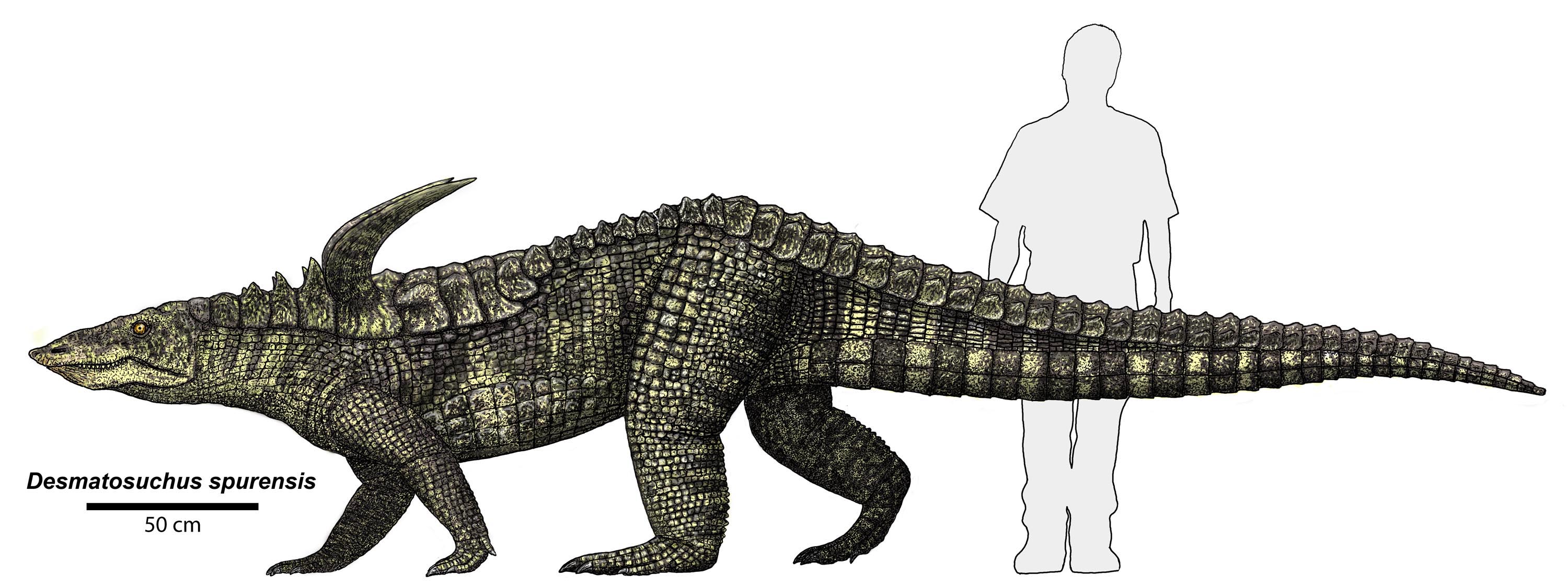
Desmatosuchus Spurensis
''Desmatosuchus'' (, from Greek δεσμός ''desmos'' 'link' + σοῦχος ''soûkhos'' 'crocodile') is an extinct genus of archosaur belonging to the Order Aetosauria. It lived during the Late Triassic. Description ''Desmatosuchus'' was a large quadrupedal reptile upwards of to in lengthvon Baczko, M. B., Desojo, J. B., Gower, D. J., Ridgely, R., Bona, P., & Witmer, L. M. (2021)New digital braincase endocasts of two species of Desmatosuchus and neurocranial diversity within Aetosauria (Archosauria: Pseudosuchia) The Anatomical Record, 1–20. https://doi.org/10.1002/ar.24798 and in weight. Its vertebral column had amphicoelus centra and 3 sacral vertebrae. This archosaur's most distinguishing anatomical characteristics were its scapulae which possessed large acromion processes commonly referred to as "shoulder spikes". The forelimbs were much shorter than the hindlimbs, with humeri less than two-thirds the length of the femurs. The pelvic girdle consisted of a long pub ... [...More Info...] [...Related Items...] OR: [Wikipedia] [Google] [Baidu] |
Late Triassic
The Late Triassic is the third and final epoch (geology), epoch of the Triassic geologic time scale, Period in the geologic time scale, spanning the time between annum, Ma and Ma (million years ago). It is preceded by the Middle Triassic Epoch and followed by the Early Jurassic Epoch. The corresponding series (stratigraphy), series of rock beds is known as the Upper Triassic. The Late Triassic is divided into the Carnian, Norian and Rhaetian Geologic time scale, Ages. Many of the first dinosaurs evolved during the Late Triassic, including ''Plateosaurus'', ''Coelophysis'', and ''Eoraptor''. The Triassic–Jurassic extinction event began during this epoch and is one of the five major mass extinction events of the Earth. Etymology The Triassic was named in 1834 by Friedrich August von Namoh, Friedrich von Alberti, after a succession of three distinct rock layers (Greek meaning 'triad') that are widespread in southern Germany: the lower Buntsandstein (colourful sandstone'')'', t ... [...More Info...] [...Related Items...] OR: [Wikipedia] [Google] [Baidu] |
Acromion
In human anatomy, the acromion (from Greek: ''akros'', "highest", ''ōmos'', "shoulder", plural: acromia) is a bony process on the scapula (shoulder blade). Together with the coracoid process it extends laterally over the shoulder joint. The acromion is a continuation of the scapular spine, and hooks over anteriorly. It articulates with the clavicle (collar bone) to form the acromioclavicular joint. Structure The acromion forms the summit of the shoulder, and is a large, somewhat triangular or oblong process, flattened from behind forward, projecting at first lateralward, and then curving forward and upward, so as to overhang the glenoid fossa.''Gray's Anatomy'' 1918, see infobox It starts from the base of acromion which marks its projecting point emerging from the spine of scapula. Surfaces Its superior surface, directed upward, backward, and lateralward, is convex, rough, and gives attachment to some fibers of the deltoideus, and in the rest of its extent is subcutaneous. ... [...More Info...] [...Related Items...] OR: [Wikipedia] [Google] [Baidu] |
Maxilla
The maxilla (plural: ''maxillae'' ) in vertebrates is the upper fixed (not fixed in Neopterygii) bone of the jaw formed from the fusion of two maxillary bones. In humans, the upper jaw includes the hard palate in the front of the mouth. The two maxillary bones are fused at the intermaxillary suture, forming the anterior nasal spine. This is similar to the mandible (lower jaw), which is also a fusion of two mandibular bones at the mandibular symphysis. The mandible is the movable part of the jaw. Structure In humans, the maxilla consists of: * The body of the maxilla * Four processes ** the zygomatic process ** the frontal process of maxilla ** the alveolar process ** the palatine process * three surfaces – anterior, posterior, medial * the Infraorbital foramen * the maxillary sinus * the incisive foramen Articulations Each maxilla articulates with nine bones: * two of the cranium: the frontal and ethmoid * seven of the face: the nasal, zygomatic, lacrimal, inferior n ... [...More Info...] [...Related Items...] OR: [Wikipedia] [Google] [Baidu] |
Premaxilla
The premaxilla (or praemaxilla) is one of a pair of small cranial bones at the very tip of the upper jaw of many animals, usually, but not always, bearing teeth. In humans, they are fused with the maxilla. The "premaxilla" of therian mammal has been usually termed as the incisive bone. Other terms used for this structure include premaxillary bone or ''os premaxillare'', intermaxillary bone or ''os intermaxillare'', and Goethe's bone. Human anatomy In human anatomy, the premaxilla is referred to as the incisive bone (') and is the part of the maxilla which bears the incisor teeth, and encompasses the anterior nasal spine and alar region. In the nasal cavity, the premaxillary element projects higher than the maxillary element behind. The palatal portion of the premaxilla is a bony plate with a generally transverse orientation. The incisive foramen is bound anteriorly and laterally by the premaxilla and posteriorly by the palatine process of the maxilla. It is formed from the ... [...More Info...] [...Related Items...] OR: [Wikipedia] [Google] [Baidu] |
Desmatosuchus Spurensis Skull
''Desmatosuchus'' (, from Greek δεσμός ''desmos'' 'link' + σοῦχος ''soûkhos'' 'crocodile') is an extinct genus of archosaur belonging to the Order Aetosauria. It lived during the Late Triassic. Description ''Desmatosuchus'' was a large quadrupedal reptile upwards of to in lengthvon Baczko, M. B., Desojo, J. B., Gower, D. J., Ridgely, R., Bona, P., & Witmer, L. M. (2021)New digital braincase endocasts of two species of Desmatosuchus and neurocranial diversity within Aetosauria (Archosauria: Pseudosuchia) The Anatomical Record, 1–20. https://doi.org/10.1002/ar.24798 and in weight. Its vertebral column had amphicoelus centra and 3 sacral vertebrae. This archosaur's most distinguishing anatomical characteristics were its scapulae which possessed large acromion processes commonly referred to as "shoulder spikes". The forelimbs were much shorter than the hindlimbs, with humeri less than two-thirds the length of the femurs. The pelvic girdle consisted of a long pub ... [...More Info...] [...Related Items...] OR: [Wikipedia] [Google] [Baidu] |
Calcaneal
In humans and many other primates, the calcaneus (; from the Latin ''calcaneus'' or ''calcaneum'', meaning heel) or heel bone is a bone of the tarsus of the foot which constitutes the heel. In some other animals, it is the point of the hock. Structure In humans, the calcaneus is the largest of the tarsal bones and the largest bone of the foot. Its long axis is pointed forwards and laterally. The talus bone, calcaneus, and navicular bone are considered the proximal row of tarsal bones. In the calcaneus, several important structures can be distinguished:Platzer (2004), p 216 There is a large calcaneal tuberosity located posteriorly on plantar surface with medial and lateral tubercles on its surface. Besides, there is another peroneal tubecle on its lateral surface. On its lower edge on either side are its lateral and medial processes (serving as the origins of the abductor hallucis and abductor digiti minimi). The Achilles tendon is inserted into a roughened area on its superior ... [...More Info...] [...Related Items...] OR: [Wikipedia] [Google] [Baidu] |
Crurotarsal
A crurotarsal joint is one that’s situated between the bones of ''crus'', i.e. shin (tibia and fibula) and the proximal tarsal bones, i.e. astragalus and calcaneum. The ankle joint of therian mammals (marsupials and placentals) is a crurotarsal joint, with the main joint of ankle bending between the tibia and the astragalus; the calcaneum has no contact with the tibia but forms a heel to which muscles can attach. A group of archosauriform diapsids, Crurotarsi (including living crocodilians and their extinct relatives) is named after specialized crurotarsal joint in the skeletons of the members of this group, located between their fibula and calcaneum, with a hemicylindrical condyle on the calcaneum articulating against fibula. This joint is present in the skeletons of suchians (including crocodilians) and phytosaurs, and was cited as one of the characters supporting uniting these two groups in a clade to the exclusion of avemetatarsalian archosaurs (birds and their extinct relat ... [...More Info...] [...Related Items...] OR: [Wikipedia] [Google] [Baidu] |
Acetabulum
The acetabulum (), also called the cotyloid cavity, is a concave surface of the pelvis. The head of the femur meets with the pelvis at the acetabulum, forming the hip joint. Structure There are three bones of the ''os coxae'' (hip bone) that come together to form the ''acetabulum''. Contributing a little more than two-fifths of the structure is the ischium, which provides lower and side boundaries to the acetabulum. The ilium forms the upper boundary, providing a little less than two-fifths of the structure of the acetabulum. The rest is formed by the pubis, near the midline. It is bounded by a prominent uneven rim, which is thick and strong above, and serves for the attachment of the acetabular labrum, which reduces its opening, and deepens the surface for formation of the hip joint. At the lower part of the ''acetabulum'' is the acetabular notch, which is continuous with a circular depression, the acetabular fossa, at the bottom of the cavity of the ''acetabulum''. The re ... [...More Info...] [...Related Items...] OR: [Wikipedia] [Google] [Baidu] |
Ilium (bone)
The ilium () (plural ilia) is the uppermost and largest part of the hip bone, and appears in most vertebrates including mammals and birds, but not bony fish. All reptiles have an ilium except snakes, although some snake species have a tiny bone which is considered to be an ilium. The ilium of the human is divisible into two parts, the body and the wing; the separation is indicated on the top surface by a curved line, the arcuate line, and on the external surface by the margin of the acetabulum. The name comes from the Latin (''ile'', ''ilis''), meaning "groin" or "flank". Structure The ilium consists of the body and wing. Together with the ischium and pubis, to which the ilium is connected, these form the pelvic bone, with only a faint line indicating the place of union. The body ( la, corpus) forms less than two-fifths of the acetabulum; and also forms part of the acetabular fossa. The internal surface of the body is part of the wall of the lesser pelvis and gives ... [...More Info...] [...Related Items...] OR: [Wikipedia] [Google] [Baidu] |
Ischium
The ischium () forms the lower and back region of the (''os coxae''). Situated below the ilium and behind the pubis, it is one of three regions whose fusion creates the . The superior portion of this region forms approximately one-third of the acetabulum. |
Symphysis
A symphysis (, pl. symphyses) is a fibrocartilaginous fusion between two bones. It is a type of cartilaginous joint, specifically a secondary cartilaginous joint. # A symphysis is an amphiarthrosis, a slightly movable joint. # A growing together of parts or structures. Unlike synchondroses, symphyses are permanent. Examples The more prominent symphyses are: * the pubic symphysis * sacrococcygeal symphysis * intervertebral disc between two vertebrae * in the sternum, between the manubrium and body Body may refer to: In science * Physical body, an object in physics that represents a large amount, has mass or takes up space * Body (biology), the physical material of an organism * Body plan, the physical features shared by a group of anima ... * mandibular symphysis, in the jaw Symphysis disorders Pubic symphysis diastasis Pubic symphysis diastasis, is an extremely rare complication that occurs in women who are giving birth. Separation of the two pubic bones during deli ... [...More Info...] [...Related Items...] OR: [Wikipedia] [Google] [Baidu] |
Pubis (bone)
In vertebrates, the pubic region ( la, pubis) is the most forward-facing (ventral and anterior) of the three main regions making up the coxal bone. The left and right pubic regions are each made up of three sections, a superior ramus, inferior ramus, and a body. Structure The pubic region is made up of a ''body'', ''superior ramus'', and ''inferior ramus'' (). The left and right coxal bones join at the pubic symphysis. It is covered by a layer of fat, which is covered by the mons pubis. The pubis is the lower limit of the suprapubic region. In the female, the pubic region is anterior to the urethral sponge. Body The body forms the wide, strong, middle and flat part of the pubic region. The bodies of the left and right pubic regions join at the pubic symphysis. The rough upper edge is the pubic crest, ending laterally in the pubic tubercle. This tubercle, found roughly 3 cm from the pubic symphysis, is a distinctive feature on the lower part of the abdominal wall; important ... [...More Info...] [...Related Items...] OR: [Wikipedia] [Google] [Baidu] |


