|
Depressed Skull Fracture
A skull fracture is a break in one or more of the eight bones that form the cranial portion of the skull, usually occurring as a result of blunt force trauma. If the force of the impact is excessive, the bone may fracture at or near the site of the impact and cause damage to the underlying structures within the skull such as the membranes, blood vessels, and brain. While an uncomplicated skull fracture can occur without associated physical or neurological damage and is in itself usually not clinically significant, a fracture in healthy bone indicates that a substantial amount of force has been applied and increases the possibility of associated injury. Any significant blow to the head results in a concussion, with or without loss of consciousness. A fracture in conjunction with an overlying laceration that tears the epidermis and the meninges, or runs through the paranasal sinuses and the middle ear structures, bringing the outside environment into contact with the cranial cavit ... [...More Info...] [...Related Items...] OR: [Wikipedia] [Google] [Baidu] |
Skull
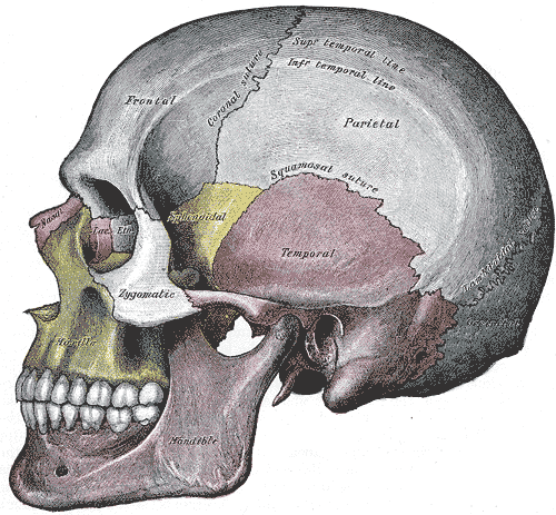
The skull is a bone protective cavity for the brain. The skull is composed of four types of bone i.e., cranial bones, facial bones, ear ossicles and hyoid bone. However two parts are more prominent: the cranium and the mandible. In humans, these two parts are the neurocranium and the viscerocranium ( facial skeleton) that includes the mandible as its largest bone. The skull forms the anterior-most portion of the skeleton and is a product of cephalisation—housing the brain, and several sensory structures such as the eyes, ears, nose, and mouth. In humans these sensory structures are part of the facial skeleton. Functions of the skull include protection of the brain, fixing the distance between the eyes to allow stereoscopic vision, and fixing the position of the ears to enable sound localisation of the direction and distance of sounds. In some animals, such as horned ungulates (mammals with hooves), the skull also has a defensive function by providing the mount (on the front ... [...More Info...] [...Related Items...] OR: [Wikipedia] [Google] [Baidu] |
Suture (joint)
In anatomy, fibrous joints are joints connected by fibrous tissue, consisting mainly of collagen. These are fixed joints where bones are united by a layer of white fibrous tissue of varying thickness. In the skull the joints between the bones are called sutures. Such immovable joints are also referred to as synarthroses. Types Most fibrous joints are also called "fixed" or "immovable". These joints have no joint cavity and are connected via fibrous connective tissue. The skull bones are connected by fibrous joints called '' sutures''. In fetal skulls the sutures are wide to allow slight movement during birth. They later become rigid ( synarthrodial). Some of the long bones in the body such as the radius and ulna in the forearm are joined by a ''syndesmosis'' (along the interosseous membrane). Syndemoses are slightly moveable ( amphiarthrodial). The distal tibiofibular joint is another example. A ''gomphosis'' is a joint between the root of a tooth and the socket in the max ... [...More Info...] [...Related Items...] OR: [Wikipedia] [Google] [Baidu] |
Osteogenesis Imperfecta
Osteogenesis imperfecta (; OI), colloquially known as brittle bone disease, is a group of genetic disorders that all result in bones that break easily. The range of symptoms—on the skeleton as well as on the body's other organs—may be mild to severe. Symptoms found in various types of OI include whites of the eye (sclerae) that are blue instead, short stature, loose joints, hearing loss, breathing problems and problems with the teeth (dentinogenesis imperfecta). Potentially life-threatening complications, all of which become more common in more severe OI, include: tearing ( dissection) of the major arteries, such as the aorta; pulmonary valve insufficiency secondary to distortion of the ribcage; and basilar invagination. The underlying mechanism is usually a problem with connective tissue due to a lack of, or poorly formed, type I collagen. In more than 90% of cases, OI occurs due to mutations in the ''COL1A1'' or ''COL1A2'' genes. These mutations may be inherited ... [...More Info...] [...Related Items...] OR: [Wikipedia] [Google] [Baidu] |
Cleidocranial Dysplasia
Cleidocranial dysostosis (CCD), also called cleidocranial dysplasia, is a birth defect that mostly affects the bones and teeth. The collarbones are typically either poorly developed or absent, which allows the shoulders to be brought close together. The front of the skull often does not close until later, and those affected are often shorter than average. Other symptoms may include a prominent forehead, wide set eyes, abnormal teeth, and a flat nose. Symptoms vary among people; however, intelligence is typically unaffected. The condition is either inherited from a person's parents or occurs as a new mutation. It is inherited in an autosomal dominant manner. It is due to a defect in the RUNX2 gene which is involved in bone formation. Diagnosis is suspected based on symptoms and X-rays with confirmation by genetic testing. Other conditions that can produce similar symptoms include mandibuloacral dysplasia, pyknodysostosis, osteogenesis imperfecta, and Hajdu-Cheney syndrome. Tre ... [...More Info...] [...Related Items...] OR: [Wikipedia] [Google] [Baidu] |
Lambdoidal Suture
The lambdoid suture (or lambdoidal suture) is a dense, fibrous connective tissue joint on the posterior aspect of the skull that connects the parietal bones with the occipital bone. It is continuous with the occipitomastoid suture. Structure The lambdoid suture is between the paired parietal bones and the occipital bone of the skull. It runs from the asterion on each side. Nerve supply The lambdoid suture may be supplied by a branch of the supraorbital nerve, a branch of the frontal branch of the trigeminal nerve. Clinical significance At birth, the bones of the skull do not meet. If certain bones of the skull grow too fast, then craniosynostosis (premature closure of the sutures) may occur. This can result in skull deformities. If the lambdoid suture closes too soon on one side, the skull will appear twisted and asymmetrical, a condition called "plagiocephaly". Plagiocephaly refers to the shape and not the condition. The condition is craniosynostosis. The lambdoid sutu ... [...More Info...] [...Related Items...] OR: [Wikipedia] [Google] [Baidu] |
Cleidocranial2
Cleidocranial dysostosis (CCD), also called cleidocranial dysplasia, is a birth defect that mostly affects the bones and teeth. The collarbones are typically either poorly developed or absent, which allows the shoulders to be brought close together. The front of the skull often does not close until later, and those affected are often shorter than average. Other symptoms may include a prominent forehead, wide set eyes, abnormal teeth, and a flat nose. Symptoms vary among people; however, intelligence is typically unaffected. The condition is either inherited from a person's parents or occurs as a new mutation. It is inherited in an autosomal dominant manner. It is due to a defect in the RUNX2 gene which is involved in bone formation. Diagnosis is suspected based on symptoms and X-rays with confirmation by genetic testing. Other conditions that can produce similar symptoms include mandibuloacral dysplasia, pyknodysostosis, osteogenesis imperfecta, and Hajdu-Cheney syndrome. Tre ... [...More Info...] [...Related Items...] OR: [Wikipedia] [Google] [Baidu] |
Neurosurgery
Neurosurgery or neurological surgery, known in common parlance as brain surgery, is the medical specialty concerned with the surgical treatment of disorders which affect any portion of the nervous system including the brain, spinal cord and peripheral nervous system. Education and context In different countries, there are different requirements for an individual to legally practice neurosurgery, and there are varying methods through which they must be educated. In most countries, neurosurgeon training requires a minimum period of seven years after graduating from medical school. United States In the United States, a neurosurgeon must generally complete four years of undergraduate education, four years of medical school, and seven years of residency (PGY-1-7). Most, but not all, residency programs have some component of basic science or clinical research. Neurosurgeons may pursue additional training in the form of a fellowship after residency, or, in some cases, as a senior resid ... [...More Info...] [...Related Items...] OR: [Wikipedia] [Google] [Baidu] |
Dura Mater
In neuroanatomy, dura mater is a thick membrane made of dense irregular connective tissue that surrounds the brain and spinal cord. It is the outermost of the three layers of membrane called the meninges that protect the central nervous system. The other two meningeal layers are the arachnoid mater and the pia mater. It envelops the arachnoid mater, which is responsible for keeping in the cerebrospinal fluid. It is derived primarily from the neural crest cell population, with postnatal contributions of the paraxial mesoderm. Structure The dura mater has several functions and layers. The dura mater is a membrane that envelops the arachnoid mater. It surrounds and supports the dural sinuses (also called dural venous sinuses, cerebral sinuses, or cranial sinuses) and carries blood from the brain toward the heart. Cranial dura mater has two layers called ''lamellae'', a superficial layer (also called the periosteal layer), which serves as the skull's inner periosteum, called the ... [...More Info...] [...Related Items...] OR: [Wikipedia] [Google] [Baidu] |
Intracranial Pressure
Intracranial pressure (ICP) is the pressure exerted by fluids such as cerebrospinal fluid (CSF) inside the skull and on the brain tissue. ICP is measured in millimeters of mercury (mmHg) and at rest, is normally 7–15 Millimeter of mercury, mmHg for a Supine position, supine adult. The body has various mechanisms by which it keeps the ICP stable, with CSF pressures varying by about 1 mmHg in normal adults through shifts in production and absorption of CSF. Changes in ICP are attributed to volume changes in one or more of the constituents contained in the cranium. CSF pressure has been shown to be influenced by abrupt changes in intrathoracic pressure during coughing (which is induced by contraction of the diaphragm and abdominal wall muscles, the latter of which also increases intra-abdominal pressure), the valsalva maneuver, and communication with the vasculature (venous and arterial systems). Intracranial hypertension (IH), also called increased ICP (IICP) or raised intracrani ... [...More Info...] [...Related Items...] OR: [Wikipedia] [Google] [Baidu] |
Depressed Skull Fracture2
Depression may refer to: Mental health * Depression (mood), a state of low mood and aversion to activity * Mood disorders characterized by depression are commonly referred to as simply ''depression'', including: ** Dysthymia, also known as persistent depressive disorder ** Major depressive disorder, also known as clinical depression Economics * Economic depression, a sustained, long-term downturn in economic activity in one or more economies ** Great Depression, a severe economic depression during the 1930s, commonly referred to as simply ''the Depression'' ** Long Depression, an economic depression during 1873–96, known at the time as the ''Great Depression'' Biology * Depression (kinesiology), an anatomical term of motion, refers to downward movement, the opposite of elevation * Depression (physiology), a reduction in a biological variable or the function of an organ * Central nervous system depression, physiological depression of the central nervous system that can result ... [...More Info...] [...Related Items...] OR: [Wikipedia] [Google] [Baidu] |
Parietal Bone
The parietal bones () are two bones in the Human skull, skull which, when joined at a fibrous joint, form the sides and roof of the Human skull, cranium. In humans, each bone is roughly quadrilateral in form, and has two surfaces, four borders, and four angles. It is named from the Latin ''paries'' (''-ietis''), wall. Surfaces External The external surface [Fig. 1] is convex, smooth, and marked near the center by an eminence, the parietal eminence (''tuber parietale''), which indicates the point where ossification commenced. Crossing the middle of the bone in an arched direction are two curved lines, the superior and inferior temporal lines; the former gives attachment to the temporal fascia, and the latter indicates the upper limit of the muscular origin of the temporal muscle. Above these lines the bone is covered by a tough layer of fibrous tissue – the epicranial aponeurosis; below them it forms part of the temporal fossa, and affords attachment to the temporal muscle. ... [...More Info...] [...Related Items...] OR: [Wikipedia] [Google] [Baidu] |


