|
Deep Branch Of Medial Circumflex Femoral Artery
The deep branch runs obliquely upward upon the tendon of the obturator externus and in front of the quadratus femoris toward the trochanteric fossa, where it anastomoses with twigs from the superior gluteal artery and inferior gluteal artery The inferior gluteal artery (sciatic artery), the smaller of the two terminal branches of the anterior trunk of the internal iliac artery, is distributed chiefly to the buttock and back of the thigh. It passes down on the sacral plexus of nerves .... References Arteries of the lower limb {{circulatory-stub ... [...More Info...] [...Related Items...] OR: [Wikipedia] [Google] [Baidu] |
Medial Circumflex Femoral Artery
The medial circumflex femoral artery (internal circumflex artery, medial femoral circumflex artery) is an artery in the upper thigh that arises from the profunda femoris artery''.'' Damage to the artery following a femoral neck fracture may lead to avascular necrosis (ischemic) of the femoral neck/head. Structure Origin The medial femoral circumflex artery arises from the posterior medial aspect of the profunda femoris artery''.'' The medial femoral circumflex artery may occasionally arise directly from the femoral artery. Course and relations It winds around the medial side of the femur, passing first between the pectineus and iliopsoas muscles, and then between the obturator externus and the adductor brevis muscles. Branches At the upper border of the adductor brevis it gives off two branches: * The '' ascending branch'' * The ''descending branch'' descends beneath the adductor brevis, to supply it and the adductor magnus; the continuation of the vessel passes backward ... [...More Info...] [...Related Items...] OR: [Wikipedia] [Google] [Baidu] |
Obturator Externus
The external obturator muscle, obturator externus muscle (; OE) is a flat, triangular muscle, which covers the outer surface of the anterior wall of the pelvis. It is sometimes considered part of the medial compartment of thigh, and sometimes considered part of the gluteal region. Structure It arises from the margin of bone immediately around the medial side of the obturator membrane and surrounding bone, viz., from the inferior pubic ramus, and the ramus of the ischium; it also arises from the medial two-thirds of the outer surface of the obturator membrane, and from the tendinous arch which completes the canal for the passage of the obturator vessels and nerves. The fibers springing from the pubic arch extend on to the inner surface of the bone, where they obtain a narrow origin between the margin of the foramen and the attachment of the obturator membrane. The fibers converge and pass posterolateral and upward, and end in a tendon which runs across the back of the neck of ... [...More Info...] [...Related Items...] OR: [Wikipedia] [Google] [Baidu] |
Quadratus Femoris
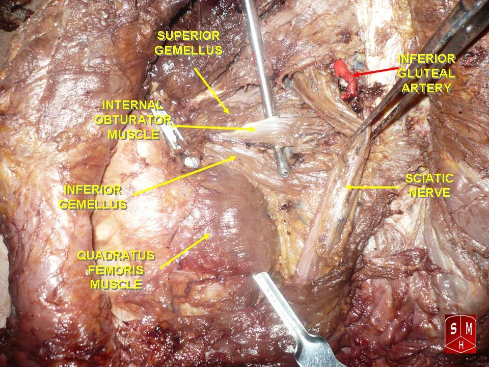
The quadratus femoris is a flat, quadrilateral skeletal muscle. Located on the posterior side of the hip joint, it is a strong external rotator and adductor of the thigh, but also acts to stabilize the femoral head in the acetabulum. Quadratus femoris use in the Meyer's muscle pedicle grafting to prevent avascular necrosis of femur head. Course It originates on the lateral border of the ischial tuberosity of the ischium of the pelvis. From there, it passes laterally to its insertion on the posterior side of the head of the femur: the quadrate tubercle on the intertrochanteric crest and along the quadrate line, the vertical line which runs downward to bisect the lesser trochanter on the medial side of the femur. Along its course, quadratus is aligned edge to edge with the inferior gemellus above and the adductor magnus below, so that its upper and lower borders run horizontal and parallel. At its origin, the upper margin of the adductor magnus is separated from it by the te ... [...More Info...] [...Related Items...] OR: [Wikipedia] [Google] [Baidu] |
Trochanteric Fossa
In mammals including humans, the medial surface of the greater trochanter has at its base a deep depression bounded posteriorly by the intertrochanteric crest, called the trochanteric fossa. This fossa is the point of insertion of four muscles. Moving from the inferior-most to the superior-most, they are: the tendon of the obturator externus muscle, the obturator internus, the superior gemellus and inferior gemellus. The width and depth of the trochanteric fossa varies taxonomically.Gray's AnatomyNetter 2003Romer 1956 In reptiliomorphs such as Seymouria or Diadectes and basal reptiles such as Pareiasaurus, the trochanteric fossa (also known as the intertrochanteric fossa) is a very large depression on the ventral/posterior side of the femur. It is bounded medially by the internal trochanter (also known as the lesser trochanter), laterally by the posterior branch of the ventral ridge, and inferiorly by the convergence of the anterior and posterior branches of the ventral ridge and ... [...More Info...] [...Related Items...] OR: [Wikipedia] [Google] [Baidu] |
Superior Gluteal Artery
The superior gluteal artery is the largest and final branch of the internal iliac artery. It is the continuation of the posterior division of that vessel. It is a short artery which runs backward between the lumbosacral trunk and the first sacral nerve. It divides into a superficial and a deep branch after passing out of the pelvis above the upper border of the piriformis muscle. Within the pelvis, it gives off branches to the iliacus, piriformis, and obturator internus muscles. Just previous to exiting the pelvic cavity, it also gives off a nutrient artery which enters the ilium. Structure The superior gluteal artery is the largest and final branch of the internal iliac artery. It branches from the posterior division of the internal iliac artery. It exits the pelvis through the greater sciatic foramen. It divides into a superficial and a deep branch. Superficial branch The superficial branch passes over the piriformis muscle. It enters the deep surface of the gluteus maximus mus ... [...More Info...] [...Related Items...] OR: [Wikipedia] [Google] [Baidu] |
Inferior Gluteal Artery
The inferior gluteal artery (sciatic artery), the smaller of the two terminal branches of the anterior trunk of the internal iliac artery, is distributed chiefly to the buttock and back of the thigh. It passes down on the sacral plexus of nerves and the piriformis muscle, behind the internal pudendal artery. It passes through the lower part of the greater sciatic foramen. It escapes from the pelvis between piriformis muscle and coccygeus muscle. It then descends in the interval between the greater trochanter of the femur and tuberosity of the ischium. It is accompanied by the sciatic nerve and the posterior femoral cutaneous nerves, and covered by the gluteus maximus. It continues down the back of the thigh, supplying the skin, and anastomosing with branches of the perforating arteries. Additional images File:Gray544.png, The arteries of the gluteal and posterior femoral regions. File:Gray829.png, Dissection of side wall of pelvis showing sacral and pudendal plexuses. See a ... [...More Info...] [...Related Items...] OR: [Wikipedia] [Google] [Baidu] |
