|
Decerebrate Posturing
Abnormal posturing is an involuntary flexion or extension of the arms and legs, indicating severe brain injury. It occurs when one set of muscles becomes incapacitated while the opposing set is not, and an external stimulus such as pain causes the working set of muscles to contract.AllRefer.com. 200"Decorticate Posture". Retrieved January 15, 2007. The posturing may also occur without a stimulus.WrongDiagnosis.com(Alarming Signs and Symptoms: Lippincott Manual of Nursing Practice Series). Retrieved on September 15, 2007. Since posturing is an important indicator of the amount of damage that has occurred to the brain, it is used by medical professionals to measure the severity of a coma with the Glasgow Coma Scale (for adults) and the Pediatric Glasgow Coma Scale (for infants). The presence of abnormal posturing indicates a severe medical emergency requiring immediate medical attention. Decerebrate and decorticate posturing are strongly associated with poor outcome in a varie ... [...More Info...] [...Related Items...] OR: [Wikipedia] [Google] [Baidu] |
Neurology
Neurology (from , "string, nerve" and the suffix wikt:-logia, -logia, "study of") is the branch of specialty (medicine) , medicine dealing with the diagnosis and treatment of all categories of conditions and disease involving the nervous system, which comprises the Human brain, brain, the spinal cord and the peripheral nervous system , peripheral nerves. Neurological practice relies heavily on the field of neuroscience, the scientific study of the nervous system, using various techniques of neurotherapy. IEEE Brain (2019). "Neurotherapy: Treating Disorders by Retraining the Brain". ''The Future Neural Therapeutics White Paper''. Retrieved 23.01.2025 from: https://brain.ieee.org/topics/neurotherapy-treating-disorders-by-retraining-the-brain/#:~:text=Neurotherapy%20trains%20a%20patient's%20brain,wave%20activity%20through%20positive%20reinforcement International Neuromodulation Society, Retrieved 23 January 2025 from: https://www.neuromodulation.com/ Val Danilov I (2023). "The O ... [...More Info...] [...Related Items...] OR: [Wikipedia] [Google] [Baidu] |
Corticospinal Tract
The corticospinal tract is a white matter motor pathway starting at the cerebral cortex that terminates on lower motor neurons and interneurons in the spinal cord, controlling movements of the limbs and trunk. There are more than one million neurons in the corticospinal tract, and they become myelinated usually in the first two years of life. The corticospinal tract is one of the pyramidal tracts, the other being the corticobulbar tract The corticobulbar (or corticonuclear) tract is a two-neuron white matter motor pathway connecting the motor cortex in the cerebral cortex to the Medullary pyramids (brainstem), medullary pyramids, which are part of the brainstem's medulla oblonga .... Anatomy The corticospinal tract originates in several parts of the brain, including not just the motor areas, but also the primary somatosensory cortex and premotor areas. Most of the neurons originate in either the primary motor cortex (precentral gyrus, Brodmann area 4) or the premotor fron ... [...More Info...] [...Related Items...] OR: [Wikipedia] [Google] [Baidu] |
Cerebellum
The cerebellum (: cerebella or cerebellums; Latin for 'little brain') is a major feature of the hindbrain of all vertebrates. Although usually smaller than the cerebrum, in some animals such as the mormyrid fishes it may be as large as it or even larger. In humans, the cerebellum plays an important role in motor control and cognition, cognitive functions such as attention and language as well as emotion, emotional control such as regulating fear and pleasure responses, but its movement-related functions are the most solidly established. The human cerebellum does not initiate movement, but contributes to motor coordination, coordination, precision, and accurate timing: it receives input from sensory systems of the spinal cord and from other parts of the brain, and integrates these inputs to fine-tune motor activity. Cerebellar damage produces disorders in fine motor skill, fine movement, sense of balance, equilibrium, list of human positions, posture, and motor learning in humans. ... [...More Info...] [...Related Items...] OR: [Wikipedia] [Google] [Baidu] |
Midbrain
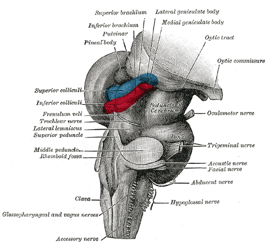
The midbrain or mesencephalon is the uppermost portion of the brainstem connecting the diencephalon and cerebrum with the pons. It consists of the cerebral peduncles, tegmentum, and tectum. It is functionally associated with vision, hearing, motor control, sleep and wakefulness, arousal (alertness), and temperature regulation.Breedlove, Watson, & Rosenzweig. Biological Psychology, 6th Edition, 2010, pp. 45-46 The name ''mesencephalon'' comes from the Greek ''mesos'', "middle", and ''enkephalos'', "brain". Structure The midbrain is the shortest segment of the brainstem, measuring less than 2cm in length. It is situated mostly in the posterior cranial fossa, with its superior part extending above the tentorial notch. The principal regions of the midbrain are the tectum, the cerebral aqueduct, tegmentum, and the cerebral peduncles. Rostral and caudal, Rostrally the midbrain adjoins the diencephalon (thalamus, hypothalamus, etc.), while Rostral and caudal, cau ... [...More Info...] [...Related Items...] OR: [Wikipedia] [Google] [Baidu] |
Brain Stem
The brainstem (or brain stem) is the posterior stalk-like part of the brain that connects the cerebrum with the spinal cord. In the human brain the brainstem is composed of the midbrain, the pons, and the medulla oblongata. The midbrain is continuous with the thalamus of the diencephalon through the tentorial notch, and sometimes the diencephalon is included in the brainstem. The brainstem is very small, making up around only 2.6 percent of the brain's total weight. It has the critical roles of regulating heart and respiratory function, helping to control heart rate and breathing rate. It also provides the main motor and sensory nerve supply to the face and neck via the cranial nerves. Ten pairs of cranial nerves come from the brainstem. Other roles include the regulation of the central nervous system and the body's sleep cycle. It is also of prime importance in the conveyance of motor and sensory pathways from the rest of the brain to the body, and from the body back ... [...More Info...] [...Related Items...] OR: [Wikipedia] [Google] [Baidu] |
Upper Limb
The upper Limb (anatomy), limbs or upper extremities are the forelimbs of an upright posture, upright-postured tetrapod vertebrate, extending from the scapulae and clavicles down to and including the digit (anatomy), digits, including all the musculatures and ligaments involved with the shoulder, elbow, wrist and knuckle joints. In humans, each upper limb is divided into the shoulder, arm, elbow, forearm, wrist and hand, and is primarily used for climbing, manual handling of loads, lifting and dexterity, manipulating objects. In anatomy, just as arm refers to the upper arm, leg refers to the lower leg. Definition In formal usage, the term "arm" only refers to the structures from the shoulder to the elbow, explicitly excluding the forearm, and thus "upper limb" and "arm" are not synonymous. However, in casual usage, the terms are often used interchangeably. The term "upper arm" is redundant in anatomy, but in informal usage is used to distinguish between the two terms. Structure I ... [...More Info...] [...Related Items...] OR: [Wikipedia] [Google] [Baidu] |
Decerebrate
Decerebration is the elimination of cerebral brain function in an animal by removing the cerebrum, cutting across the brain stem, or severing certain arteries in the brain stem. As a result, the animal loses certain reflexes that are integrated in different parts of the brain. Furthermore, the reflexes which are functional will be hyperreactive (and therefore very accentuated) due to the removal of inhibiting higher- brain centers (e.g. the facilitatory area of the reticular formation will not receive regulating input from cerebellum, basal ganglia and the cortex). Methods Decerebration describes the ligation along the neural axis in distinct parts of the brain in experimental animals. Generally: * lower decerebration (the cut is made above the upper border of the pons) * middle decerebration (cut is made through the red nucleus) * upper decerebration (cut is made so the cortical area is removed) Lower decerebration results in a "bulbospinal" animal: reflexes which are integ ... [...More Info...] [...Related Items...] OR: [Wikipedia] [Google] [Baidu] |
Brain Damage
Brain injury (BI) is the destruction or degeneration of brain cells. Brain injuries occur due to a wide range of internal and external factors. In general, brain damage refers to significant, undiscriminating trauma-induced damage. A common category with the greatest number of injuries is traumatic brain injury (TBI) following physical trauma or head injury from an outside source, and the term acquired brain injury (ABI) is used in appropriate circles to differentiate brain injuries occurring after birth from injury, from a genetic disorder (GBI), or from a congenital disorder (CBI). Primary and secondary brain injuries identify the processes involved, while focal and diffuse brain injury describe the severity and localization. Impaired function of affected areas can be compensated through neuroplasticity by forming new neural connections. Signs and symptoms Symptoms of brain injuries vary based on the severity of the injury or how much of the brain is affected. The fou ... [...More Info...] [...Related Items...] OR: [Wikipedia] [Google] [Baidu] |
Mesencephalon
The midbrain or mesencephalon is the uppermost portion of the brainstem connecting the diencephalon and cerebrum with the pons. It consists of the cerebral peduncles, tegmentum, and tectum. It is functionally associated with vision, hearing, motor control, sleep and wakefulness, arousal (alertness), and temperature regulation.Breedlove, Watson, & Rosenzweig. Biological Psychology, 6th Edition, 2010, pp. 45-46 The name ''mesencephalon'' comes from the Greek ''mesos'', "middle", and ''enkephalos'', "brain". Structure The midbrain is the shortest segment of the brainstem, measuring less than 2cm in length. It is situated mostly in the posterior cranial fossa, with its superior part extending above the tentorial notch. The principal regions of the midbrain are the tectum, the cerebral aqueduct, tegmentum, and the cerebral peduncles. Rostral and caudal, Rostrally the midbrain adjoins the diencephalon (thalamus, hypothalamus, etc.), while Rostral and caudal, cau ... [...More Info...] [...Related Items...] OR: [Wikipedia] [Google] [Baidu] |
Thalamus
The thalamus (: thalami; from Greek language, Greek Wikt:θάλαμος, θάλαμος, "chamber") is a large mass of gray matter on the lateral wall of the third ventricle forming the wikt:dorsal, dorsal part of the diencephalon (a division of the forebrain). Nerve fibers project out of the thalamus to the cerebral cortex in all directions, known as the thalamocortical radiations, allowing hub (network science), hub-like exchanges of information. It has several functions, such as the relaying of sensory neuron, sensory and motor neuron, motor signals to the cerebral cortex and the regulation of consciousness, sleep, and alertness. Anatomically, the thalami are paramedian symmetrical structures (left and right), within the vertebrate brain, situated between the cerebral cortex and the midbrain. It forms during embryonic development as the main product of the diencephalon, as first recognized by the Swiss embryologist and anatomist Wilhelm His Sr. in 1893. Anatomy The thalami ar ... [...More Info...] [...Related Items...] OR: [Wikipedia] [Google] [Baidu] |
Internal Capsule
The internal capsule is a paired white matter structure, as a two-way nerve tract, tract, carrying afferent nerve fiber, ascending and efferent nerve fiber, descending axon, fibers, to and from the cerebral cortex. The internal capsule is situated in the Anatomical terms of location#Medial and lateral, inferomedial part of each cerebral hemisphere of the brain. It carries information past the subcortical basal ganglia. As it courses it separates the caudate nucleus and the thalamus from the putamen and the globus pallidus. It also separates the caudate nucleus and the putamen in the dorsal striatum, a brain region involved in motor and reward pathways. The internal capsule is V-shaped in transection forming an anterior and posterior limb, with the angle between them called the genu. The corticospinal tract constitutes a large part of the internal capsule, carrying motor information from the primary motor cortex to the lower motor neurons in the spinal cord. Above the basal gangli ... [...More Info...] [...Related Items...] OR: [Wikipedia] [Google] [Baidu] |
Cerebral Hemisphere
The vertebrate cerebrum (brain) is formed by two cerebral hemispheres that are separated by a groove, the longitudinal fissure. The brain can thus be described as being divided into left and right cerebral hemispheres. Each of these hemispheres has an outer layer of grey matter, the cerebral cortex, that is supported by an inner layer of white matter. In eutherian (placental) mammals, the hemispheres are linked by the corpus callosum, a very large bundle of axon, nerve fibers. Smaller commissures, including the anterior commissure, the posterior commissure and the fornix (neuroanatomy), fornix, also join the hemispheres and these are also present in other vertebrates. These commissures transfer information between the two hemispheres to coordinate localized functions. There are three known poles of the cerebral hemispheres: the ''occipital lobe, occipital pole'', the ''frontal lobe, frontal pole'', and the ''temporal lobe, temporal pole''. The central sulcus is a prominent fissu ... [...More Info...] [...Related Items...] OR: [Wikipedia] [Google] [Baidu] |


