|
Cri Du Chat Syndrome
Cri du chat syndrome is a rare genetic disorder due to a partial chromosome deletion on chromosome 5. Its name is a French term ("cat-cry" or " call of the cat") referring to the characteristic cat-like cry of affected children. It was first described by Jérôme Lejeune in 1963. The condition affects an estimated 1 in 50,000 live births across all ethnicities and is more common in females by a 4:3 ratio. Signs and symptoms The syndrome gets its name from the characteristic cry of affected infants, which is similar to that of a meowing kitten, due to problems with the larynx and nervous system. About one third of children lose the cry by age of 2 years. Other symptoms of cri du chat syndrome may include: * feeding problems because of difficulty in swallowing and sucking; * mutism; * low birth weight and poor growth; * severe cognitive, speech and motor disabilities; * behavioural problems such as hyperactivity, aggression, outbursts and repetitive movements; * unusual facial fe ... [...More Info...] [...Related Items...] OR: [Wikipedia] [Google] [Baidu] |
Medical Genetics
Medical genetics is the branch tics in that human genetics is a field of scientific research that may or may not apply to medicine, while medical genetics refers to the application of genetics to medical care. For example, research on the causes and inheritance of genetic disorders would be considered within both human genetics and medical genetics, while the diagnosis, management, and counselling people with genetic disorders would be considered part of medical genetics. In contrast, the study of typically non-medical phenotypes such as the genetics of eye color would be considered part of human genetics, but not necessarily relevant to medical genetics (except in situations such as albinism). ''Genetic medicine'' is a newer term for medical genetics and incorporates areas such as gene therapy, personalized medicine, and the rapidly emerging new medical specialty, predictive medicine. Scope Medical genetics encompasses many different areas, including clinical practice of ... [...More Info...] [...Related Items...] OR: [Wikipedia] [Google] [Baidu] |
Hypertelorism
Hypertelorism is an abnormally increased distance between two organs or bodily parts, usually referring to an increased distance between the orbits (eyes), or orbital hypertelorism. In this condition the distance between the inner eye corners as well as the distance between the pupils is greater than normal. Hypertelorism should not be confused with telecanthus, in which the distance between the inner eye corners is increased but the distances between the outer eye corners and the pupils remain unchanged.Michael L. Bentz: ''Pediatric Plastic Surgery''; Chapter 9 Hypertelorism by Renato Ocampo, Jr., MD/ John A. Persing, MD Hypertelorism is a symptom in a variety of syndromes, including Edwards syndrome (trisomy 18), 1q21.1 duplication syndrome, basal cell nevus syndrome, DiGeorge syndrome and Loeys–Dietz syndrome. Hypertelorism can also be seen in Apert syndrome, Autism spectrum disorder, craniofrontonasal dysplasia, Noonan syndrome, neurofibromatosis, LEOPARD syndrome, Crouzon ... [...More Info...] [...Related Items...] OR: [Wikipedia] [Google] [Baidu] |
Tetralogy Of Fallot
Tetralogy of Fallot (TOF), formerly known as Steno-Fallot tetralogy, is a congenital heart defect characterized by four specific cardiac defects. Classically, the four defects are: *pulmonary stenosis, which is narrowing of the exit from the right ventricle; * a ventricular septal defect, which is a hole allowing blood to flow between the two ventricles; * right ventricular hypertrophy, which is thickening of the right ventricular muscle; and * an overriding aorta, which is where the aorta expands to allow blood from both ventricles to enter. At birth, children may be asymptomatic or present with many severe symptoms. Later in infancy, there are typically episodes of bluish colour to the skin due to a lack of sufficient oxygenation, known as cyanosis. When affected babies cry or have a bowel movement, they may undergo a "tet spell" where they turn cyanotic, have difficulty breathing, become limp, and occasionally lose consciousness. Other symptoms may include a heart murmur, ... [...More Info...] [...Related Items...] OR: [Wikipedia] [Google] [Baidu] |
Patent Ductus Arteriosus
''Patent ductus arteriosus'' (PDA) is a medical condition in which the ''ductus arteriosus'' fails to close after birth: this allows a portion of oxygenated blood from the left heart to flow back to the lungs by flowing from the aorta, which has a higher pressure, to the pulmonary artery. Symptoms are uncommon at birth and shortly thereafter, but later in the first year of life there is often the onset of an increased work of breathing and failure to gain weight at a normal rate. With time, an uncorrected PDA usually leads to pulmonary hypertension followed by right-sided heart failure. The ''ductus arteriosus'' is a fetal blood vessel that normally closes soon after birth. In a PDA, the vessel does not close, but remains ''patent'' (open), resulting in an abnormal transmission of blood from the aorta to the pulmonary artery. PDA is common in newborns with persistent respiratory problems such as hypoxia, and has a high occurrence in premature newborns. Premature newborns are ... [...More Info...] [...Related Items...] OR: [Wikipedia] [Google] [Baidu] |
Atrial Septal Defect
Atrial septal defect (ASD) is a congenital heart defect in which blood flows between the atria (upper chambers) of the heart. Some flow is a normal condition both pre-birth and immediately post-birth via the foramen ovale; however, when this does not naturally close after birth it is referred to as a patent (open) foramen ovale (PFO). It is common in patients with a congenital atrial septal aneurysm (ASA). After PFO closure the atria normally are separated by a dividing wall, the interatrial septum. If this septum is defective or absent, then oxygen-rich blood can flow directly from the left side of the heart to mix with the oxygen-poor blood in the right side of the heart; or the opposite, depending on whether the left or right atrium has the higher blood pressure. In the absence of other heart defects, the left atrium has the higher pressure. This can lead to lower-than-normal oxygen levels in the arterial blood that supplies the brain, organs, and tissues. However, an ASD m ... [...More Info...] [...Related Items...] OR: [Wikipedia] [Google] [Baidu] |
Ventricular Septal Defect
A ventricular septal defect (VSD) is a defect in the ventricular septum, the wall dividing the left and right ventricles of the heart. The extent of the opening may vary from pin size to complete absence of the ventricular septum, creating one common ventricle. The ventricular septum consists of an inferior muscular and superior membranous portion and is extensively innervated with conducting cardiomyocytes. The membranous portion, which is close to the atrioventricular node, is most commonly affected in adults and older children in the United States. It is also the type that will most commonly require surgical intervention, comprising over 80% of cases. Membranous ventricular septal defects are more common than muscular ventricular septal defects, and are the most common congenital cardiac anomaly. Signs and symptoms Ventricular septal defect is usually symptomless at birth. It usually manifests a few weeks after birth. VSD is an acyanotic congenital heart defect, aka a lef ... [...More Info...] [...Related Items...] OR: [Wikipedia] [Google] [Baidu] |
Single Palmar Crease
In humans, a single transverse palmar crease is a single crease that extends across the palm of the hand, formed by the fusion of the two palmar creases (known in palmistry as the "heart line" and the "head line"). Although it is found more frequently in persons with several abnormal medical conditions, it is not predictive of any of these conditions since it is also found in perfectly healthy persons. It is found in 1.5% of the world population in at least one hand. Former name Because it resembles the usual condition of non-human simians, it was, in the past, called the simian crease or simian line. These terms have widely fallen out of favor due to their pejorative connotation. Medical significance The presence of a single transverse palmar crease has no medical significance. It is found in 1.5% of all people, and though it is found at a higher frequency in people with abnormal medical conditions, in every one of these conditions many people do not have a single transverse palme ... [...More Info...] [...Related Items...] OR: [Wikipedia] [Google] [Baidu] |
Brachydactyly
Brachydactyly (Greek βραχύς = "short" plus δάκτυλος = "finger"), is a medical term which literally means "short finger". The shortness is relative to the length of other long bones and other parts of the body. Brachydactyly is an inherited, dominant trait. It most often occurs as an isolated dysmelia, but can also occur with other anomalies as part of many congenital syndromes. Brachydactyly may also be a signal that one is at risk for congenital heart disease due to the association between congenital heart disease and carpenter's syndrome and the link between carpenter's syndrome and brachydactyly Nomograms for normal values of finger length as a ratio to other body measurements have been published. In clinical genetics, the most commonly used index of digit length is the dimensionless ratio of the length of the third (middle) finger to the hand length. Both are expressed in the same units (centimeters, for example) and are measured in an open hand from the finger ... [...More Info...] [...Related Items...] OR: [Wikipedia] [Google] [Baidu] |
Low-set Ears
Low-set ears are a clinical feature in which the ears are positioned lower on the head than usual. They are present in many congenital conditions. Low-set ears are defined as outer ears positioned two or more standard deviations lower than the population average. Clinically, if the point at which the helix of the outer ear meets the cranium is at or below the line connecting the inner canthi of eyes(bicanthal plane), the ears are considered low set. Low-set ears can be associated with conditions such as: *Down syndrome *Turner syndrome *Noonan syndrome *Patau syndrome *DiGeorge syndrome *Cri du chat syndrome *Edwards syndrome *Fragile X syndrome *Okamoto syndrome It is usually bilateral, but it can be unilateral in Goldenhar syndrome. See also *LEOPARD syndrome Noonan syndrome with multiple lentigines (NSML) which is part of a group called Ras/MAPK pathway syndromes, is a rare autosomal dominant, multisystem disease caused by a mutation in the protein tyrosine phosphatase, non-r ... [...More Info...] [...Related Items...] OR: [Wikipedia] [Google] [Baidu] |
Nasal Bridge
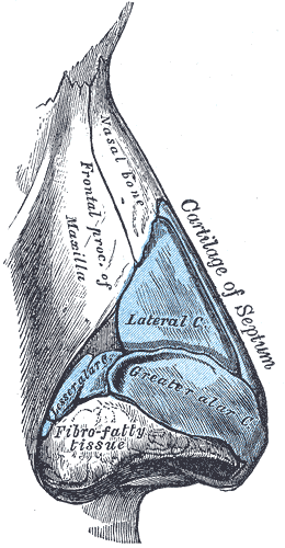
The nasal bridge is the upper, bony part of the human nose, which overlies the nasal bones. Association with epicanthic folds Low-rooted nasal bridges are closely associated with epicanthic folds. A lower nasal bridge is more likely to cause an epicanthic fold, and vice versa. Dysmorphology A lower or higher than average nasal bridge can be a sign of various genetic disorders, such as fetal alcohol syndrome. A flat nasal bridge can be a sign of Down syndrome (Trisomy 21), Fragile X syndrome, 48,XXXY variant Klinefelter syndrome, or Bartarlla-Scott syndrome. An appearance of a widened nasal bridge can be seen with dystopia canthorum, which is a lateral displacement of the inner canthi of the eyes. from UTMB, Dept. of Otolaryngology. DATE: March 17, 2004. RESIDENT PHYSICIAN: Jing Shen. FACUL ... [...More Info...] [...Related Items...] OR: [Wikipedia] [Google] [Baidu] |
Strabismus
Strabismus is a vision disorder in which the eyes do not properly align with each other when looking at an object. The eye that is focused on an object can alternate. The condition may be present occasionally or constantly. If present during a large part of childhood, it may result in amblyopia, or lazy eyes, and loss of depth perception. If onset is during adulthood, it is more likely to result in double vision. Strabismus can occur due to muscle dysfunction, farsightedness, problems in the brain, trauma or infections. Risk factors include premature birth, cerebral palsy and a family history of the condition. Types include esotropia, where the eyes are crossed ("cross eyed"); exotropia, where the eyes diverge ("lazy eyed" or "wall eyed"); and hypertropia or hypotropia where they are vertically misaligned. They can also be classified by whether the problem is present in all directions a person looks (comitant) or varies by direction (incomitant). Diagnosis may be made by obser ... [...More Info...] [...Related Items...] OR: [Wikipedia] [Google] [Baidu] |
Palpebral Fissure
The palpebral fissure is the elliptic space between the medial and lateral canthi of the two open eyelids. In simple terms, it is the opening between the eyelids. In adult humans, this measures about 10 mm vertically and 30 mm horizontally. Variations Congenital dysmorphisms It can be reduced (short, "narrow") in horizontal size by fetal alcohol syndrome and in Williams syndrome. The chromosomal conditions trisomy 9 and trisomy 21 (Down syndrome) can cause the palpebral fissures to be upslanted, whereas Marfan syndrome can cause a downslant. An increase in vertical height can be seen in genetic disorders such as cri-du-chat syndrome. Acquired The fissure may be increased in vertical height in Graves' disease, which is manifested as Dalrymple's sign. It is seen in disorders such as cri-du-chat syndrome. In animal studies using four times the therapeutic concentration of the ophthalmic solution latanoprost, the size of the palpebral fissure can be increased. The condition ... [...More Info...] [...Related Items...] OR: [Wikipedia] [Google] [Baidu] |


