|
Costoclavicular Ligament
The costoclavicular ligament, also known as the rhomboid ligament or Halsted's ligament, is a ligament of the shoulder girdle. It is short, flat, and rhomboid in form. It is the major stabilizing factor of the sternoclavicular joint and is the axis of movement of the joint, especially during elevation of the clavicle. Attached below to the upper and medial part of the cartilage of the first rib, it ascends at an angle posteriorly and laterally, and is fixed above to the costal tuberosity on the inferior aspect of the clavicle. It is in relation, in front, with the tendon of origin of the subclavius; behind, with the subclavian vein The subclavian vein is a paired large vein, one on either side of the body, that is responsible for draining blood from the upper extremities, allowing this blood to return to the heart. The left subclavian vein plays a key role in the absorption .... References {{DEFAULTSORT:Costoclavicular Ligament Ligaments of the upper limb ... [...More Info...] [...Related Items...] OR: [Wikipedia] [Google] [Baidu] |
Sternoclavicular Articulation
The sternoclavicular joint or sternoclavicular articulation is a synovial saddle joint between the manubrium of the sternum, and the clavicle, as well as the first rib. The joint possesses a joint capsule, and an articular disk, and is reinforced by multiple ligaments. Structure The joint is structurally classed as a synovial plane joint and functionally classed as a diarthrosis and multiaxial joint. It is composed of two portions separated by an articular disc of fibrocartilage. The joint is formed by the sternal end of the clavicle, the clavicular notch (the superior and lateral part of the sternum), and (the superior surface of) the cartilage of the first rib (visible from the outside as the suprasternal notch). The articular surface of the clavicle is larger than that of the sternum, and is invested with a layer of cartilage, which is considerably thicker than that of the sternum. The joint receives arterial supply via branches of the internal thoracic artery, and of th ... [...More Info...] [...Related Items...] OR: [Wikipedia] [Google] [Baidu] |
First Rib
The rib cage, as an enclosure that comprises the ribs, vertebral column and sternum in the thorax of most vertebrates, protects vital organs such as the heart, lungs and great vessels. The sternum, together known as the thoracic cage, is a semi-rigid bony and cartilaginous structure which surrounds the thoracic cavity and supports the shoulder girdle to form the core part of the human skeleton. A typical human thoracic cage consists of 12 pairs of ribs and the adjoining costal cartilages, the sternum (along with the manubrium and xiphoid process), and the 12 thoracic vertebrae articulating with the ribs. Together with the skin and associated fascia and muscles, the thoracic cage makes up the thoracic wall and provides attachments for extrinsic skeletal muscles of the neck, upper limbs, upper abdomen and back. The rib cage intrinsically holds the muscles of respiration ( diaphragm, intercostal muscles, etc.) that are crucial for active inhalation and forced exhalation, and there ... [...More Info...] [...Related Items...] OR: [Wikipedia] [Google] [Baidu] |
Clavicle
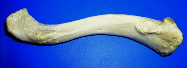
The clavicle, or collarbone, is a slender, S-shaped long bone approximately 6 inches (15 cm) long that serves as a strut between the shoulder blade and the sternum (breastbone). There are two clavicles, one on the left and one on the right. The clavicle is the only long bone in the body that lies horizontally. Together with the shoulder blade, it makes up the shoulder girdle. It is a palpable bone and, in people who have less fat in this region, the location of the bone is clearly visible. It receives its name from the Latin ''clavicula'' ("little key"), because the bone rotates along its axis like a key when the shoulder is abducted. The clavicle is the most commonly fractured bone. It can easily be fractured by impacts to the shoulder from the force of falling on outstretched arms or by a direct hit. Structure The collarbone is a thin doubly curved long bone that connects the arm to the trunk of the body. Located directly above the first rib, it acts as a strut to k ... [...More Info...] [...Related Items...] OR: [Wikipedia] [Google] [Baidu] |
Costal Tuberosity Of Clavicle
On the medial part of the clavicle is a broad rough surface, the costal tuberosity (impression for costoclavicular ligament), rather more than 2 cm. in length, for the attachment of the costoclavicular ligament The costoclavicular ligament, also known as the rhomboid ligament or Halsted's ligament, is a ligament of the shoulder girdle. It is short, flat, and rhomboid in form. It is the major stabilizing factor of the sternoclavicular joint and is the a .... References Shoulder Skeletal system Upper limb anatomy Clavicle {{musculoskeletal-stub ... [...More Info...] [...Related Items...] OR: [Wikipedia] [Google] [Baidu] |
Ligament
A ligament is the fibrous connective tissue that connects bones to other bones. It is also known as ''articular ligament'', ''articular larua'', ''fibrous ligament'', or ''true ligament''. Other ligaments in the body include the: * Peritoneal ligament: a fold of peritoneum or other membranes. * Fetal remnant ligament: the remnants of a fetal tubular structure. * Periodontal ligament: a group of fibers that attach the cementum of teeth to the surrounding alveolar bone. Ligaments are similar to tendons and fasciae as they are all made of connective tissue. The differences among them are in the connections that they make: ligaments connect one bone to another bone, tendons connect muscle to bone, and fasciae connect muscles to other muscles. These are all found in the skeletal system of the human body. Ligaments cannot usually be regenerated naturally; however, there are periodontal ligament stem cells located near the periodontal ligament which are involved in the adult regener ... [...More Info...] [...Related Items...] OR: [Wikipedia] [Google] [Baidu] |
Shoulder Girdle
The shoulder girdle or pectoral girdle is the set of bones in the appendicular skeleton which connects to the arm on each side. In humans it consists of the clavicle and scapula; in those species with three bones in the shoulder, it consists of the clavicle, scapula, and coracoid. Some mammalian species (such as the dog and the horse) have only the scapula. The pectoral girdles are to the upper limbs as the pelvic girdle is to the lower limbs; the girdles are the parts of the appendicular skeleton that anchor the appendages to the axial skeleton. In humans, the only true anatomical joints between the shoulder girdle and the axial skeleton are the sternoclavicular joints on each side. No anatomical joint exists between each scapula and the rib cage; instead the muscular connection or physiological joint between the two permits great mobility of the shoulder girdle compared to the compact pelvic girdle; because the upper limb is not usually involved in weight bearing, its stabilit ... [...More Info...] [...Related Items...] OR: [Wikipedia] [Google] [Baidu] |
Rhomboid
Traditionally, in two-dimensional geometry, a rhomboid is a parallelogram in which adjacent sides are of unequal lengths and angles are non-right angled. A parallelogram with sides of equal length (equilateral) is a rhombus but not a rhomboid. A parallelogram with right angled corners is a rectangle but not a rhomboid. The term ''rhomboid'' is now more often used for a rhombohedron or a more general parallelepiped, a solid figure with six faces in which each face is a parallelogram and pairs of opposite faces lie in parallel planes. Some crystals are formed in three-dimensional rhomboids. This solid is also sometimes called a rhombic prism. The term occurs frequently in science terminology referring to both its two- and three-dimensional meaning. History Euclid introduced the term in his '' Elements'' in Book I, Definition 22, Euclid never used the definition of rhomboid again and introduced the word parallelogram in Proposition 34 of Book I; ''"In parallelogrammic areas the ... [...More Info...] [...Related Items...] OR: [Wikipedia] [Google] [Baidu] |
Sternoclavicular Joint
The sternoclavicular joint or sternoclavicular articulation is a synovial saddle joint between the manubrium of the sternum, and the clavicle, as well as the first rib. The joint possesses a joint capsule, and an articular disk, and is reinforced by multiple ligaments. Structure The joint is structurally classed as a synovial plane joint and functionally classed as a diarthrosis and multiaxial joint. It is composed of two portions separated by an articular disc of fibrocartilage. The joint is formed by the sternal end of the clavicle, the clavicular notch (the superior and lateral part of the sternum), and (the superior surface of) the cartilage of the first rib (visible from the outside as the suprasternal notch). The articular surface of the clavicle is larger than that of the sternum, and is invested with a layer of cartilage, which is considerably thicker than that of the sternum. The joint receives arterial supply via branches of the internal thoracic artery, and of th ... [...More Info...] [...Related Items...] OR: [Wikipedia] [Google] [Baidu] |
Cartilage
Cartilage is a resilient and smooth type of connective tissue. In tetrapods, it covers and protects the ends of long bones at the joints as articular cartilage, and is a structural component of many body parts including the rib cage, the neck and the bronchial tubes, and the intervertebral discs. In other taxa, such as chondrichthyans, but also in cyclostomes, it may constitute a much greater proportion of the skeleton. It is not as hard and rigid as bone, but it is much stiffer and much less flexible than muscle. The matrix of cartilage is made up of glycosaminoglycans, proteoglycans, collagen fibers and, sometimes, elastin. Because of its rigidity, cartilage often serves the purpose of holding tubes open in the body. Examples include the rings of the trachea, such as the cricoid cartilage and carina. Cartilage is composed of specialized cells called chondrocytes that produce a large amount of collagenous extracellular matrix, abundant ground substance that is rich in pro ... [...More Info...] [...Related Items...] OR: [Wikipedia] [Google] [Baidu] |
Costal Tuberosity
On the medial part of the clavicle is a broad rough surface, the costal tuberosity (impression for costoclavicular ligament), rather more than 2 cm. in length, for the attachment of the costoclavicular ligament The costoclavicular ligament, also known as the rhomboid ligament or Halsted's ligament, is a ligament of the shoulder girdle. It is short, flat, and rhomboid in form. It is the major stabilizing factor of the sternoclavicular joint and is the a .... References Shoulder Skeletal system Upper limb anatomy Clavicle {{musculoskeletal-stub ... [...More Info...] [...Related Items...] OR: [Wikipedia] [Google] [Baidu] |
Subclavius
The subclavius is a small triangular muscle, placed between the clavicle and the first rib. Along with the pectoralis major and pectoralis minor muscles, the subclavius muscle makes up the anterior axioappendicular muscles, also known as anterior wall of the axilla. Structure It arises by a short, thick tendon from the first rib and its cartilage at their junction, in front of the costoclavicular ligament. The fleshy fibers proceed obliquely superolaterally, to be inserted into the groove on the under surface of the clavicle. Innervation The nerve to subclavius (or subclavian nerve) innervates the muscle. This arises from the junction of the fifth and sixth cervical nerves, from the superior/upper trunk of the brachial plexus. Variation Insertion into coracoid process instead of clavicle or into both clavicle and coracoid process. Sternoscapular fasciculus to the upper border of scapula. Sternoclavicularis from manubrium to clavicle between pectoralis major and coracoclavicular ... [...More Info...] [...Related Items...] OR: [Wikipedia] [Google] [Baidu] |
Subclavian Vein
The subclavian vein is a paired large vein, one on either side of the body, that is responsible for draining blood from the upper extremities, allowing this blood to return to the heart. The left subclavian vein plays a key role in the absorption of lipids, by allowing products that have been carried by lymph in the thoracic duct to enter the bloodstream. The diameter of the subclavian veins is approximately 1–2 cm, depending on the individual. Structure Each subclavian vein is a continuation of the axillary vein and runs from the outer border of the first rib to the medial border of anterior scalene muscle. From here it joins with the internal jugular vein to form the brachiocephalic vein (also known as "innominate vein"). The angle of union is termed the venous angle. The subclavian vein follows the subclavian artery and is separated from the subclavian artery by the insertion of anterior scalene. Thus, the subclavian vein lies anterior to the anterior scalene while the su ... [...More Info...] [...Related Items...] OR: [Wikipedia] [Google] [Baidu] |


