|
Corneal Epithelium
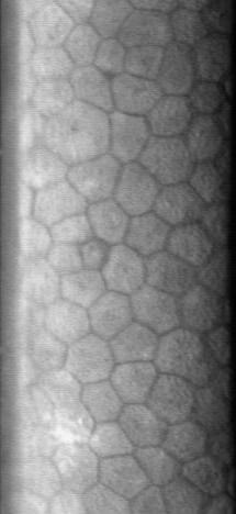
The corneal epithelium (epithelium corneæ anterior layer) is made up of epithelial tissue and covers the front of the cornea. It acts as a barrier to protect the cornea, resisting the free flow of fluids from the tears, and prevents bacteria from entering the epithelium and corneal stroma. Anatomy The corneal epithelium consists of several layers of cells. The cells of the deepest layer are columnar, known as basal cells which are attached by multiprotein complexes known as hemidesmosomes to an underlying basement membrane. Then follow two or three layers of polyhedral cells, commonly known as wing cells. The majority of these are prickle cells, similar to those found in the stratum mucosum of the cuticle. Lastly, there are three or four layers of squamous cells, with flattened nuclei. The layers of the epithelium are constantly undergoing mitosis. Basal and wing cells migrate to the anterior of the cornea, while squamous cells age and slough off into the tear film. Central ... [...More Info...] [...Related Items...] OR: [Wikipedia] [Google] [Baidu] |
Corneal Endothelium
The corneal endothelium is a single layer of endothelial cells on the inner surface of the cornea. It faces the chamber formed between the cornea and the iris. The corneal endothelium are specialized, flattened, mitochondria-rich cells that line the posterior surface of the cornea and face the anterior chamber of the eye. The corneal endothelium governs fluid and solute transport across the posterior surface of the cornea and maintains the cornea in the slightly dehydrated state that is required for optical transparency. Embryology and anatomy The corneal endothelium is embryologically derived from the neural crest. The postnatal total endothelial cellularity of the cornea (approximately 300,000 cells per cornea) is achieved as early as the second trimester of gestation. Thereafter the endothelial cell density (but not the absolute number of cells) rapidly declines, as the fetal cornea grows in surface area, achieving a final adult density of approximately 2400 - 3200 cells ... [...More Info...] [...Related Items...] OR: [Wikipedia] [Google] [Baidu] |
Hemidesmosomes
Hemidesmosomes are very small stud-like structures found in keratinocytes of the epidermis of skin that attach to the extracellular matrix. They are similar in form to desmosomes when visualized by electron microscopy, however, desmosomes attach to adjacent cells. Hemidesmosomes are also comparable to focal adhesions, as they both attach cells to the extracellular matrix. Instead of desmogleins and desmocollins in the extracellular space, hemidesmosomes utilize integrins. Hemidesmosomes are found in epithelial cells connecting the basal epithelial cells to the lamina lucida, which is part of the basal lamina. Hemidesmosomes are also involved in signaling pathways, such as keratinocyte migration or carcinoma cell intrusion. Structure Hemidesmosomes can be categorized into two types based on their protein constituents. Type 1 hemidesmosomes are found in stratified and pseudo-stratified epithelium. Type 1 hemidesmosomes have five main elements: integrin α6 β4, plectin in its i ... [...More Info...] [...Related Items...] OR: [Wikipedia] [Google] [Baidu] |
Peripheral Ulcerative Keratitis
Peripheral Ulcerative Keratitis (PUK) is a group of destructive inflammatory diseases involving the peripheral cornea in human eyes. The symptoms of PUK include pain, redness of the eyeball, photophobia, and decreased vision accompanied by distinctive signs of crescent-shaped damage of the cornea. The causes of this disease are broad, ranging from injuries, contamination of contact lenses, to association with other systemic conditions. PUK is associated with different ocular and systemic diseases. Mooren's ulcer is a common form of PUK. The majority of PUK is mediated by local or systemic immunological processes, which can lead to inflammation and eventually tissue damage. Standard PUK diagnostic test involves reviewing the medical history and a completing physical examinations. Two major treatments are the use of medications such as corticosteroids or other immunosuppressive agents and surgical resection of the conjunctiva. The prognosis of PUK is unclear with one study providi ... [...More Info...] [...Related Items...] OR: [Wikipedia] [Google] [Baidu] |
Stratified Squamous Epithelium
A stratified squamous epithelium consists of squamous (flattened) epithelial cells arranged in layers upon a basal membrane. Only one layer is in contact with the basement membrane; the other layers adhere to one another to maintain structural integrity. Although this epithelium is referred to as squamous, many cells within the layers may not be flattened; this is due to the convention of naming epithelia according to the cell type at the surface. In the deeper layers, the cells may be columnar or cuboidal. There are no intercellular spaces. This type of epithelium is well suited to areas in the body subject to constant abrasion, as the thickest layers can be sequentially sloughed off and replaced before the basement membrane is exposed. It forms the outermost layer of the skin and the inner lining of the mouth, esophagus and vagina. In the epidermis of skin in mammals, reptiles, and birds, the layer of keratin in the outer layer of the stratified squamous epithelial surface ... [...More Info...] [...Related Items...] OR: [Wikipedia] [Google] [Baidu] |
Photorefractive Keratectomy
Photorefractive keratectomy (PRK) and laser-assisted sub-epithelial keratectomy (or laser epithelial keratomileusis) (LASEK) are laser eye surgery procedures intended to correct a person's vision, reducing dependency on glasses or contact lenses. LASEK and PRK permanently change the shape of the anterior central cornea using an excimer laser to ablate (remove by vaporization) a small amount of tissue from the corneal stroma at the front of the eye, just under the corneal epithelium. The outer layer of the cornea is removed prior to the ablation. A computer system tracks the patient's eye position 60 to 4,000 times per second, depending on the specifications of the laser that is used. The computer system redirects laser pulses for precise laser placement. Most modern lasers will automatically center on the patient's visual axis and will pause if the eye moves out of range and then resume ablating at that point after the patient's eye is re-centered. The outer layer of the cornea, ... [...More Info...] [...Related Items...] OR: [Wikipedia] [Google] [Baidu] |
Mitosis
In cell biology, mitosis () is a part of the cell cycle in which replicated chromosomes are separated into two new nuclei. Cell division by mitosis gives rise to genetically identical cells in which the total number of chromosomes is maintained. Therefore, mitosis is also known as equational division. In general, mitosis is preceded by S phase of interphase (during which DNA replication occurs) and is often followed by telophase and cytokinesis; which divides the cytoplasm, organelles and cell membrane of one cell into two new cells containing roughly equal shares of these cellular components. The different stages of mitosis altogether define the mitotic (M) phase of an animal cell cycle—the division of the mother cell into two daughter cells genetically identical to each other. The process of mitosis is divided into stages corresponding to the completion of one set of activities and the start of the next. These stages are preprophase (specific to plant cells), prophase ... [...More Info...] [...Related Items...] OR: [Wikipedia] [Google] [Baidu] |
Cell Nucleus
The cell nucleus (pl. nuclei; from Latin or , meaning ''kernel'' or ''seed'') is a membrane-bound organelle found in eukaryotic cells. Eukaryotic cells usually have a single nucleus, but a few cell types, such as mammalian red blood cells, have no nuclei, and a few others including osteoclasts have many. The main structures making up the nucleus are the nuclear envelope, a double membrane that encloses the entire organelle and isolates its contents from the cellular cytoplasm; and the nuclear matrix, a network within the nucleus that adds mechanical support. The cell nucleus contains nearly all of the cell's genome. Nuclear DNA is often organized into multiple chromosomes – long stands of DNA dotted with various proteins, such as histones, that protect and organize the DNA. The genes within these chromosomes are structured in such a way to promote cell function. The nucleus maintains the integrity of genes and controls the activities of the cell by regulating gene expres ... [...More Info...] [...Related Items...] OR: [Wikipedia] [Google] [Baidu] |
Squamous
Epithelium or epithelial tissue is one of the four basic types of animal tissue, along with connective tissue, muscle tissue and nervous tissue. It is a thin, continuous, protective layer of compactly packed cells with a little intercellular matrix. Epithelial tissues line the outer surfaces of organs and blood vessels throughout the body, as well as the inner surfaces of cavities in many internal organs. An example is the epidermis, the outermost layer of the skin. There are three principal shapes of epithelial cell: squamous (scaly), columnar, and cuboidal. These can be arranged in a singular layer of cells as simple epithelium, either squamous, columnar, or cuboidal, or in layers of two or more cells deep as stratified (layered), or ''compound'', either squamous, columnar or cuboidal. In some tissues, a layer of columnar cells may appear to be stratified due to the placement of the nuclei. This sort of tissue is called pseudostratified. All glands are made up of epithelia ... [...More Info...] [...Related Items...] OR: [Wikipedia] [Google] [Baidu] |
Stratum Mucosum
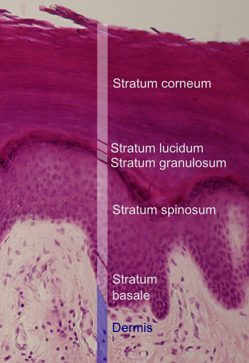
The Malpighian layer (''stratum mucosum'' or ''stratum malpighii'') of the epidermis, the outermost layer of the skin, is generally defined as both the stratum basale (basal layer) and the thicker stratum spinosum (spinous layer/prickle cell layer) immediately above it as a single unit,McGrath, J.A.; Eady, R.A.; Pope, F.M. (2004). ''Rook's Textbook of Dermatology'' (Seventh Edition). Blackwell Publishing. Pages 3.1-3.6. . although it is occasionally defined as the stratum basale specifically, or the stratum spinosum specifically. It is named after the Italian biologist and physician Marcello Malpighi. Basal cell carcinoma originates from the basal layer of the stratum malpighii. This layer is where almost all of the mitotic activity in the epidermis occurs. The activity of these cells is increased by IL-1 (interleukin-1) and epidermal growth factor. The activity is decreased by transforming growth factor beta.Mescher, A. L., Mescher, A. L., & Junqueira, L. C. U. (2016). Junqueira ... [...More Info...] [...Related Items...] OR: [Wikipedia] [Google] [Baidu] |
Prickle Cell
Spinous cells, or prickle cells, are keratin producing Epidermis (skin), epidermal cells owing their prickly appearance to their numerous intracellular connections. They make up the Stratum spinosum, stratum spinosum (prickly layer) of the epidermis and provide a continuous net-like layer of protection for underlying tissue. They are susceptible to mutations caused by sunlight and can become malignant. Location Spinous cells are found in the superficial layers of the skin. They are found in the Stratum spinosum, stratum spinosum (prickly layer, spinosum layer), which lies above the Stratum basale, stratum basale (basal layer) and below the Stratum granulosum, stratum granulosum (granular layer) of the epidermis. The spinous cells are arranged several layers thick to form a net-like covering. Origin Spinous cells originate through mitosis in the basal layer (also known as the germinative layer). They are pushed upward into the stratum spinosum by the continuous formation of n ... [...More Info...] [...Related Items...] OR: [Wikipedia] [Google] [Baidu] |
Simple Columnar Epithelium
Simple columnar epithelium is a single layer of columnar epithelial cells which are tall and slender with oval-shaped nuclei located in the basal region, attached to the basement membrane. In humans, simple columnar epithelium lines most organs of the digestive tract including the stomach, and intestines. Simple columnar epithelium also lines the uterus. Structure Simple columnar epithelium is further divided into two categories: ciliated and non-ciliated (glandular). The ciliated part of the simple columnar epithelium has tiny hairs which help move mucus and other substances up the respiratory tract. The shape of the simple columnar epithelium cells are tall and narrow giving a column like appearance. the apical surfaces of the tissue face the lumen of organs while the basal side faces the basement membrane. the nuclei are located closer along the basal side of the cell. Absorptive columnar epithelium is characterized as having a striated boarder on its apical side, this bo ... [...More Info...] [...Related Items...] OR: [Wikipedia] [Google] [Baidu] |
Bowman's Membrane
The Bowman layer (Bowman's membrane, anterior limiting lamina, anterior elastic lamina) is a smooth, acellular, nonregenerating layer, located between the superficial epithelium and the stroma in the cornea of the eye. It is composed of strong, randomly oriented collagen fibrils in which the smooth anterior surface faces the epithelial basement membrane and the posterior surface merges with the collagen lamellae of the corneal stroma proper. In adult humans, Bowman layer is 8-12 μm thick. With ageing, this layer becomes thinner. The function of the Bowman layer remains unclear and appears to have no critical function in corneal physiology. Recently, it is postulated that the layer may act as a physical barrier to protect the subepithelial nerve plexus and thereby hastens epithelial innervation and sensory recovery. Moreover, it may also serve as a barrier that prevents direct traumatic contact with the corneal stroma and hence it is highly involved in stromal wound healing ... [...More Info...] [...Related Items...] OR: [Wikipedia] [Google] [Baidu] |


