|
Corneal Endothelium
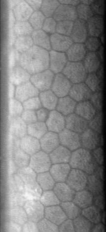
The corneal endothelium is a single layer of endothelial cells on the inner surface of the cornea. It faces the chamber formed between the cornea and the iris. The corneal endothelium are specialized, flattened, mitochondria-rich cells that line the posterior surface of the cornea and face the anterior chamber of the eye. The corneal endothelium governs fluid and solute transport across the posterior surface of the cornea and maintains the cornea in the slightly dehydrated state that is required for optical transparency. Embryology and anatomy The corneal endothelium is embryologically derived from the neural crest. The postnatal total endothelial cellularity of the cornea (approximately 300,000 cells per cornea) is achieved as early as the second trimester of gestation. Thereafter the endothelial cell density (but not the absolute number of cells) rapidly declines, as the fetal cornea grows in surface area, achieving a final adult density of approximately 2400 - 3200 cells ... [...More Info...] [...Related Items...] OR: [Wikipedia] [Google] [Baidu] |
Corneal Epithelium
The corneal epithelium (epithelium corneæ anterior layer) is made up of epithelial tissue and covers the front of the cornea. It acts as a barrier to protect the cornea, resisting the free flow of fluids from the tears, and prevents bacteria from entering the epithelium and corneal stroma. Anatomy The corneal epithelium consists of several layers of cells. The cells of the deepest layer are columnar, known as basal cells which are attached by multiprotein complexes known as hemidesmosomes to an underlying basement membrane. Then follow two or three layers of polyhedral cells, commonly known as wing cells. The majority of these are prickle cells, similar to those found in the stratum mucosum of the cuticle. Lastly, there are three or four layers of squamous cells, with flattened nuclei. The layers of the epithelium are constantly undergoing mitosis. Basal and wing cells migrate to the anterior of the cornea, while squamous cells age and slough off into the tear film. Central ... [...More Info...] [...Related Items...] OR: [Wikipedia] [Google] [Baidu] |
Cataract Surgery
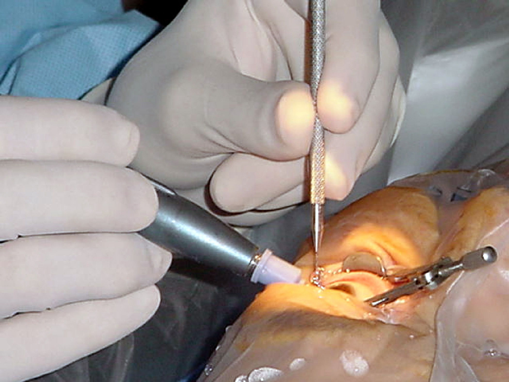
Cataract surgery, also called lens replacement surgery, is the removal of the natural lens of the eye (also called "crystalline lens") that has developed an opacification, which is referred to as a cataract, and its replacement with an intraocular lens. Metabolic changes of the crystalline lens fibers over time lead to the development of the cataract, causing impairment or loss of vision. Some infants are born with congenital cataracts, and certain environmental factors may also lead to cataract formation. Early symptoms may include strong glare from lights and small light sources at night, and reduced acuity at low light levels. During cataract surgery, a patient's cloudy natural cataract lens is removed, either by emulsification in place or by cutting it out. An artificial intraocular lens (IOL) is implanted in its place. Cataract surgery is generally performed by an ophthalmologist in an ambulatory setting at a surgical center or hospital rather than an inpatient setting. Eit ... [...More Info...] [...Related Items...] OR: [Wikipedia] [Google] [Baidu] |
Visual System
The visual system comprises the sensory organ (the eye) and parts of the central nervous system (the retina containing photoreceptor cells, the optic nerve, the optic tract and the visual cortex) which gives organisms the sense of sight (the ability to perception, detect and process visible light) as well as enabling the formation of several non-image photo response functions. It detects and interprets information from the optical spectrum perceptible to that species to "build a representation" of the surrounding environment. The visual system carries out a number of complex tasks, including the reception of light and the formation of monocular neural representations, colour vision, the neural mechanisms underlying stereopsis and assessment of distances to and between objects, the identification of a particular object of interest, motion perception, the analysis and integration of visual information, pattern recognition, accurate motor coordination under visual guidance, and mor ... [...More Info...] [...Related Items...] OR: [Wikipedia] [Google] [Baidu] |
Human Eye Anatomy
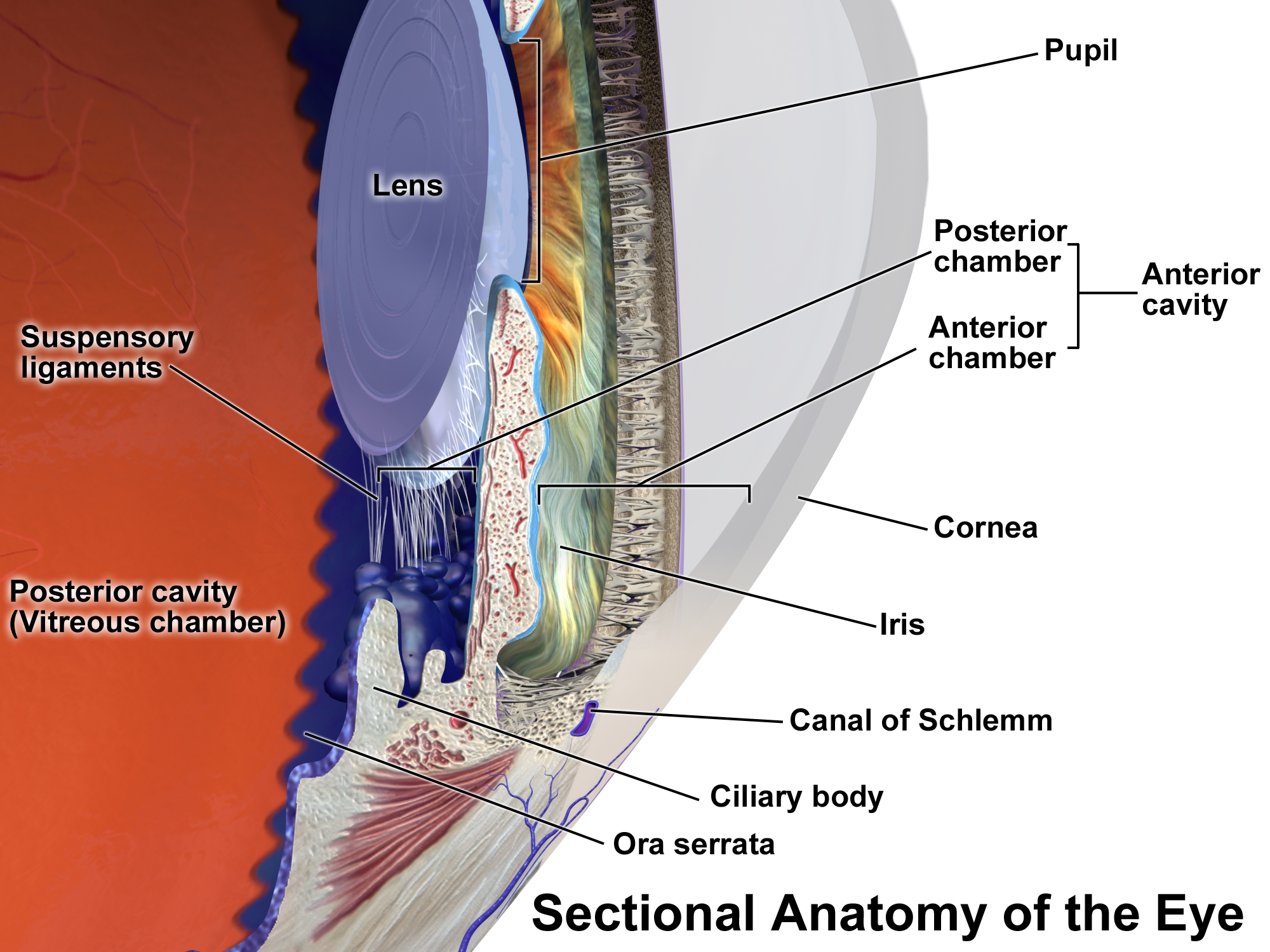
Humans (''Homo sapiens'') are the most abundant and widespread species of primate, characterized by bipedalism and exceptional cognitive skills due to a large and complex brain. This has enabled the development of advanced tools, culture, and language. Humans are highly social and tend to live in complex social structures composed of many cooperating and competing groups, from families and kinship networks to political states. Social interactions between humans have established a wide variety of values, social norms, and rituals, which bolster human society. Its intelligence and its desire to understand and influence the environment and to explain and manipulate phenomena have motivated humanity's development of science, philosophy, mythology, religion, and other fields of study. Although some scientists equate the term ''humans'' with all members of the genus ''Homo'', in common usage, it generally refers to ''Homo sapiens'', the only extant member. Anatomically modern huma ... [...More Info...] [...Related Items...] OR: [Wikipedia] [Google] [Baidu] |
Corneal Transplantation
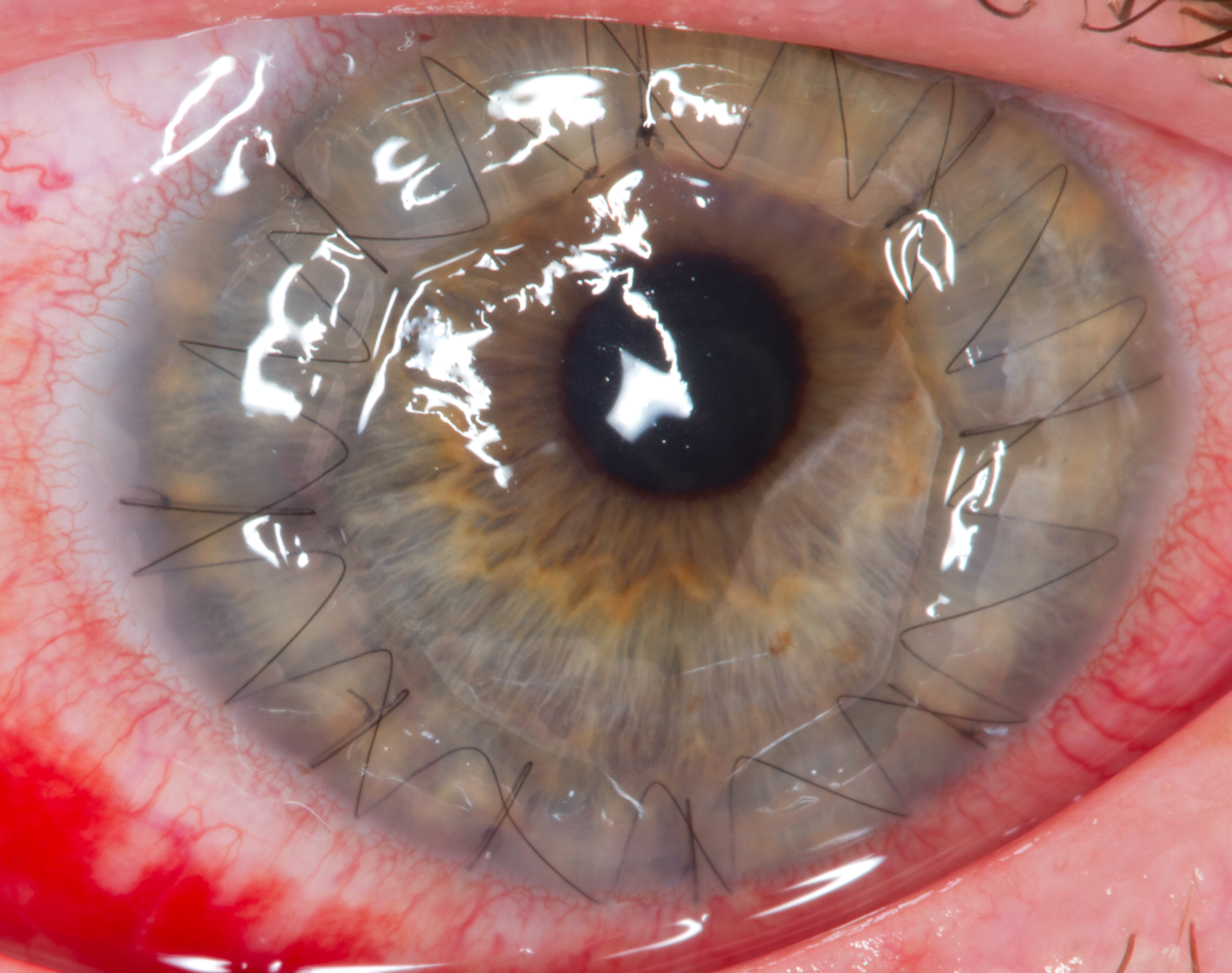
Corneal transplantation, also known as corneal grafting, is a surgical procedure where a damaged or diseased cornea is replaced by donated corneal tissue (the graft). When the entire cornea is replaced it is known as penetrating keratoplasty and when only part of the cornea is replaced it is known as lamellar keratoplasty. Keratoplasty simply means surgery to the cornea. The graft is taken from a recently deceased individual with no known diseases or other factors that may affect the chance of survival of the donated tissue or the health of the recipient. The cornea is the transparent front part of the eye that covers the iris, pupil and anterior chamber. The surgical procedure is performed by ophthalmologists, physicians who specialize in eyes, and is often done on an outpatient basis. Donors can be of any age, as is shown in the case of Janis Babson, who donated her eyes after dying at the age of 10. Corneal transplantation is performed when medicines, keratoconus conservat ... [...More Info...] [...Related Items...] OR: [Wikipedia] [Google] [Baidu] |
X-linked Endothelial Corneal Dystrophy
X-linked endothelial corneal dystrophy (XECD) is a rare form of corneal dystrophy described first in 2006, based on a 4-generation family of 60 members with 9 affected males and 35 trait carriers, which led to mapping the XECD locus to Xq25. It manifests as severe corneal opacification or clouding, sometimes congenital, in the form of a ground glass, milky corneal tissue, and moon crater-like changes of corneal endothelium The corneal endothelium is a single layer of endothelial cells on the inner surface of the cornea. It faces the chamber formed between the cornea and the iris. The corneal endothelium are specialized, flattened, mitochondria-rich cells that li .... Trait carriers manifest only endothelial alterations resembling moon craters. As of December 2014, the molecular basis for this disease remained unknown, although 181 genes were known to be within the XECD locus, of which 68 were known to be protein-coding. References External links {{Human corneal dystrophy ... [...More Info...] [...Related Items...] OR: [Wikipedia] [Google] [Baidu] |
Iritis
Uveitis () is inflammation of the uvea, the pigmented layer of the eye between the inner retina and the outer fibrous layer composed of the sclera and cornea. The uvea consists of the middle layer of pigmented vascular structures of the eye and includes the iris, ciliary body, and choroid. Uveitis is described anatomically, by the part of the eye affected, as anterior, intermediate or posterior, or panuveitic if all parts are involved. Anterior uveitis ( iridocyclytis) is the most common, with the incidence of uveitis overall affecting approximately 1:4500, most commonly those between the ages of 20-60. Symptoms include eye pain, eye redness, floaters and blurred vision, and ophthalmic examination may show dilated ciliary blood vessels and the presence of cells in the anterior chamber. Uveitis may arise spontaneously, have a genetic component, or be associated with an autoimmune disease or infection. While the eye is a relatively protected environment, its immune mechanisms may ... [...More Info...] [...Related Items...] OR: [Wikipedia] [Google] [Baidu] |
Aging
Ageing ( BE) or aging ( AE) is the process of becoming older. The term refers mainly to humans, many other animals, and fungi, whereas for example, bacteria, perennial plants and some simple animals are potentially biologically immortal. In a broader sense, ageing can refer to single cells within an organism which have ceased dividing, or to the population of a species. In humans, ageing represents the accumulation of changes in a human being over time and can encompass physical, psychological, and social changes. Reaction time, for example, may slow with age, while memories and general knowledge typically increase. Ageing increases the risk of human diseases such as cancer, Alzheimer's disease, diabetes, cardiovascular disease, stroke and many more. Of the roughly 150,000 people who die each day across the globe, about two-thirds die from age-related causes. Current ageing theories are assigned to the damage concept, whereby the accumulation of damage (such as DNA ox ... [...More Info...] [...Related Items...] OR: [Wikipedia] [Google] [Baidu] |
Glaucoma
Glaucoma is a group of eye diseases that result in damage to the optic nerve (or retina) and cause vision loss. The most common type is open-angle (wide angle, chronic simple) glaucoma, in which the drainage angle for fluid within the eye remains open, with less common types including closed-angle (narrow angle, acute congestive) glaucoma and normal-tension glaucoma. Open-angle glaucoma develops slowly over time and there is no pain. Peripheral vision may begin to decrease, followed by central vision, resulting in blindness if not treated. Closed-angle glaucoma can present gradually or suddenly. The sudden presentation may involve severe eye pain, blurred vision, mid-dilated pupil, redness of the eye, and nausea. Vision loss from glaucoma, once it has occurred, is permanent. Eyes affected by glaucoma are referred to as being glaucomatous. Risk factors for glaucoma include increasing age, high pressure in the eye, a family history of glaucoma, and use of steroid medication. F ... [...More Info...] [...Related Items...] OR: [Wikipedia] [Google] [Baidu] |
Intraocular Lens
Intraocular lens (IOL) is a lens implanted in the eye as part of a treatment for cataracts or myopia. If the natural lens is left in the eye, the IOL is known as phakic, otherwise it is a pseudophakic, or false lens. Such a lens is typically implanted during cataract surgery, after the eye's cloudy natural lens (cataract) has been removed. The pseudophakic IOL provides the same light-focusing function as the natural crystalline lens. The phakic type of IOL is placed over the existing natural lens and is used in refractive surgery to change the eye's optical power as a treatment for myopia (nearsightedness). This is an alternative to LASIK. IOLs usually consist of a small plastic lens with plastic side struts, called haptics, to hold the lens in place in the capsular bag inside the eye. IOLs were conventionally made of an inflexible material (PMMA), although this has largely been superseded by the use of flexible materials, such as silicone. Most IOLs fitted today are fixed mo ... [...More Info...] [...Related Items...] OR: [Wikipedia] [Google] [Baidu] |
Fuchs' Dystrophy
Fuchs dystrophy, also referred to as Fuchs endothelial corneal dystrophy (FECD) and Fuchs endothelial dystrophy (FED), is a slowly progressing corneal dystrophy that usually affects both eyes and is slightly more common in women than in men. Although early signs of Fuchs dystrophy are sometimes seen in people in their 30s and 40s, the disease rarely affects vision until people reach their 50s and 60s. Signs and symptoms As a progressive, chronic condition, signs and symptoms of Fuchs dystrophy gradually progress over decades of life, starting in middle age. Early symptoms include blurry vision upon wakening which improves during the morning, as fluid retained in the cornea is unable to evaporate through the surface of the eye when the lids are closed overnight. As the disease worsens, the interval of blurry morning vision extends from minutes to hours. In moderate stages of the disease, an increase in guttae and swelling in the cornea can contribute to changes in vision and decreas ... [...More Info...] [...Related Items...] OR: [Wikipedia] [Google] [Baidu] |
COL1A1
Collagen, type I, alpha 1, also known as alpha-1 type I collagen, is a protein that in humans is encoded by the gene. ''COL1A1'' encodes the major component of type I collagen, the fibrillar collagen found in most connective tissues, including cartilage. Function Collagen is a protein that strengthens and supports many tissues in the body, including cartilage, bone, tendon, skin and the white part of the eye (sclera). The gene produces a component of type I collagen, called the pro-alpha1(I) chain. This chain combines with another pro-alpha1(I) chain and also with a pro-alpha2(I) chain (produced by the gene) to make a molecule of type I procollagen. These triple-stranded, rope-like procollagen molecules must be processed by enzymes outside the cell. Once these molecules are processed, they arrange themselves into long, thin fibrils that cross-link to one another in the spaces around cells. The cross-links result in the formation of very strong mature type I collagen fibers ... [...More Info...] [...Related Items...] OR: [Wikipedia] [Google] [Baidu] |


