|
Complement Component 3
Complement component 3, often simply called C3, is a protein of the immune system. It plays a central role in the complement system and contributes to innate immunity. In humans it is encoded on chromosome 19 by a gene called ''C3''. Function C3 plays a central role in the activation of the complement system. Its activation is required for both classical and alternative complement activation pathways. People with C3 deficiency are susceptible to bacterial infection. One form of C3-convertase, also known as C4b2a, is formed by a heterodimer of activated forms of C4 and C2. It catalyzes the proteolytic cleavage of C3 into C3a and C3b, generated during activation through the classical pathway as well as the lectin pathway. C3a is an anaphylotoxin and the precursor of some cytokines such as ASP, and C3b serves as an opsonizing agent. Factor I can cleave C3b into C3c and C3d, the latter of which plays a role in enhancing B cell responses. In the alternative complement pathway, ... [...More Info...] [...Related Items...] OR: [Wikipedia] [Google] [Baidu] |
Protein
Proteins are large biomolecules and macromolecules that comprise one or more long chains of amino acid residues. Proteins perform a vast array of functions within organisms, including catalysing metabolic reactions, DNA replication, responding to stimuli, providing structure to cells and organisms, and transporting molecules from one location to another. Proteins differ from one another primarily in their sequence of amino acids, which is dictated by the nucleotide sequence of their genes, and which usually results in protein folding into a specific 3D structure that determines its activity. A linear chain of amino acid residues is called a polypeptide. A protein contains at least one long polypeptide. Short polypeptides, containing less than 20–30 residues, are rarely considered to be proteins and are commonly called peptides. The individual amino acid residues are bonded together by peptide bonds and adjacent amino acid residues. The sequence of amino acid residue ... [...More Info...] [...Related Items...] OR: [Wikipedia] [Google] [Baidu] |
B Cell
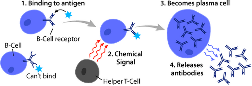
B cells, also known as B lymphocytes, are a type of white blood cell of the lymphocyte subtype. They function in the humoral immunity component of the adaptive immune system. B cells produce antibody molecules which may be either secreted or inserted into the plasma membrane where they serve as a part of B-cell receptors. When a naïve or memory B cell is activated by an antigen, it proliferates and differentiates into an antibody-secreting effector cell, known as a plasmablast or plasma cell. Additionally, B cells present antigens (they are also classified as professional antigen-presenting cells (APCs)) and secrete cytokines. In mammals, B cells mature in the bone marrow, which is at the core of most bones. In birds, B cells mature in the bursa of Fabricius, a lymphoid organ where they were first discovered by Chang and Glick, which is why the 'B' stands for bursa and not bone marrow as commonly believed. B cells, unlike the other two classes of lymphocytes, T cells and ... [...More Info...] [...Related Items...] OR: [Wikipedia] [Google] [Baidu] |
Shunt Nephritis
Shunt nephritis is a rare disease of the kidney that can occur in patients being treated for hydrocephalus with a cerebral shunt. It usually results from an infected shunt that produces a long-standing blood infection, particularly by the bacterium '' Staphylococcus epidermidis''. Kidney disease results from an immune response that deposits immune complexes in the kidney. The most common signs and symptoms of the condition are blood and protein in the urine, anemia, and high blood pressure. Diagnosis is based on these findings in the context of characteristic laboratory values. Treatment includes antibiotics and the prompt removal of the infected shunt. Over half of individuals with shunt nephritis recover completely; most of the remainder have some degree of persistent kidney disease. Presentation The clinical presentation of shunt nephritis is variable, but the most common manifestations of shunt nephritis include blood in the urine, protein in the urine, anemia, and high bloo ... [...More Info...] [...Related Items...] OR: [Wikipedia] [Google] [Baidu] |
Membranoproliferative Glomerulonephritis
Membranoproliferative glomerulonephritis (MPGN) is a type of glomerulonephritis caused by deposits in the kidney glomerular mesangium and basement membrane ( GBM) thickening, activating complement and damaging the glomeruli. MPGN accounts for approximately 4% of primary renal causes of nephrotic syndrome in children and 7% in adults. It should not be confused with membranous glomerulonephritis, a condition in which the basement membrane is thickened, but the mesangium is not. Type There are three types of MPGN, but this classification is becoming obsolete as the causes of this pattern are becoming understood. Type I Type I, the most common by far, is caused by immune complexes depositing in the kidney. It is characterised by subendothelial and mesangial immune deposits. It is believed to be associated with the classical complement pathway. Type II Also called recently as ‘C3 nephropathy’ The preferred name is "dense deposit disease." Most cases of dense deposit disease d ... [...More Info...] [...Related Items...] OR: [Wikipedia] [Google] [Baidu] |
Post-infectious Glomerulonephritis
Acute proliferative glomerulonephritis is a disorder of the small blood vessels of the kidney. It is a common complication of bacterial infections, typically skin infection by ''Streptococcus'' bacteria types 12, 4 and 1 (impetigo) but also after streptococcal pharyngitis, for which it is also known as postinfectious glomerulonephritis (PIGN) or poststreptococcal glomerulonephritis (PSGN). It can be a risk factor for future albuminuria. In adults, the signs and symptoms of infection may still be present at the time when the kidney problems develop, and the terms ''infection-related glomerulonephritis'' or ''bacterial infection-related glomerulonephritis'' are also used. Acute glomerulonephritis resulted in 19,000 deaths in 2013, down from 24,000 deaths in 1990 worldwide. Signs and symptoms Among the signs and symptoms of acute proliferative glomerulonephritis are the following: * Hematuria * Oliguria * Edema * Hypertension * Fever (headache, malaise, anorexia, nausea.) Cause ... [...More Info...] [...Related Items...] OR: [Wikipedia] [Google] [Baidu] |
Blood
Blood is a body fluid in the circulatory system of humans and other vertebrates that delivers necessary substances such as nutrients and oxygen to the cells, and transports metabolic waste products away from those same cells. Blood in the circulatory system is also known as ''peripheral blood'', and the blood cells it carries, ''peripheral blood cells''. Blood is composed of blood cells suspended in blood plasma. Plasma, which constitutes 55% of blood fluid, is mostly water (92% by volume), and contains proteins, glucose, mineral ions, hormones, carbon dioxide (plasma being the main medium for excretory product transportation), and blood cells themselves. Albumin is the main protein in plasma, and it functions to regulate the colloidal osmotic pressure of blood. The blood cells are mainly red blood cells (also called RBCs or erythrocytes), white blood cells (also called WBCs or leukocytes) and platelets (also called thrombocytes). The most abundant cells in vertebrate blo ... [...More Info...] [...Related Items...] OR: [Wikipedia] [Google] [Baidu] |
Keratinocyte
Keratinocytes are the primary type of Cell (biology), cell found in the epidermis (skin), epidermis, the outermost layer of the skin. In humans, they constitute 90% of epidermal skin cells. Basal cells in the stratum basale, basal layer (''stratum basale'') of the skin are sometimes referred to as basal keratinocytes. Keratinocytes form a barrier against environmental damage by heat, UV radiation, Dehydration, water loss, pathogenic bacteria, fungi, parasites, and viruses. A number of structural proteins, enzymes, lipids, and antimicrobial peptides contribute to maintain the important barrier function of the skin. Keratinocytes differentiate from epidermal stem cells in the lower part of the epidermis and migrate towards the surface, finally becoming corneocytes and eventually be shed off, which happens every 40 to 56 days in humans. Function The primary function of keratinocytes is the formation of a barrier against environmental damage by heat, UV radiation, Dehydration, wat ... [...More Info...] [...Related Items...] OR: [Wikipedia] [Google] [Baidu] |
Hepatocyte
A hepatocyte is a cell of the main parenchymal tissue of the liver. Hepatocytes make up 80% of the liver's mass. These cells are involved in: * Protein synthesis * Protein storage * Transformation of carbohydrates * Synthesis of cholesterol, bile salts and phospholipids * Detoxification, modification, and excretion of exogenous and endogenous substances * Initiation of formation and secretion of bile Structure The typical hepatocyte is cubical with sides of 20-30 μm, (in comparison, a human hair has a diameter of 17 to 180 μm).The diameter of human hair ranges from 17 to 181 μm. The typical volume of a hepatocyte is 3.4 x 10⁻⁹ cm3. Smooth endoplasmic reticulum is abundant in hepatocytes, in contrast to most other cell types. Microanatomy Hepatocytes display an eosinophilic cytoplasm, reflecting numerous mitochondria, and basophilic stippling due to large amounts of smooth endoplasmic reticulum and free ribosomes. Brown lipofuscin granules are also observed ... [...More Info...] [...Related Items...] OR: [Wikipedia] [Google] [Baidu] |
Factor H
Factor H is a member of the regulators of complement activation family and is a complement control protein. It is a large (155 kilodaltons), soluble glycoprotein that circulates in human plasma (at typical concentrations of 200–300 micrograms per milliliter). Its principal function is to regulate the alternative pathway of the complement system, ensuring that the complement system is directed towards pathogens or other dangerous material and does not damage host tissue. Factor H regulates complement activation on self cells and surfaces by possessing both cofactor activity for the Factor I mediated C3b cleavage, and decay accelerating activity against the alternative pathway C3-convertase, C3bBb. Factor H exerts its protective action on self cells and self surfaces but not on the surfaces of bacteria or viruses. This is thought to be the result of Factor H having the ability to adopt conformations with lower or higher activities as a cofactor for C3 cleavage or decay accelerat ... [...More Info...] [...Related Items...] OR: [Wikipedia] [Google] [Baidu] |
Cofactor (biochemistry)
A cofactor is a non-protein chemical compound or metallic ion that is required for an enzyme's role as a catalyst (a catalyst is a substance that increases the rate of a chemical reaction). Cofactors can be considered "helper molecules" that assist in biochemical transformations. The rates at which these happen are characterized in an area of study called enzyme kinetics. Cofactors typically differ from ligands in that they often derive their function by remaining bound. Cofactors can be divided into two types: inorganic ions and complex organic molecules called coenzymes. Coenzymes are mostly derived from vitamins and other organic essential nutrients in small amounts. (Note that some scientists limit the use of the term "cofactor" for inorganic substances; both types are included here.) Coenzymes are further divided into two types. The first is called a "prosthetic group", which consists of a coenzyme that is tightly (or even covalently) and permanently bound to a protein. ... [...More Info...] [...Related Items...] OR: [Wikipedia] [Google] [Baidu] |
Decay-accelerating Factor
Complement decay-accelerating factor, also known as CD55 or DAF, is a protein that, in humans, is encoded by the ''CD55'' gene. DAF regulates the complement system on the cell surface. It recognizes C4b and C3b fragments that are created during activation of C4 ( classical or lectin pathway) or C3 (alternative pathway). Interaction of DAF with cell-associated C4b of the classical and lectin pathways interferes with the conversion of C2 to C2b, thereby preventing formation of the C4b2a C3-convertase, and interaction of DAF with C3b of the alternative pathway interferes with the conversion of factor B to Bb by factor D, thereby preventing formation of the C3bBb C3 convertase of the alternative pathway. Thus, by limiting the amplification convertases of the complement cascade, DAF indirectly blocks the formation of the membrane attack complex. This glycoprotein is broadly distributed among hematopoietic and non-hematopoietic cells. It is a determinant for the Cromer blood group syste ... [...More Info...] [...Related Items...] OR: [Wikipedia] [Google] [Baidu] |
Nucleophile
In chemistry, a nucleophile is a chemical species that forms bonds by donating an electron pair. All molecules and ions with a free pair of electrons or at least one pi bond can act as nucleophiles. Because nucleophiles donate electrons, they are Lewis bases. ''Nucleophilic'' describes the affinity of a nucleophile to bond with positively charged atomic nuclei. Nucleophilicity, sometimes referred to as nucleophile strength, refers to a substance's nucleophilic character and is often used to compare the affinity of atoms. Neutral nucleophilic reactions with solvents such as alcohols and water are named solvolysis. Nucleophiles may take part in nucleophilic substitution, whereby a nucleophile becomes attracted to a full or partial positive charge, and nucleophilic addition. Nucleophilicity is closely related to basicity. History The terms ''nucleophile'' and ''electrophile'' were introduced by Christopher Kelk Ingold in 1933, replacing the terms ''anionoid'' and ''cationoid' ... [...More Info...] [...Related Items...] OR: [Wikipedia] [Google] [Baidu] |


