|
Common Plantar Digital Nerves Of Medial Plantar Nerve
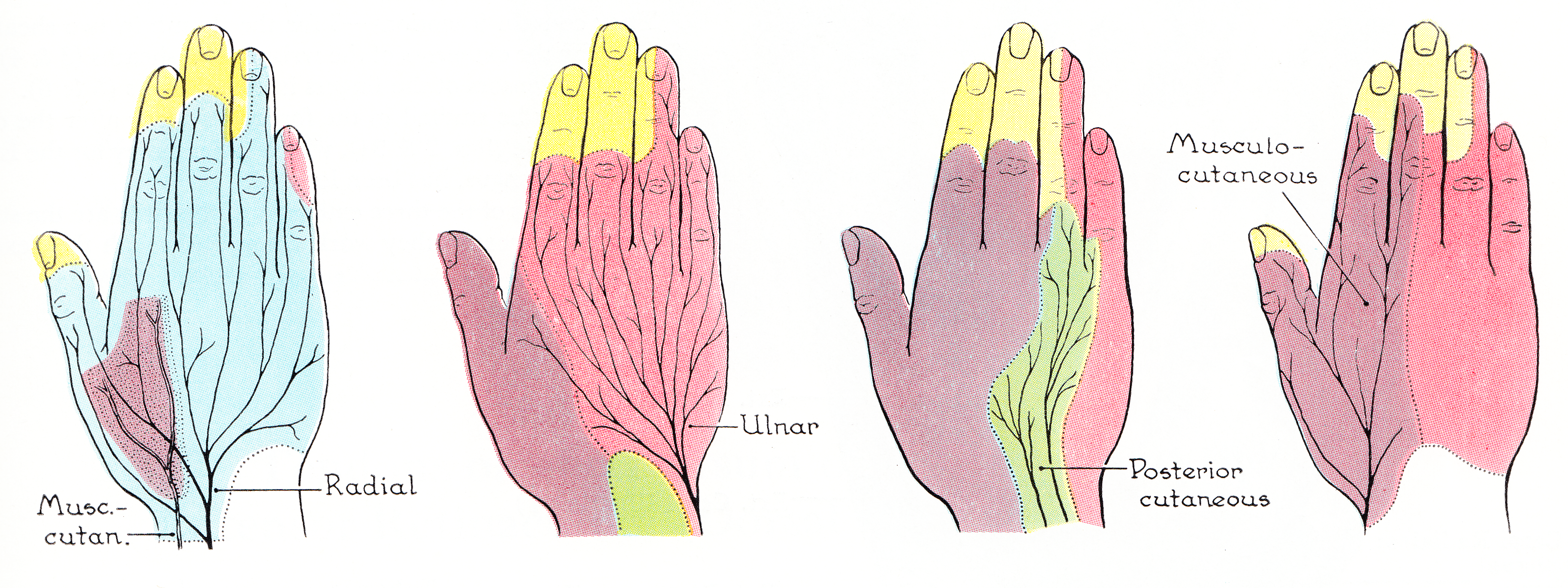
The common plantar digital nerves of medial plantar nerve are nerves of the foot. The three common digital nerves (nn. digitales plantares communes) pass between the divisions of the plantar aponeurosis, and each splits into two proper digital nerves: * Those of the first common digital nerve supply the adjacent sides of the great and second toes; * Those of the second, the adjacent sides of the second and third toes; and those of the third, the adjacent sides of the third and fourth toes. * The third common digital nerve receives a communicating branch from the lateral plantar nerve; the first gives a twig to the first Lumbricalis. Each proper digital nerve gives off cutaneous and articular filaments; and opposite the last phalanx sends upward a dorsal branch, which supplies the structures around the nail, the continuation of the nerve being distributed to the ball of the toe. It will be observed that these digital nerves are similar in their distribution to those of the median ... [...More Info...] [...Related Items...] OR: [Wikipedia] [Google] [Baidu] |
Medial Plantar Nerve
The medial plantar nerve (internal plantar nerve) is the larger of the two terminal divisions of the tibial nerve (medial and lateral plantar nerve), which accompanies the medial plantar artery. From its origin under the laciniate ligament it passes under cover of the abductor hallucis muscle, and, appearing between this muscle and the flexor digitorum brevis, gives off a proper digital plantar nerve and finally divides opposite the bases of the metatarsal bones into three common digital plantar nerves. Branches The branches of the medial plantar nerve are: (1) cutaneous, (2) muscular, (3) articular, (4) a proper digital nerve to the medial side of the great toe, and (5) three common digital nerves. Cutaneous branches The cutaneous branches pierce the plantar aponeurosis between the abductor hallucis and the flexor digitorum brevis and are distributed to the skin of the sole of the foot. Muscular branches The muscular branches supply muscles on the medial side of the sole, incl ... [...More Info...] [...Related Items...] OR: [Wikipedia] [Google] [Baidu] |
Lumbricals Of The Foot
The lumbricals are four small skeletal muscles, accessory to the tendons of the flexor digitorum longus muscle. They are numbered from the medial side of the foot. Structure The lumbricals arise from the tendons of the flexor digitorum longus muscle, as far back as their angles of division, each springing from two tendons, except the first. The first lumbrical is unipennate, while the second, third and fourth are bipennate. The muscles end in tendons, which pass forward on the medial sides of the four lesser toes, and are inserted into the expansions of the tendons of the extensor digitorum longus muscle on the dorsal surfaces of the proximal phalanges. All four lumbricals insert into extensor hoods of the phalanges, thus creating extension at the inter-phalangeal (PIP and DIP) joints. However, as the tendons also pass inferior to the metatarsal phalangeal (MTP) joints it creates flexion at this joint. Innervation The most medial lumbrical is innervated by the medial pl ... [...More Info...] [...Related Items...] OR: [Wikipedia] [Google] [Baidu] |
Median Nerve
The median nerve is a nerve in humans and other animals in the upper limb. It is one of the five main nerves originating from the brachial plexus. The median nerve originates from the lateral and medial cords of the brachial plexus, and has contributions from ventral roots of C5-C7 (lateral cord) and C8 and T1 (medial cord). The median nerve is the only nerve that passes through the carpal tunnel. Carpal tunnel syndrome is the disability that results from the median nerve being pressed in the carpal tunnel. Structure The median nerve arises from the branches from lateral and medial cords of the brachial plexus, courses through the anterior part of arm, forearm, and hand, and terminates by supplying the muscles of the hand. Arm After receiving inputs from both the lateral and medial cords of the brachial plexus, the median nerve enters the arm from the axilla at the inferior margin of the teres major muscle. It then passes vertically down and courses lateral to the brachial ar ... [...More Info...] [...Related Items...] OR: [Wikipedia] [Google] [Baidu] |
Common Plantar Digital Arteries
The common plantar digital arteries are arteries of the foot. See also * Common plantar digital nerves of medial plantar nerve * Common plantar digital nerves of lateral plantar nerve External links * http://www.dartmouth.edu/~humananatomy/figures/chapter_17/17-3.HTM Arteries of the lower limb {{circulatory-stub ... [...More Info...] [...Related Items...] OR: [Wikipedia] [Google] [Baidu] |
