|
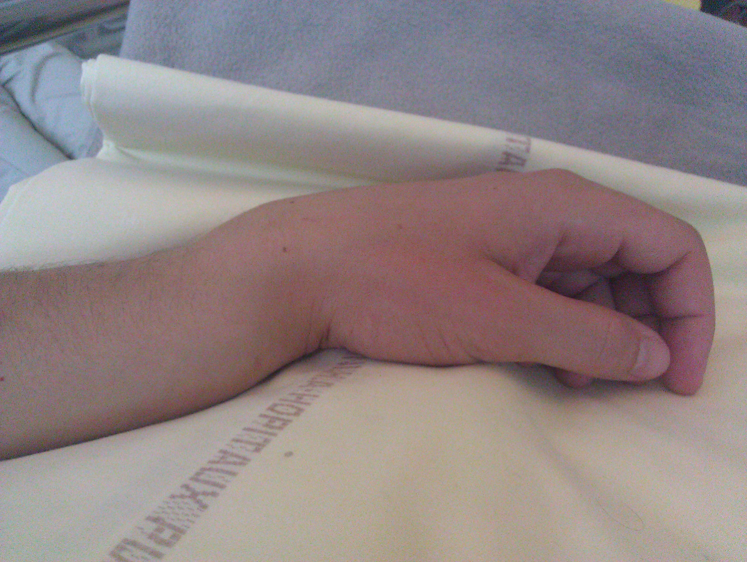
Colles' Fracture
A Colles' fracture is a type of fracture of the distal forearm in which the broken end of the radius is bent backwards. Symptoms may include pain, swelling, deformity, and bruising. Complications may include damage to the median nerve. It typically occurs as a result of a fall on an outstretched hand. Risk factors include osteoporosis. The diagnosis may be confirmed via X-rays. The tip of the ulna may also be broken. Treatment may include casting or surgery. Surgical reduction and casting is possible in the majority of cases in people over the age of 50. Pain management can be achieved during the reduction with procedural sedation and analgesia or a hematoma block. A year or two may be required for healing to occur. About 15% of people have a Colles' fracture at some point in their life. They occur more commonly in young adults and older people than in children and middle-aged adults. Women are more frequently affected than men. The fracture is named after Abraham Colles ... [...More Info...] [...Related Items...] OR: [Wikipedia] [Google] [Baidu] |
Emergency Medicine
Emergency medicine is the medical speciality concerned with the care of illnesses or injuries requiring immediate medical attention. Emergency physicians (often called “ER doctors” in the United States) continuously learn to care for unscheduled and undifferentiated patients of all ages. As first-line providers, in coordination with Emergency Medical Services, they are primarily responsible for initiating resuscitation and stabilization and performing the initial investigations and interventions necessary to diagnose and treat illnesses or injuries in the acute phase. Emergency physicians generally practise in hospital emergency departments, pre-hospital settings via emergency medical services, and intensive care units. Still, they may also work in primary care settings such as urgent care clinics. Sub-specializations of emergency medicine include; disaster medicine, medical toxicology, point-of-care ultrasonography, critical care medicine, emergency medical service ... [...More Info...] [...Related Items...] OR: [Wikipedia] [Google] [Baidu] |
Smith's Fracture
A Smith's fracture, is a fracture of the distal radius. Although it can also be caused by a direct blow to the dorsal forearm or by a fall with the wrist flexed, the most common mechanism of injury for Smith's fracture occurs in a palmar fall with the wrist joint slightly dorsiflexed. Smith's fractures are less common than Colles' fractures. The distal fracture fragment is displaced volarly (ventrally), as opposed to a Colles' fracture which the fragment is displaced dorsally. Depending on the severity of the impact, there may be one or many fragments and it may or may not involve the articular surface of the wrist joint. Classification A commonly used classification of distal radial fractures is the Frykman Classification: * Type I: Extra-articular * Type II: Type I, with fracture of distal ulna * Type III: Radiocarpal joint involvement * Type IV: Type III with fracture of distal ulna * Type V: Distal radioulnar joint involved. * Type VI: Type V with fracture of distal ulna * ... [...More Info...] [...Related Items...] OR: [Wikipedia] [Google] [Baidu] |
Gartland & Werley Classification
Gartland & Werley classification is a system of categorizing Colles' fractures. In the Gartland & Werley classification system there are three types of fractures. The classification system is based on metaphysical comminution, intra-articular extension and displacement, and was first published in 1951. Classification * Type 1: Extra-articular, displaced * Type 2: Intra-articular, no displacement * Type 3: Intra-articular, displaced See also * Frykman classification Frykman classification is a system of categorizing Colles' fractures. In the Frykman classification system there are four types of fractures. Classification Though the Frykman classification system has traditionally been used, there is little valu ... * Lidström classification * Nissen-Lie classification * Older's classification References {{DEFAULTSORT:Gartland and Werley classification Orthopedic classifications ... [...More Info...] [...Related Items...] OR: [Wikipedia] [Google] [Baidu] |
Frykman Classification
Frykman classification is a system of categorizing Colles' fractures. In the Frykman classification system there are four types of fractures. Classification Though the Frykman classification system has traditionally been used, there is little value in its use because it does not help direct treatment. The classification is as follows: See also * Gartland & Werley classification Gartland & Werley classification is a system of categorizing Colles' fractures. In the Gartland & Werley classification system there are three types of fractures. The classification system is based on metaphysical comminution, intra-articular exte ... * Lidström classification * Nissen-Lie classification * Older's classification References {{Reflist Orthopedic classifications ... [...More Info...] [...Related Items...] OR: [Wikipedia] [Google] [Baidu] |
Bayonet
A bayonet (from French ) is a knife, dagger, sword, or spike-shaped weapon designed to fit on the end of the muzzle of a rifle, musket or similar firearm, allowing it to be used as a spear-like weapon.Brayley, Martin, ''Bayonets: An Illustrated History'', Iola, WI: Krause Publications, , (2004), pp. 9–10, 83–85. From the 17th century to World War I, it was a weapon for infantry attacks. Today it is considered an ancillary weapon or a weapon of last resort. History The term ''bayonette'' itself dates back to the mid-to-late 16th century, but it is not clear whether bayonets at the time were knives that could be fitted to the ends of firearms, or simply a type of knife. For example, Cotgrave's 1611 ''Dictionarie'' describes the bayonet as "a kind of small flat pocket dagger, furnished with knives; or a great knife to hang at the girdle". Likewise, Pierre Borel wrote in 1655 that a kind of long-knife called a ''bayonette'' was made in Bayonne but does not give any ... [...More Info...] [...Related Items...] OR: [Wikipedia] [Google] [Baidu] |
Dinner Fork
In cutlery or kitchenware, a fork (from la, furca 'pitchfork') is a utensil, now usually made of metal, whose long handle terminates in a head that branches into several narrow and often slightly curved tine (structural), tines with which one can spear foods either to hold them to cut with a Table knife, knife or to lift them to the mouth. History Bone forks have been found in archaeological sites of the Bronze Age Qijia culture (2400–1900 BC), the Shang dynasty (c. 1600–c. 1050 BC), as well as later Chinese dynasties.Needham (2000). ''Science and Civilisation in China. Volume 6: Biology and biological technology. Part V: Fermentations and food science.'' Cambridge University Press. Pages 105–110. A stone carving from an Eastern Han tomb (in Ta-kua-liang, Suide County, Shaanxi) depicts three hanging two-pronged forks in a dining scene. Similar forks have also been depicted on top of a stove in a scene at another Eastern Han tomb (in Suide County, Shaanxi). In Ancient Eg ... [...More Info...] [...Related Items...] OR: [Wikipedia] [Google] [Baidu] |
Metacarpus
In human anatomy, the metacarpal bones or metacarpus form the intermediate part of the skeletal hand located between the phalanges of the fingers and the carpal bones of the wrist, which forms the connection to the forearm. The metacarpal bones are analogous to the metatarsal bones in the foot. Structure The metacarpals form a transverse arch to which the rigid row of distal carpal bones are fixed. The peripheral metacarpals (those of the thumb and little finger) form the sides of the cup of the palmar gutter and as they are brought together they deepen this concavity. The index metacarpal is the most firmly fixed, while the thumb metacarpal articulates with the trapezium and acts independently from the others. The middle metacarpals are tightly united to the carpus by intrinsic interlocking bone elements at their bases. The ring metacarpal is somewhat more mobile while the fifth metacarpal is semi-independent.Tubiana ''et al'' 1998, p 11 Each metacarpal bone consists of a bod ... [...More Info...] [...Related Items...] OR: [Wikipedia] [Google] [Baidu] |
Carpal Bones
The carpal bones are the eight small bones that make up the wrist (or carpus) that connects the hand to the forearm. The term "carpus" is derived from the Latin carpus and the Greek καρπός (karpós), meaning "wrist". In human anatomy, the main role of the wrist is to facilitate effective positioning of the hand and powerful use of the extensors and flexors of the forearm, and the mobility of individual carpal bones increase the freedom of movements at the wrist.Kingston 2000, pp 126-127 In tetrapods, the carpus is the sole cluster of bones in the wrist between the radius and ulna and the metacarpus. The bones of the carpus do not belong to individual fingers (or toes in quadrupeds), whereas those of the metacarpus do. The corresponding part of the foot is the tarsus. The carpal bones allow the wrist to move and rotate vertically. Structure Bones The eight carpal bones may be conceptually organized as either two transverse rows, or three longitudinal columns. Wh ... [...More Info...] [...Related Items...] OR: [Wikipedia] [Google] [Baidu] |
Displacement (orthopedic Surgery)
A bone fracture (abbreviated FRX or Fx, Fx, or #) is a medical condition in which there is a partial or complete break in the continuity of any bone in the body. In more severe cases, the bone may be broken into several fragments, known as a ''comminuted fracture''. A bone fracture may be the result of high force impact or stress, or a minimal trauma injury as a result of certain medical conditions that weaken the bones, such as osteoporosis, osteopenia, bone cancer, or osteogenesis imperfecta, where the fracture is then properly termed a pathologic fracture. Signs and symptoms Although bone tissue contains no pain receptors, a bone fracture is painful for several reasons: * Breaking in the continuity of the periosteum, with or without similar discontinuity in endosteum, as both contain multiple pain receptors. * Edema and hematoma of nearby soft tissues caused by ruptured bone marrow evokes pressure pain. * Involuntary muscle spasms trying to hold bone fragments in pl ... [...More Info...] [...Related Items...] OR: [Wikipedia] [Google] [Baidu] |
Dorsum (biology)
Standard anatomical terms of location are used to unambiguously describe the anatomy of animals, including humans. The terms, typically derived from Latin or Greek roots, describe something in its standard anatomical position. This position provides a definition of what is at the front ("anterior"), behind ("posterior") and so on. As part of defining and describing terms, the body is described through the use of anatomical planes and anatomical axes. The meaning of terms that are used can change depending on whether an organism is bipedal or quadrupedal. Additionally, for some animals such as invertebrates, some terms may not have any meaning at all; for example, an animal that is radially symmetrical will have no anterior surface, but can still have a description that a part is close to the middle ("proximal") or further from the middle ("distal"). International organisations have determined vocabularies that are often used as standard vocabularies for subdisciplines of an ... [...More Info...] [...Related Items...] OR: [Wikipedia] [Google] [Baidu] |
Anatomical Terms Of Location
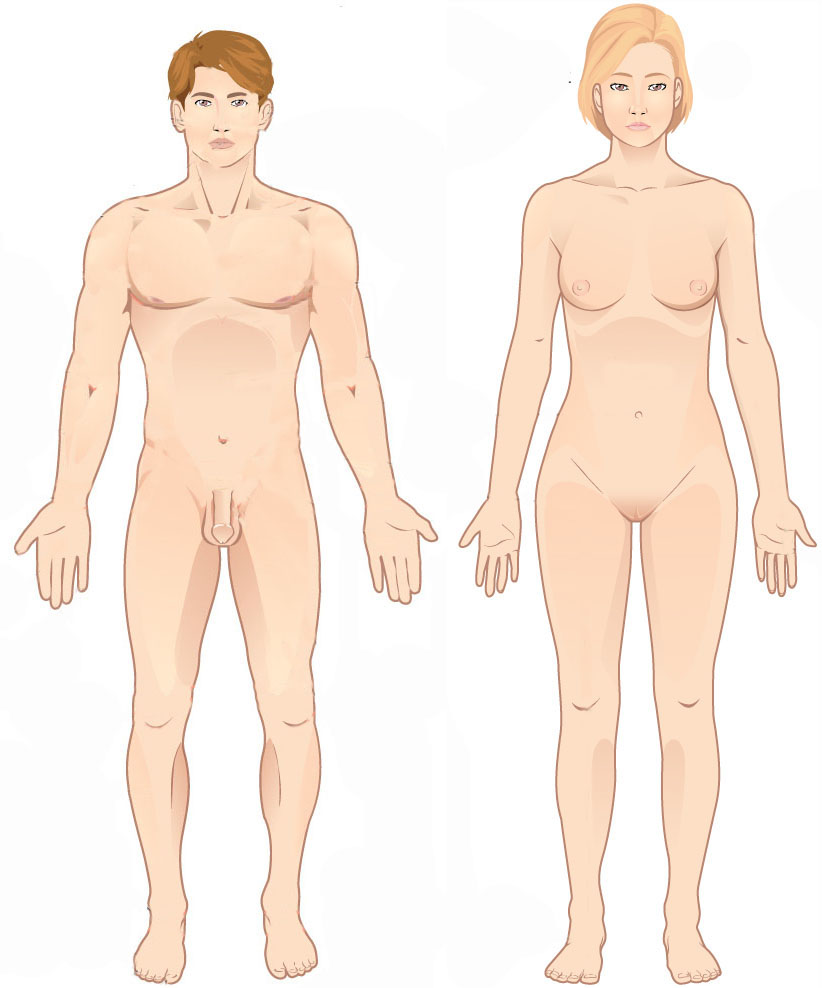
Standard anatomical terms of location are used to unambiguously describe the anatomy of animals, including humans. The terms, typically derived from Latin or Greek roots, describe something in its standard anatomical position. This position provides a definition of what is at the front ("anterior"), behind ("posterior") and so on. As part of defining and describing terms, the body is described through the use of anatomical planes and anatomical axes. The meaning of terms that are used can change depending on whether an organism is bipedal or quadrupedal. Additionally, for some animals such as invertebrates, some terms may not have any meaning at all; for example, an animal that is radially symmetrical will have no anterior surface, but can still have a description that a part is close to the middle ("proximal") or further from the middle ("distal"). International organisations have determined vocabularies that are often used as standard vocabularies for subdisciplines o ... [...More Info...] [...Related Items...] OR: [Wikipedia] [Google] [Baidu] |
Transverse Plane
The transverse plane (also known as the horizontal plane, axial plane and transaxial plane) is an anatomical plane that divides the body into superior and inferior sections. It is perpendicular to the coronal and sagittal planes. List of clinically relevant anatomical planes * Transverse ''thoracic plane'' * '' Xiphosternal plane'' (or xiphosternal junction) * ''Transpyloric plane'' * ''Subcostal plane'' * '' Umbilical plane'' (or transumbilical plane) * ''Supracristal plane'' * ''Intertubercular plane'' (or transtubercular plane) * ''Interspinous plane'' Clinically relevant anatomical planes with associated structures * The transverse ''thoracic plane'' ** Plane through T4 & T5 vertebral junction and sternal angle of Louis. ** Marks the: *** Attachment of costal cartilage of rib 2 at the sternal angle; *** Aortic arch (beginning and end); *** Upper margin of SVC; *** Thoracic duct crossing; *** Tracheal bifurcation; *** Pulmonary trunk bifurcation; * The '' xiphosternal p ... [...More Info...] [...Related Items...] OR: [Wikipedia] [Google] [Baidu] |


_dorsal_view.png)


