|
Cochin Forest Cane Turtle
__NOTOC__ The Cochin forest cane turtle (''Vijayachelys silvatica''), also known as Kavalai forest turtle, forest cane turtle or simply cane turtle, is a rare turtle from the Western Ghats of India. Described in 1912, its type locality is given as "Near Kavalai in the Cochin State Forests, inhabiting dense forest, at an elevation of about 1500 feet above sea level".Henderson (1912) Only two specimens were found at that time, and no scientist saw this turtle in the next 70 years. It was finally rediscovered in 1982, and since then a number of specimens have been found and some studies have been conducted about its affiliation and habits.Praschag et al. (2006) Like its relatives, it belongs to the subfamily Geoemydinae of the family Geoemydidae, formerly known as Bataguridae. It was once placed in the genus ''Geoemyda'' and subsequently moved to ''Heosemys''. But as it seems, the Cochin forest cane turtle forms a quite distinct lineage closely related to ''Melanochelys''. Thus, ... [...More Info...] [...Related Items...] OR: [Wikipedia] [Google] [Baidu] |
Turtle
Turtles are an order of reptiles known as Testudines, characterized by a special shell developed mainly from their ribs. Modern turtles are divided into two major groups, the Pleurodira (side necked turtles) and Cryptodira (hidden necked turtles), which differ in the way the head retracts. There are 360 living and recently extinct species of turtles, including land-dwelling tortoises and freshwater terrapins. They are found on most continents, some islands and, in the case of sea turtles, much of the ocean. Like other amniotes (reptiles, birds, and mammals) they breathe air and do not lay eggs underwater, although many species live in or around water. Turtle shells are made mostly of bone; the upper part is the domed carapace, while the underside is the flatter plastron or belly-plate. Its outer surface is covered in scales made of keratin, the material of hair, horns, and claws. The carapace bones develop from ribs that grow sideways and develop into broad flat plates th ... [...More Info...] [...Related Items...] OR: [Wikipedia] [Google] [Baidu] |
Carapace
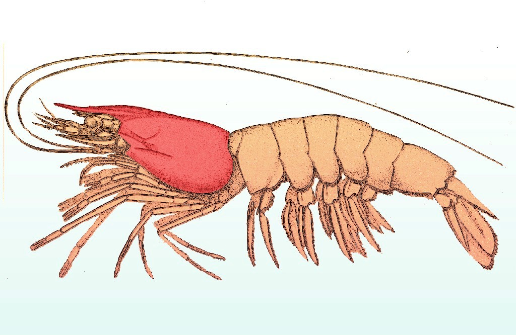
A carapace is a Dorsum (biology), dorsal (upper) section of the exoskeleton or shell in a number of animal groups, including arthropods, such as crustaceans and arachnids, as well as vertebrates, such as turtles and tortoises. In turtles and tortoises, the underside is called the plastron. Crustaceans In crustaceans, the carapace functions as a protective cover over the cephalothorax (i.e., the fused head and thorax, as distinct from the abdomen behind). Where it projects forward beyond the eyes, this projection is called a rostrum (anatomy), rostrum. The carapace is Calcification, calcified to varying degrees in different crustaceans. Zooplankton within the phylum Crustacea also have a carapace. These include Cladocera, ostracods, and Isopoda, isopods, but isopods only have a developed "cephalic shield" carapace covering the head. Arachnids In arachnids, the carapace is formed by the fusion of prosomal tergites into a single Plate (animal anatomy), plate which carries the e ... [...More Info...] [...Related Items...] OR: [Wikipedia] [Google] [Baidu] |
Skull
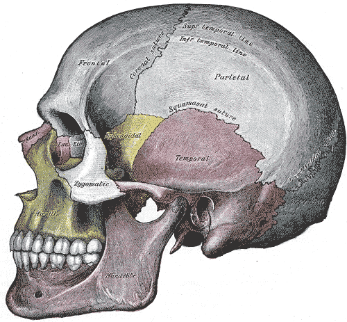
The skull is a bone protective cavity for the brain. The skull is composed of four types of bone i.e., cranial bones, facial bones, ear ossicles and hyoid bone. However two parts are more prominent: the cranium and the mandible. In humans, these two parts are the neurocranium and the viscerocranium ( facial skeleton) that includes the mandible as its largest bone. The skull forms the anterior-most portion of the skeleton and is a product of cephalisation—housing the brain, and several sensory structures such as the eyes, ears, nose, and mouth. In humans these sensory structures are part of the facial skeleton. Functions of the skull include protection of the brain, fixing the distance between the eyes to allow stereoscopic vision, and fixing the position of the ears to enable sound localisation of the direction and distance of sounds. In some animals, such as horned ungulates (mammals with hooves), the skull also has a defensive function by providing the mount (on the front ... [...More Info...] [...Related Items...] OR: [Wikipedia] [Google] [Baidu] |
Symphysis
A symphysis (, pl. symphyses) is a fibrocartilaginous fusion between two bones. It is a type of cartilaginous joint, specifically a secondary cartilaginous joint. # A symphysis is an amphiarthrosis, a slightly movable joint. # A growing together of parts or structures. Unlike synchondroses, symphyses are permanent. Examples The more prominent symphyses are: * the pubic symphysis * sacrococcygeal symphysis * intervertebral disc between two vertebrae * in the sternum, between the manubrium and body Body may refer to: In science * Physical body, an object in physics that represents a large amount, has mass or takes up space * Body (biology), the physical material of an organism * Body plan, the physical features shared by a group of anima ... * mandibular symphysis, in the jaw Symphysis disorders Pubic symphysis diastasis Pubic symphysis diastasis, is an extremely rare complication that occurs in women who are giving birth. Separation of the two pubic bones during deli ... [...More Info...] [...Related Items...] OR: [Wikipedia] [Google] [Baidu] |
Mandible
In anatomy, the mandible, lower jaw or jawbone is the largest, strongest and lowest bone in the human facial skeleton. It forms the lower jaw and holds the lower tooth, teeth in place. The mandible sits beneath the maxilla. It is the only movable bone of the skull (discounting the ossicles of the middle ear). It is connected to the temporal bones by the temporomandibular joints. The bone is formed prenatal development, in the fetus from a fusion of the left and right mandibular prominences, and the point where these sides join, the mandibular symphysis, is still visible as a faint ridge in the midline. Like other symphyses in the body, this is a midline articulation where the bones are joined by fibrocartilage, but this articulation fuses together in early childhood.Illustrated Anatomy of the Head and Neck, Fehrenbach and Herring, Elsevier, 2012, p. 59 The word "mandible" derives from the Latin word ''mandibula'', "jawbone" (literally "one used for chewing"), from ''wikt:mandere ... [...More Info...] [...Related Items...] OR: [Wikipedia] [Google] [Baidu] |
Premaxilla
The premaxilla (or praemaxilla) is one of a pair of small cranial bones at the very tip of the upper jaw of many animals, usually, but not always, bearing teeth. In humans, they are fused with the maxilla. The "premaxilla" of therian mammal has been usually termed as the incisive bone. Other terms used for this structure include premaxillary bone or ''os premaxillare'', intermaxillary bone or ''os intermaxillare'', and Goethe's bone. Human anatomy In human anatomy, the premaxilla is referred to as the incisive bone (') and is the part of the maxilla which bears the incisor teeth, and encompasses the anterior nasal spine and alar region. In the nasal cavity, the premaxillary element projects higher than the maxillary element behind. The palatal portion of the premaxilla is a bony plate with a generally transverse orientation. The incisive foramen is bound anteriorly and laterally by the premaxilla and posteriorly by the palatine process of the maxilla. It is formed from the ... [...More Info...] [...Related Items...] OR: [Wikipedia] [Google] [Baidu] |
Orbit (anatomy)
In anatomy, the orbit is the cavity or socket of the skull in which the eye and its appendages are situated. "Orbit" can refer to the bony socket, or it can also be used to imply the contents. In the adult human, the volume of the orbit is , of which the eye occupies . The orbital contents comprise the eye, the orbital and retrobulbar fascia, extraocular muscles, cranial nerves II, III, IV, V, and VI, blood vessels, fat, the lacrimal gland with its sac and duct, the eyelids, medial and lateral palpebral ligaments, cheek ligaments, the suspensory ligament, septum, ciliary ganglion and short ciliary nerves. Structure The orbits are conical or four-sided pyramidal cavities, which open into the midline of the face and point back into the head. Each consists of a base, an apex and four walls."eye, human."Encyclopædia Britannica from Encyclopædia Britannica 2006 Ultimate Reference Suite DVD 2009 Openings There are two important foramina, or windows, two important fissu ... [...More Info...] [...Related Items...] OR: [Wikipedia] [Google] [Baidu] |
Suture (anatomy)
In anatomy, a suture is a fairly rigid joint between two or more hard elements of an organism, with or without significant overlap of the elements. Sutures are found in the skeletons or exoskeletons of a wide range of animals, in both invertebrates and vertebrates. Sutures are found in animals with hard parts from the Cambrian period to the present day. Sutures were and are formed by several different methods, and they exist between hard parts that are made from several different materials. Vertebrate skeletons The skeletons of vertebrate animals (fish, amphibians, reptiles, birds, and mammals) are made of bone, in which the main rigid ingredient is calcium phosphate. Cranial sutures The skulls of most vertebrates consist of sets of bony plates held together by cranial sutures. These sutures are held together mainly by Sharpey's fibers which grow from each bone into the adjoining one. Sutures in the ankles of land vertebrates In the type of crurotarsal ankle which is found ... [...More Info...] [...Related Items...] OR: [Wikipedia] [Google] [Baidu] |
Median (other)
Median may refer to: Mathematics and statistics * Median (statistics), in statistics, a number that separates the lowest- and highest-value halves * Median (geometry), in geometry, a line joining a vertex of a triangle to the midpoint of the opposite side * Median (graph theory), a vertex m(a,b,c) that belongs to shortest paths between each pair of a, b, and c * Median algebra, an algebraic triple product generalising the algebraic properties of the majority function * Median graph, undirected graph in which every three vertices a, b, and c have a unique median * Geometric median, a point minimizing the sum of distances to a given set of points People * Median (rapper), a rapper from the U.S. city of Raleigh, North Carolina Science and technology * Median (biology), an anatomical term of location, meaning at or towards the central plane of a bilaterally symmetrical organism or structure * Median filter, a nonlinear digital filtering technique used to reduce noise in images * M ... [...More Info...] [...Related Items...] OR: [Wikipedia] [Google] [Baidu] |
Ossification
Ossification (also called osteogenesis or bone mineralization) in bone remodeling is the process of laying down new bone material by cells named osteoblasts. It is synonymous with bone tissue formation. There are two processes resulting in the formation of normal, healthy bone tissue: Intramembranous ossification is the direct laying down of bone into the primitive connective tissue ( mesenchyme), while endochondral ossification involves cartilage as a precursor. In fracture healing, endochondral osteogenesis is the most commonly occurring process, for example in fractures of long bones treated by plaster of Paris, whereas fractures treated by open reduction and internal fixation with metal plates, screws, pins, rods and nails may heal by intramembranous osteogenesis. Heterotopic ossification is a process resulting in the formation of bone tissue that is often atypical, at an extraskeletal location. Calcification is often confused with ossification. Calcification is sy ... [...More Info...] [...Related Items...] OR: [Wikipedia] [Google] [Baidu] |
Ligament
A ligament is the fibrous connective tissue that connects bones to other bones. It is also known as ''articular ligament'', ''articular larua'', ''fibrous ligament'', or ''true ligament''. Other ligaments in the body include the: * Peritoneal ligament: a fold of peritoneum or other membranes. * Fetal remnant ligament: the remnants of a fetal tubular structure. * Periodontal ligament: a group of fibers that attach the cementum of teeth to the surrounding alveolar bone. Ligaments are similar to tendons and fasciae as they are all made of connective tissue. The differences among them are in the connections that they make: ligaments connect one bone to another bone, tendons connect muscle to bone, and fasciae connect muscles to other muscles. These are all found in the skeletal system of the human body. Ligaments cannot usually be regenerated naturally; however, there are periodontal ligament stem cells located near the periodontal ligament which are involved in the adult regener ... [...More Info...] [...Related Items...] OR: [Wikipedia] [Google] [Baidu] |


.jpg)
