|
Claustrum
The claustrum (Latin, meaning "to close" or "to shut") is a thin, bilateral collection of neurons and supporting glial cells, that connects to cortical (e.g., the pre-frontal cortex) and subcortical regions (e.g., the thalamus) of the brain. It is located between the insula medially and the putamen laterally, separated by the extreme and external capsules respectively. The blood supply to the claustrum is fulfilled via the middle cerebral artery. It is considered to be the most densely connected structure in the brain, allowing for integration of various cortical inputs (e.g., colour, sound and touch) into one experience rather than singular events. The claustrum is difficult to study given the limited number of individuals with claustral lesions and the poor resolution of neuroimaging. The claustrum is made up of various cell types differing in size, shape and neurochemical composition. Five cell types exist, and a majority of these cells resemble pyramidal neurons found in ... [...More Info...] [...Related Items...] OR: [Wikipedia] [Google] [Baidu] |
Extreme Capsule
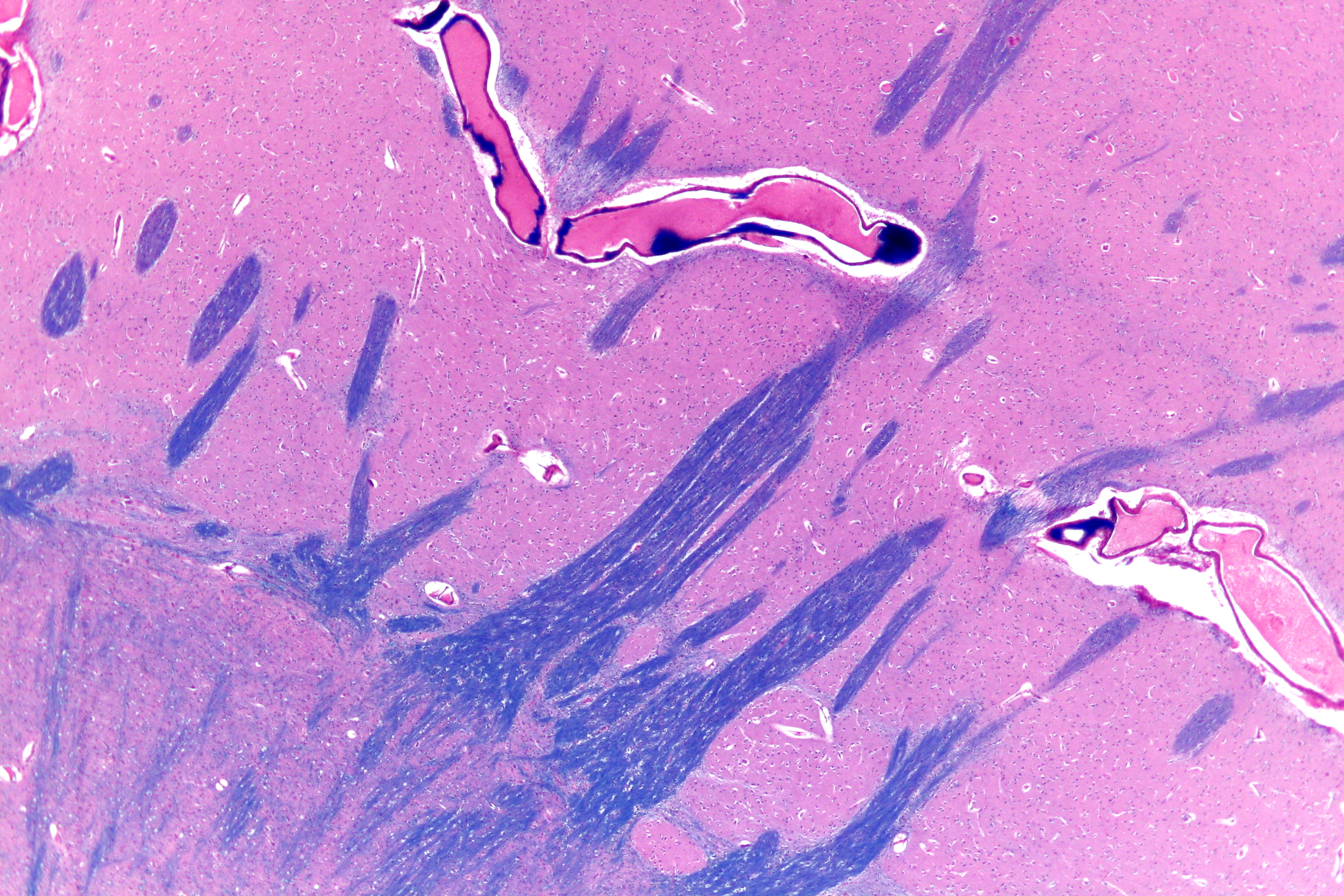
The extreme capsule (Latin: capsula extrema) is a series of nerve tracts between the claustrum and the insular cortex. It is also described as a thin capsule of white matter as association fibres. The extreme capsule is separated from the external capsule by the claustrum, and the extreme capsule separates the claustrum from the insular cortex, and all these lie lateral to the corpus striatum components. From the midline of the brain to the side, the extreme capsule is the outermost from the external capsule and the inner internal capsule. It is most easily visible in a horizontal section, just lateral to the claustrum. Additional images File:Slide6gg.JPG, Extreme capsule File:Slide6kk.JPG, Extreme capsule References External links * Image at neuropat.dote.hu Cerebrum {{neuroanatomy-stub ... [...More Info...] [...Related Items...] OR: [Wikipedia] [Google] [Baidu] |
Human Brain
The human brain is the central organ of the human nervous system, and with the spinal cord makes up the central nervous system. The brain consists of the cerebrum, the brainstem and the cerebellum. It controls most of the activities of the body, processing, integrating, and coordinating the information it receives from the sense organs, and making decisions as to the instructions sent to the rest of the body. The brain is contained in, and protected by, the skull bones of the head. The cerebrum, the largest part of the human brain, consists of two cerebral hemispheres. Each hemisphere has an inner core composed of white matter, and an outer surface – the cerebral cortex – composed of grey matter. The cortex has an outer layer, the neocortex, and an inner allocortex. The neocortex is made up of six neuronal layers, while the allocortex has three or four. Each hemisphere is conventionally divided into four lobes – the frontal, temporal, parietal, and occipital lo ... [...More Info...] [...Related Items...] OR: [Wikipedia] [Google] [Baidu] |
External Capsules
The external capsule is a series of white matter fiber tracts in the brain. These fibers run between the most lateral (toward the side of the head) segment of the lentiform nucleus (more specifically the putamen) and the claustrum. The white matter of the external capsule contains fibers known as corticocortical association fibers. These fibers are responsible for connecting the cerebral cortex to another cortical area. The capsule itself appears as a thin white sheet of white matter. The external capsule is a route for cholinergic fibers from the basal forebrain to the cerebral cortex. The putamen separates the external capsule from the internal capsule medially and the claustrum separates it from the extreme capsule laterally. But the external capsule eventually joins the internal capsule The internal capsule is a white matter structure situated in the inferomedial part of each cerebral hemisphere of the brain. It carries information past the basal ganglia, separating the cau ... [...More Info...] [...Related Items...] OR: [Wikipedia] [Google] [Baidu] |
Putamen
The putamen (; from Latin, meaning "nutshell") is a round structure located at the base of the forebrain (telencephalon). The putamen and caudate nucleus together form the dorsal striatum. It is also one of the structures that compose the basal nuclei. Through various pathways, the putamen is connected to the substantia nigra, the globus pallidus, the claustrum, and the thalamus, in addition to many regions of the cerebral cortex. A primary function of the putamen is to regulate movements at various stages (e.g. preparation and execution) and influence various types of learning. It employs GABA, acetylcholine, and enkephalin to perform its functions. The putamen also plays a role in degenerative neurological disorders, such as Parkinson's disease. History The word "putamen" is from Latin, referring to that which "falls off in pruning", from "putare", meaning "to prune, to think, or to consider". Until recently, most MRI research focused broadly on the basal ganglia as a who ... [...More Info...] [...Related Items...] OR: [Wikipedia] [Google] [Baidu] |
Precuneus
In neuroanatomy, the precuneus is the portion of the superior parietal lobule on the medial surface of each brain hemisphere. It is located in front of the cuneus (the upper portion of the occipital lobe). The precuneus is bounded in front by the marginal branch of the cingulate sulcus, at the rear by the parieto-occipital sulcus, and underneath by the subparietal sulcus. It is involved with episodic memory, visuospatial processing, reflections upon self, and aspects of consciousness. The location of the precuneus makes it difficult to study. Furthermore, it is rarely subject to isolated injury due to strokes, or trauma such as gunshot wounds. This has resulted in it being "one of the less accurately mapped areas of the whole cortical surface". While originally described as homogeneous by Korbinian Brodmann, it is now appreciated to contain three subdivisions. It is also known after Achille-Louis Foville as the ''quadrate lobule of Foville''. The Latin form of was first used ... [...More Info...] [...Related Items...] OR: [Wikipedia] [Google] [Baidu] |
Coronal Section
The coronal plane (also known as the frontal plane) is an anatomical plane that divides the body into Anatomical terms of location#Dorsal and ventral, dorsal and ventral sections. It is perpendicular to the sagittal plane, sagittal and transverse plane, transverse planes. Details The coronal plane is an example of a Anatomical terms of location#General usage, longitudinal plane. For a human, the mid-coronal plane would transect a standing body into two halves (front and back, or anterior and posterior) in an imaginary line that cuts through both shoulders. The description of the coronal plane applies to most animals as well as humans even though humans walk upright and the various planes are usually shown in the vertical orientation. The sternal plane (''planum sternale'') is a coronal plane which transects the front of the Human sternum, sternum. Etymology The term is derived from Latin ''corona'' ('garland, crown'), from Ancient Greek κορώνη (''korōnē'', 'garland, wrea ... [...More Info...] [...Related Items...] OR: [Wikipedia] [Google] [Baidu] |
Cingulate Cortex
The cingulate cortex is a part of the brain situated in the medial aspect of the cerebral cortex. The cingulate cortex includes the entire cingulate gyrus, which lies immediately above the corpus callosum, and the continuation of this in the cingulate sulcus. The cingulate cortex is usually considered part of the limbic lobe. It receives inputs from the thalamus and the neocortex, and projects to the entorhinal cortex via the cingulum. It is an integral part of the limbic system, which is involved with emotion formation and processing, learning, and memory. The combination of these three functions makes the cingulate gyrus highly influential in linking motivational outcomes to behavior (e.g. a certain action induced a positive emotional response, which results in learning). This role makes the cingulate cortex highly important in disorders such as depression and schizophrenia. It also plays a role in executive function and respiratory control. Etymology The term ''cingul ... [...More Info...] [...Related Items...] OR: [Wikipedia] [Google] [Baidu] |
Attention
Attention is the behavioral and cognitive process of selectively concentrating on a discrete aspect of information, whether considered subjective or objective, while ignoring other perceivable information. William James (1890) wrote that "Attention is the taking possession by the mind, in clear and vivid form, of one out of what seem several simultaneously possible objects or trains of thought. Focalization, concentration, of consciousness are of its essence." Attention has also been described as the allocation of limited cognitive processing resources. Attention is manifested by an attentional bottleneck, in terms of the amount of data the brain can process each second; for example, in human vision, only less than 1% of the visual input data (at around one megabyte per second) can enter the bottleneck, leading to inattentional blindness. Attention remains a crucial area of investigation within education, psychology, neuroscience, cognitive neuroscience, and neuropsychology. ... [...More Info...] [...Related Items...] OR: [Wikipedia] [Google] [Baidu] |
Grey Matter
Grey matter is a major component of the central nervous system, consisting of neuronal cell bodies, neuropil (dendrites and unmyelinated axons), glial cells (astrocytes and oligodendrocytes), synapses, and capillaries. Grey matter is distinguished from white matter in that it contains numerous cell bodies and relatively few myelinated axons, while white matter contains relatively few cell bodies and is composed chiefly of long-range myelinated axons. The colour difference arises mainly from the whiteness of myelin. In living tissue, grey matter actually has a very light grey colour with yellowish or pinkish hues, which come from capillary blood vessels and neuronal cell bodies. Structure Grey matter refers to unmyelinated neurons and other cells of the central nervous system. It is present in the brain, brainstem and cerebellum, and present throughout the spinal cord. Grey matter is distributed at the surface of the cerebral hemispheres (cerebral cortex) and of the cerebellu ... [...More Info...] [...Related Items...] OR: [Wikipedia] [Google] [Baidu] |
Lateralization Of Brain Function
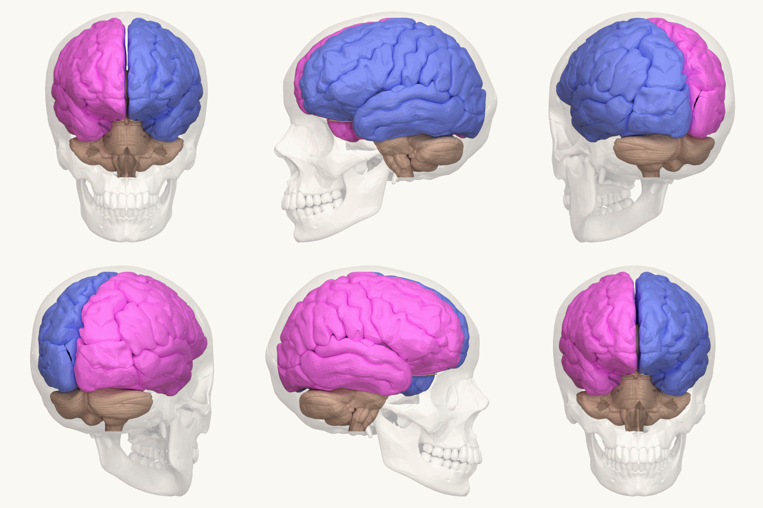
The lateralization of brain function is the tendency for some neural functions or cognitive processes to be specialized to one side of the brain or the other. The median longitudinal fissure separates the human brain into two distinct cerebral hemispheres, connected by the corpus callosum. Although the macrostructure of the two hemispheres appears to be almost identical, different composition of neuronal networks allows for specialized function that is different in each hemisphere. Lateralization of brain structures is based on general trends expressed in healthy patients; however, there are numerous counterexamples to each generalization. Each human's brain develops differently, leading to unique lateralization in individuals. This is different from specialization, as lateralization refers only to the function of one structure divided between two hemispheres. Specialization is much easier to observe as a trend, since it has a stronger anthropological history. The best examp ... [...More Info...] [...Related Items...] OR: [Wikipedia] [Google] [Baidu] |
Sensory Nervous System
The sensory nervous system is a part of the nervous system responsible for processing sensory information. A sensory system consists of sensory neurons (including the sensory receptor cells), neural pathways, and parts of the brain involved in sensory perception. Commonly recognized sensory systems are those for vision, hearing, touch, taste, smell, and balance. Senses are transducers from the physical world to the realm of the mind where people interpret the information, creating their perception of the world around them. The receptive field is the area of the body or environment to which a receptor organ and receptor cells respond. For instance, the part of the world an eye can see, is its receptive field; the light that each rod or cone can see, is its receptive field. Receptive fields have been identified for the visual system, auditory system and somatosensory system. Stimulus :Organisms need information to solve at least three kinds of problems: (a) to maintain an a ... [...More Info...] [...Related Items...] OR: [Wikipedia] [Google] [Baidu] |
Electrophysiology
Electrophysiology (from Greek , ''ēlektron'', "amber" etymology of "electron"">Electron#Etymology">etymology of "electron" , ''physis'', "nature, origin"; and , '' -logia'') is the branch of physiology that studies the electrical properties of biological cells and tissues. It involves measurements of voltage changes or electric current or manipulations on a wide variety of scales from single ion channel proteins to whole organs like the heart. In neuroscience, it includes measurements of the electrical activity of neurons, and, in particular, action potential activity. Recordings of large-scale electric signals from the nervous system, such as electroencephalography, may also be referred to as electrophysiological recordings. They are useful for electrodiagnosis and monitoring. Definition and scope Classical electrophysiological techniques Principle and mechanisms Electrophysiology is the branch of physiology that pertains broadly to the flow of ions (ion current) in biologi ... [...More Info...] [...Related Items...] OR: [Wikipedia] [Google] [Baidu] |
_(18167720666).jpg)


